| SKU | A00284-1 |
|---|---|
| Application | Western Blot |
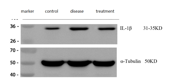
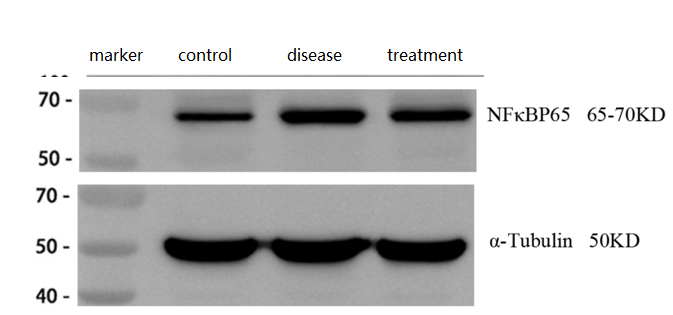
| Sample | mouse brain tissue |
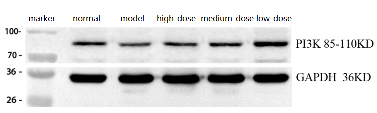
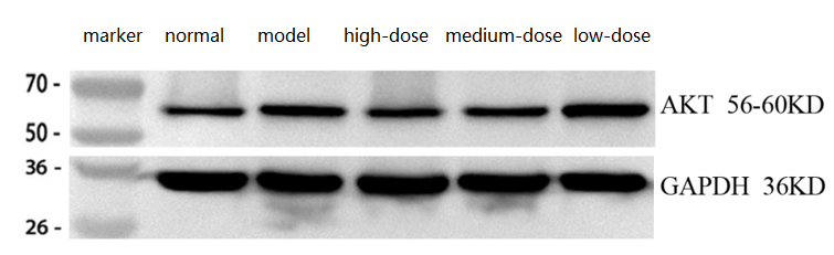
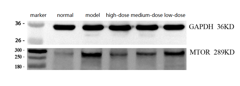
| Sample Processing Description | ① Normal mouse hippocampal tissue, ② Hippocampal tissue from Alzheimer’s disease model mouse, ③ Hippocampal tissue from Alzheimer’s disease model mouse treated with a self-developed drug. Total protein was extracted from all samples. |
| Other Reagents | RIPA lysis buffer, Protease inhibitor, Running buffer, Transfer buffer, Blocking buffer |
| Primary Antibody | NF-kB p65/RELA Antibody Picoband® |
| Primary Incubation | 1:2000, overnight at 4 ℃ |
| Secondary Antibody | HRP Conjugated AffiniPure Goat Anti-Rabbit IgG (H+L) (BA1054) |
| Secondary Incubation | 1:10000, 1 h in RT |
| Detection | Substrate: ECL substrate, Image system: ChemiDoc MP |
| Results Summary | NF-κB p65 (RelA) is one of the most classical and essential subunits of the NF-κB transcription factor family. In Alzheimer’s disease, activation of p65 can initiate neuroprotective programs in neurons. The results showed that p65 expression was significantly increased in the brains of Alzheimer’s disease model mice and decreased after treatment, consistent with the expected outcomes. |