| SKU | PB9026 |
|---|---|
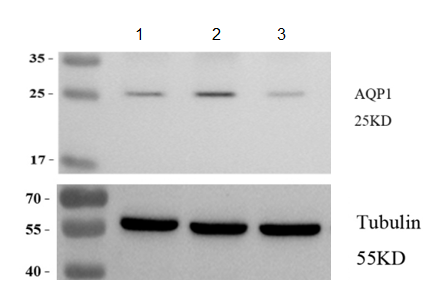
| Application | Western Blot |
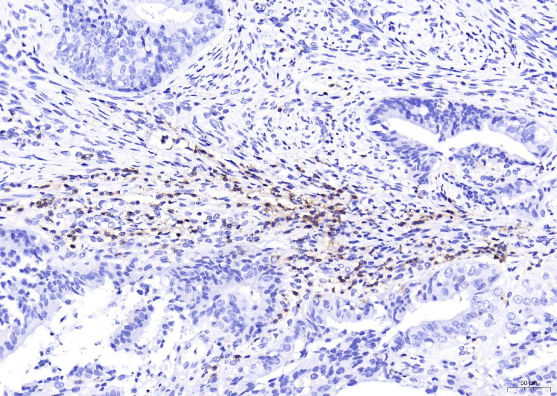
| Sample | Nude mouse tumor tissue |
| Sample Processing Description | Paraffin-embedded tumor tissue sections. |
| Primary Antibody | Ki67/MKI67 Antibody Picoband® |
| Primary Incubation | 1:1000, overnight at 4 ℃ |
| Secondary Antibody | HRP-conjugated Anti-Rabbit IgG Secondary Antibody |
| Secondary Incubation | 1 hour in room temperature |
| Detection | Substrate: ECL reagent, Imaging system:ChemiDoc MP |
| Results Summary | This antibody is highly specific and efficient, with a clean background and no nonspecific bands. The target band has sharp and well-defined edges. |