| SKU | M00566 |
|---|---|
| Application | Western Blot |
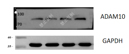
| Sample | Human A549 cells, HCC1833 cells, LU65 cells, PC-9 cells |
| Sample Processing Description | After normal cell culture, total proteins were extracted using RIPA lysis buffer with protease inhibitors. Protein concentration was determined by BCA assay, and then the four samples were adjusted to a uniform concentration of 3 mg/mL using loading buffer. |
| Other Reagents | RIPA lysis buffer, Protease inhibitor, Electrophoresis buffer, Transfer buffer, Blocking buffer |
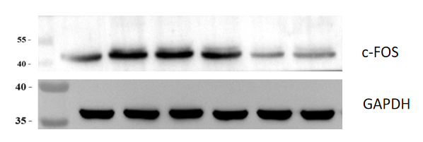
| Primary Antibody | c-Fos Rabbit Monoclonal Antibody |
| Primary Incubation | 1:2000, overnight at 4 ℃ |
| Secondary Antibody | HRP Goat Anti-Rabbit IgG |
| Secondary Incubation | 1:10000, 1 hour in room temperature |
| Detection | Substrate: ECL, Imaging system:ChemiDoc MP |
| Results Summary | The purpose of the experiment is to examine the differences in ADAM10 protein expression among different types of human lung cancer cells, in order to select suitable cells for subsequent experiments. |