| SKU | PA1054 |
|---|---|
| Application | Immunohistochemistry |
| Sample | mouse skin tissue |
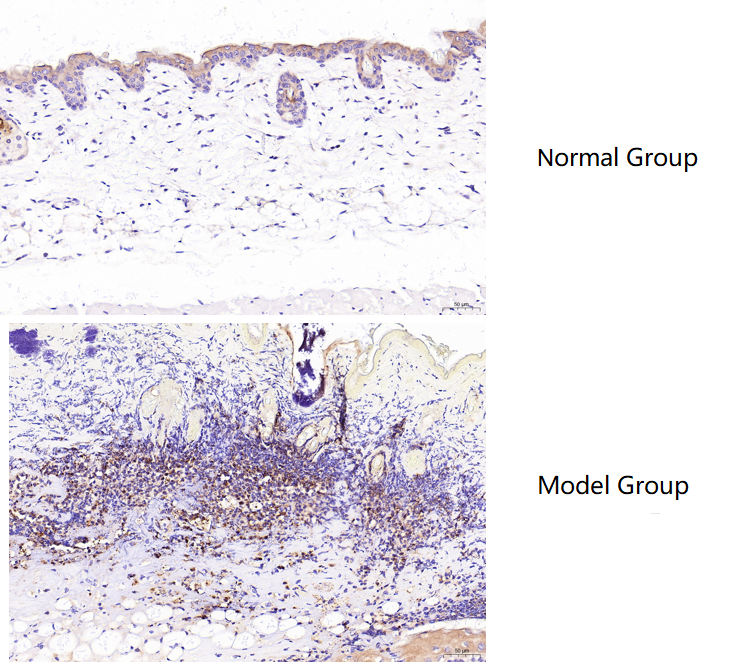
| Sample Processing Description | Male BALB/c mice aged 6–8 weeks were used. (1) Normal dorsal skin was collected from untreated mice. (2) Burn injury was induced on the dorsal skin using a burn device. After one week of housing, hair was removed with depilatory cream. Skin samples were collected from normal mice (dorsal skin) and burned mice (burned area), fixed in formalin for 72 hours, and embedded in paraffin for sectioning./td> |
| Other Reagents | Tris-EDTA Antigen Retrieval Buffer (50×, pH 9.0), DAB Chromogen Kit |
| Primary Antibody | Myeloperoxidase/MPO Antibody Picoband® |
| Primary Incubation | 1:100, overnight at 4 ℃ |
| Secondary Antibody | Polymer Anti-Rabbit IgG–HRP Immunohistochemistry Kit |
| Detection | Imaging system:Leica DM2500 |
| Results Summary | MPO (myeloperoxidase) is a lysosomal enzyme mainly present in neutrophils and monocytes/macrophages. Its core function is to generate reactive oxygen species, and therefore it plays a “double-edged sword” role in innate immune defense and inflammation-related diseases. MPO is present at very low levels in normal skin, but in burned skin, a large number of macrophages accumulate around the injured tissue, accompanied by a significant increase in MPO expression. Based on the immunohistochemical results, the staining is clear and the findings are consistent with expectations. |