| SKU | M00865 |
|---|---|
| Application | Western blot |
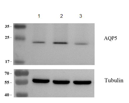
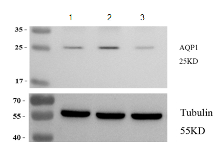
| Sample | MADB106 rat mammary carcinoma cells |
| Sample Processing Description | MADB106 cells under normal culture conditions , MADB106 cells treated with agonist , MADB106 cells treated with inhibitor |
| Other Reagents | RIPA Lysis Buffer, Protease Inhibitor, Resolving Gel Solution ,Transfer Buffer ,Blocking Buffer |
| Primary Antibody | AQP1 Rabbit Monoclonal Antibody |
| Primary Incubation | 1:3000, overnight at 4 ℃ |
| Secondary Antibody | 1:10,000, HRP-conjugated Goat Anti-Rabbit IgG |
| Secondary Incubation | 1h at RT |
| Detection | Substrate: ECL substrate, Imaging system:ChemiDoc MP |
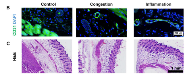
| Results Summary | AQP1 (Aquaporin 1) is the first discovered water channel protein, whose primary function is efficient water transport. Recent studies have revealed that AQP1 plays a critical role in cancer progression, promoting tumor growth in breast cancer by enhancing angiogenesis and cell migration. In this experiment, AQP1 protein levels were markedly increased in cells treated with the agonist and significantly decreased in cells treated with the inhibitor. |