| SKU | M02226 |
|---|---|
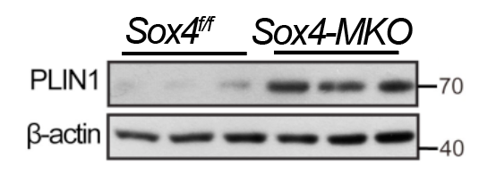
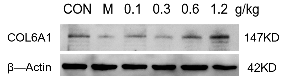
| Application | Western Blot |
| Sample | Mouse hippocampus tissue |
| Sample Processing Description | The mouse hippocampus was lysed in RIPA buffer supplemented with a protease inhibitor cocktail. After protein quantification, samples were mixed with 5× protein loading buffer and denatured by heating at 100°C for 10 minutes. Five microliters of each protein sample were loaded per lane onto SDS-PAGE. |
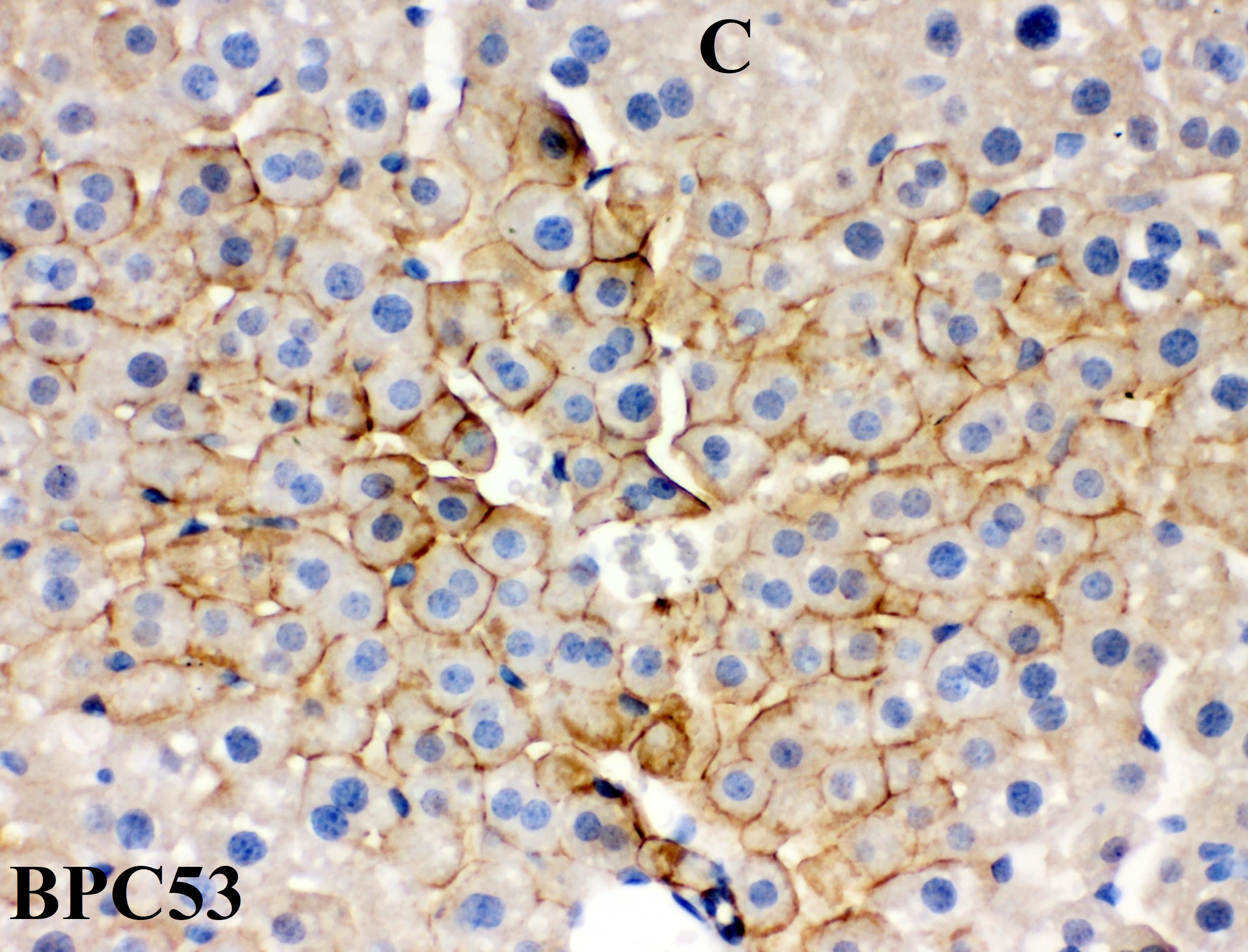
| Primary Antibody | Anti-Collagen VI COL6A1 Rabbit Monoclonal Antibody |
| Primary Incubation | overnight at 4 ℃ |
| Secondary Antibody | HRP-conjugated Anti-Rabbit IgG Secondary Antibody |
| Secondary Incubation | 1 hour in room temperature |
| Detection | Substrate: Ultra-sensitive ECL luminescent reagent (Cat# AR1191), Imaging system:ChemiDoc MP |
| Results Summary | Western blot analysis was performed using the COL6A1 antibody to detect COL6A1 protein expression in the mouse hippocampus. Although minor non-specific bands were observed, they did not affect the trend analysis, indicating that the antibody is suitable for detecting the target protein in this tissue. |