| SKU | M00371 |
|---|---|
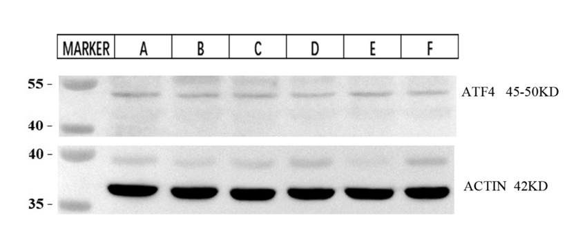
| Application | Western Blot |
| Sample | Porcine intestinal tissue |
| Sample Processing Description | Intestinal tissues from different segments of pigs infected with swine fever. |
| Other Reagents | RIPA lysis buffer, Protease inhibitor, Electrophoresis buffer, Transfer buffer, Blocking buffer |
| Primary Antibody | ATF4 Rabbit Monoclonal Antibody |
| Primary Incubation | 1:2000, overnight at 4 ℃ |
| Secondary Antibody | HRP Goat Anti-Rabbit IgG |
| Secondary Incubation | 1:10000, 1 hour in room temperature |
| Detection | Substrate: ECL, Imaging system:ChemiDoc MP |
| Results Summary | ATF4 is a key regulator of the integrated stress response. When cells encounter conditions such as oxidative stress or endoplasmic reticulum stress, the expression of ATF4 is rapidly induced and upregulated. The aim of this experiment is to determine the expression levels of ATF4 protein in different intestinal segments infected with classical swine fever. |


