| SKU | PB9449 |
|---|---|
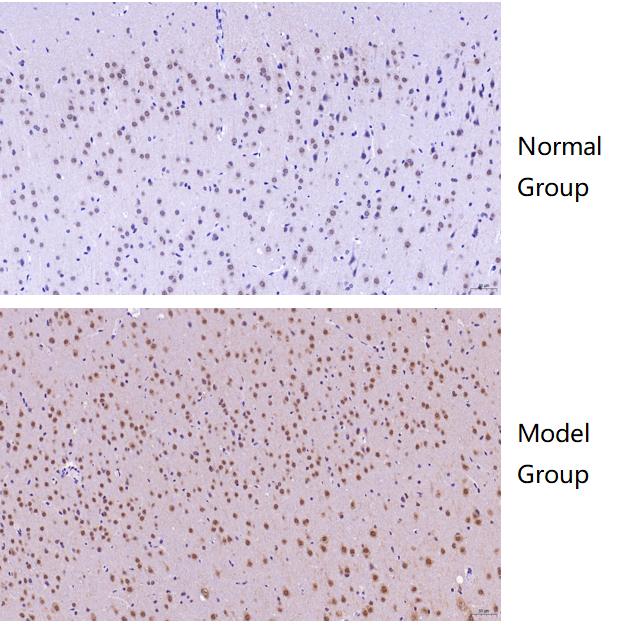
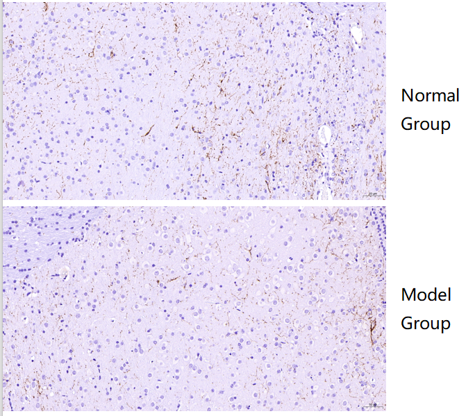
| Application | Immunohistochemistry |
| Sample | Normal mouse brain and Alzheimer’s model mouse brain tissue |
| Sample Processing Description | Paraffin-embedded normal mouse brain and Alzheimer’s model mouse brain. |
| Other Reagents | Goat serum, DAB |
| Primary Antibody | Tyrosine Hydroxylase/TH Antibody Picoband® |
| Primary Incubation | 1:500, overnight at 4 ℃ |
| Secondary Antibody | Two-step IHC kit |
| Secondary Incubation | 37 minutes in 37 ℃ |
| Detection | Imaging system:Leica DM2500 |
| Results Summary | TH is a marker of catecholaminergic neurons, specifically labeling dopaminergic, noradrenergic, and adrenergic neurons. IHC results showed that TH expression in the medulla was markedly lower in Alzheimer’s mouse compared to normal mouse. |