| SKU | PB9233 |
|---|---|
| Application | Western Blot |
| Sample | mouse uterine tissue |
| Sample Processing Description | Mouse uterine tissues were minced and further disrupted by sonication, then lysed on ice for 1 hour using RIPA buffer. After centrifugation, the supernatant was collected and quantified using the BCA method. The appropriate amount of loading buffer was added, and the samples were boiled in a water bath to denature the proteins. Finally, 15 μL of each protein sample was loaded into each lane of the SDS-PAGE gel. |
| Other Reagents | 5% Non-fat milk |
| Primary Antibody | Transferrin Receptor/TFRC Antibody |
| Primary Incubation | 1:1000, overnight at 4 ℃ |
| Secondary Antibody | HRP-conjugated Anti-Rabbit IgG Secondary Antibody |
| Secondary Incubation | 1 hour in room temperature |
| Detection | Substrate: ECL reagent, Imaging system:Tanon |
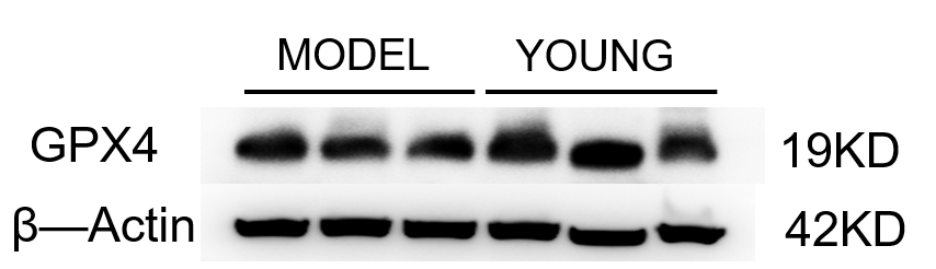
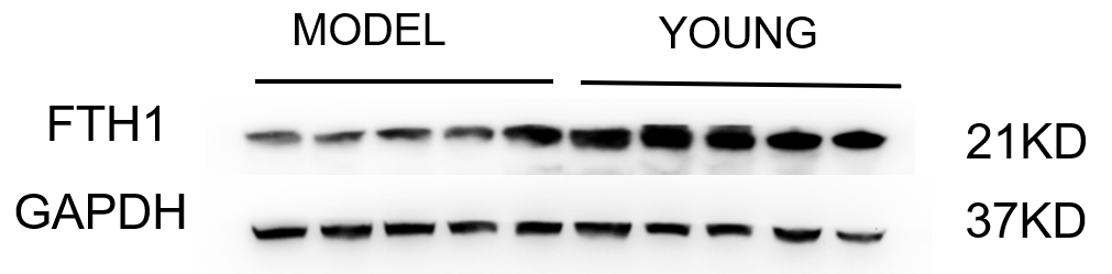
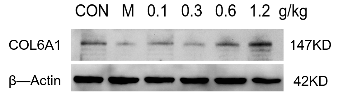
| Results Summary | The CD71 antibody was used to detect protein expression in mouse uterine tissue. The bands were clear with no nonspecific signals, and after recovery and reuse, the antibody still showed good performance. |