| SKU | M00656 |
|---|---|
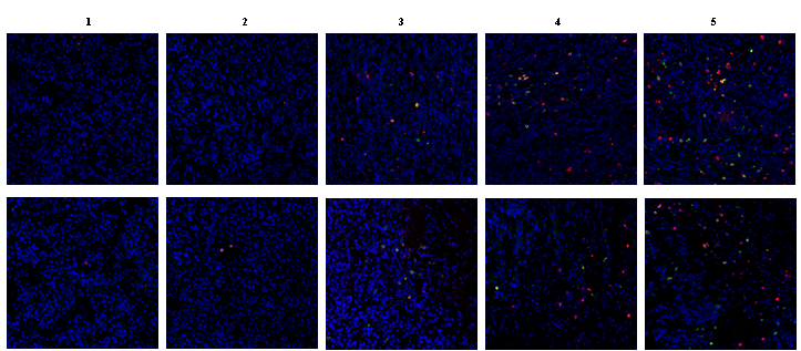
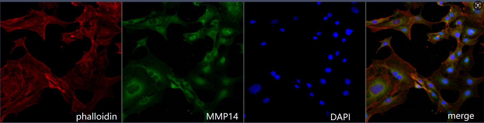
| Application | Immunofluorenscence |
| Sample | Mouse MC-8 cells |
| Sample Processing Description | 4% paraformaldehyde for 15 minutes |
| Primary Antibody | Anti-MMP14/Mt1 Mmp Rabbit Monoclonal Antibody |
| Primary Incubation | 1:200, overnight at 4 ℃ |
| Blocking Agent | Goat serum |
| Secondary Antibody | DyLight 488-conjugated Goat Anti-Rabbit IgG. |
| Secondary Incubation | Incubate at room temperature for 1 hour |
| Detection | Laser confocal microscopy |
| Results Summary | I will purchase Boster products again and recommend them to my classmates and colleagues. |