| SKU | DZ41074 |
|---|---|
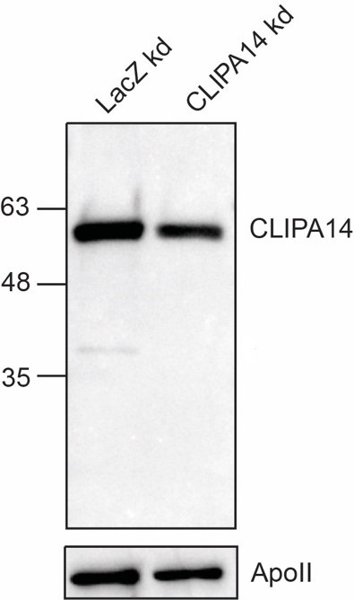
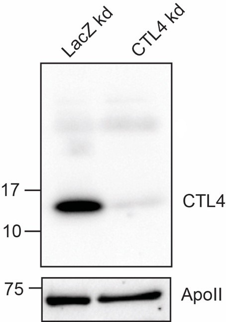
| Application | Western Blot |
| Sample | β-galactosidase gene knockdown mosquito hemolymph, CTL4 knockdown mosquito hemolymph |
| Primary Incubation | +4°C overnight |
| Secondary Incubation | For 1 hour at room temperature |
| Tertiary Incubation | 1:1000 |
| Detection | Clarity Max western ECL substrate |
| Results Summary | Image showing the specificity of CTL4 antibody. Lane 1 includes hemolymph extracted from mosquitoes silenced for the bacterial β-galactosidase gene. Lane 2 contains hemolymph extracted from mosquitoes silenced for the CTL4 gene. |