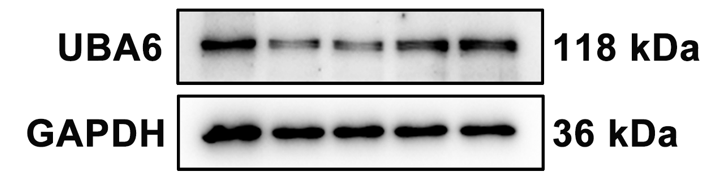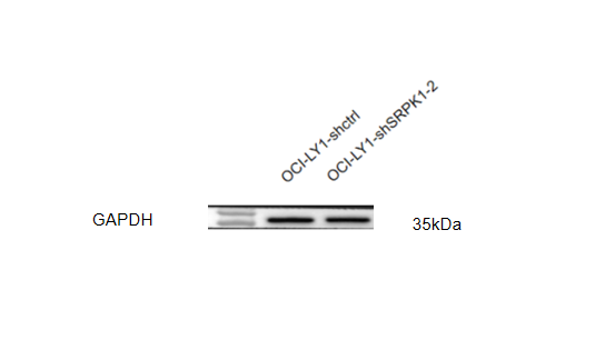
| SKU | A00227-1 |
|---|---|
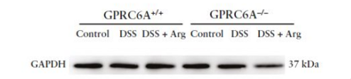
| Application | Western Blot |
| Sample | human OCI-LY1 cellls |
| Sample Processing Description | Cell samples were lysed by sonication in RIPA buffer containing protease and phosphatase inhibitors, followed by centrifugation for 10 minutes. The supernatant was mixed with loading buffer at a 4:1 ratio, boiled for 10 minutes, and 15 μL of protein was loaded per well. |
| Other Reagents | 5% non-fat milk |
| Primary Antibody | GAPDH Antibody Picoband® |
| Primary Incubation | 1:5000, overnight at 4 ℃ |
| Secondary Antibody | HRP Conjugated AffiniPure Goat Anti-Rabbit IgG (H+L) |
| Secondary Incubation | 1 h in RT |
| Detection | Substrate: ECL substrate, Image system:ChemiDoc MP |
| Results Summary | In OCI-LY1 cells treated with different concentrations of SPHINX31 for 24 h, GAPDH expression remained stable with no significant differences, showing clear bands and a clean background without nonspecific signals. |