| SKU | MA1045 |
|---|---|
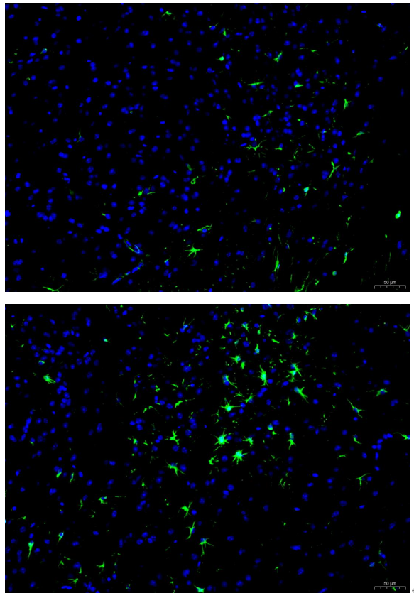
| Application | Immunofluorescence |
| Sample | rat spinal tissue |
| Sample Processing Description | Paraffin-embedded transverse sections of rat spinal cord were prepared after formalin fixation. |
| Other Reagents | Tris-EDTA Antigen Retrieval Buffer (50×, pH 9.0), DAPI |
| Primary Antibody | GFAP Antibody (Monoclonal, G-A-5) |
| Primary Incubation | 1:200, overnight at 4 ℃ |
| Secondary Antibody | Goat Anti-Mouse IgG (H+L) Secondary Antibody, Fluoro488 Conjugated |
| Secondary Incubation | 45 minutes in 37℃ |
| Detection | Imaging system:Leica DM2500 |
| Results Summary | GFAP is a marker of astrocytes. In this experiment, immunostaining for GFAP was used to label astrocytes in the gray matter of the spinal cord to observe their distribution, density, and morphology. The results showed that the labeled protoplasmic astrocytes in the gray matter had short, thick, and highly branched processes with rough surfaces, forming a dense “bushy” network tightly surrounding neuronal cell bodies and synapses, consistent with theoretical expectations and demonstrating excellent staining. |