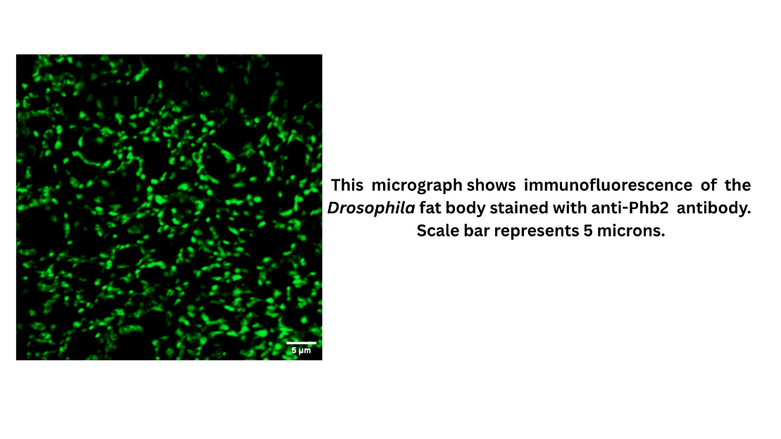
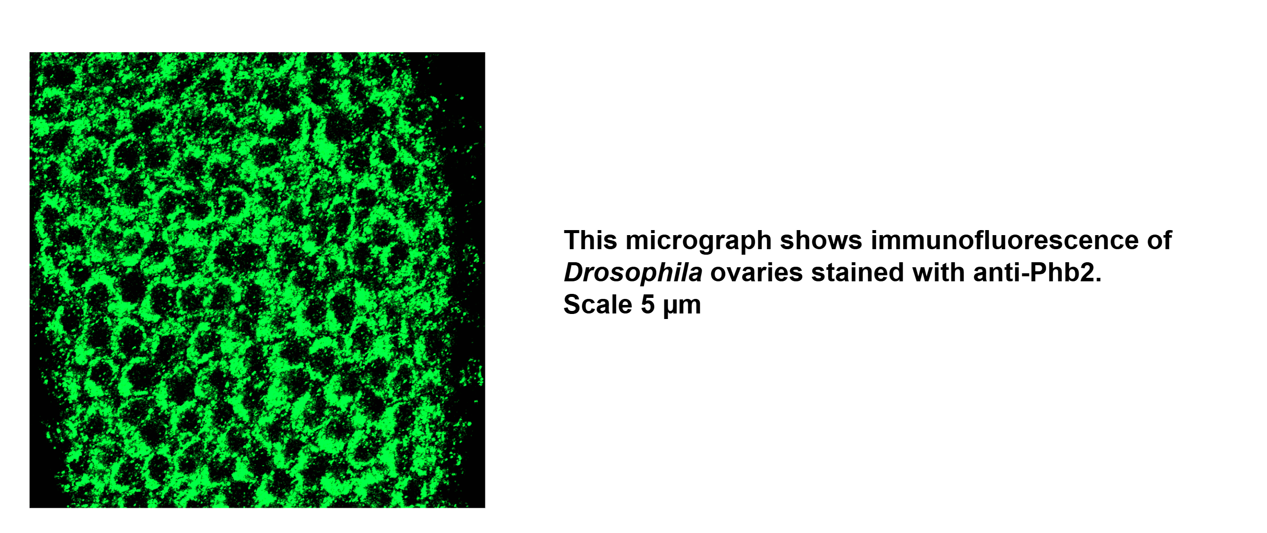
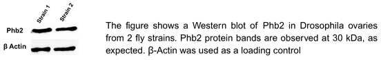
Customer Testimonials

Peer Reviews from Real Labs

Browse peer-submitted results linked to specific products—ratings, images, and practical notes included. Contribute your own result to strengthen the evidence base and claim rewards after verification.

Video Interviews

See real workflows and decision points—what researchers tried, what worked, and why. Share your story in a short interview and claim rewards!

Get Interview Rewards

Filter by category, reactivity, and host to find the most relevant peer results.

Western blot analysis of Drosophila ovaries from three fly strains showed comparable Phb2 protein levels across all samples. Consistent bands at 30 kDa confirmed expected molecular weight, with β-Actin used as a loading control to verify equal protein loa

Excellent, submitted by on
SKU DZ41317
Application Western Blot
Sample Drosophila Ovaries
Sample Processing Description Dissection, Homogenization, Sample boiling in 1X Lamelli, SDS-PAGE, Standard Western Blotting
Primary Antibody Anti-Fruit fly Phb2 Antibody
Primary Incubation 1:1000 in 5% BSA and 0.1% PBST. Either 2 hours at RT or Overnight at 4 Degrees
Secondary Antibody 1:10000 Anti-rabbit/mouse IgG horseradish peroxidase-conjugated
Secondary Incubation 1 hour at room temperature
Detection Biorad ChemiDoc
Results Summary Western blot analysis of Drosophila ovaries from three fly strains showed comparable Phb2 protein levels across all samples. Consistent bands at 30 kDa confirmed expected molecular weight, with β-Actin used as a loading control to verify equal protein loading.

Drosophila fatbody showed good staining of Phb2.

Excellent, submitted by on
SKU DZ41317
Application Immunofluorescence
Sample Drosophila Fat body and Ovaries
Sample Processing Description Dissection, Fixation in 4%PFA, Permeabilization in PBS-TX, Blocking, Primary, Washes,Secondary, Washes,Nuclei Staining, Slide Preparation
Primary Antibody Anti-Fruit fly Phb2 Antibody
Primary Incubation 1:200 in 2%BSA and 0.5% PBS-TX for Ovaries, 1:100 in 2%BSA and 0.1% PBS-TX for Fat body
Secondary Antibody 1:1000 Anti-Rabbit/Mouse Alexa Fluor 488/Cy3/Cy5
Secondary Incubation 1 hour at room temperature
Detection Zeiss LSM 900 Confocal Microscope with Airyscan 2
Results Summary Drosophila fatbody showed good staining of Phb2.

This was only optimization experiment and still needs validation through necessary experiments.

Excellent, submitted by on
SKU DZ41315
Application Immunofluorescence
Sample Drosophila Fat body and Ovaries
Sample Processing Description Dissection, Fixation in 4%PFA, Permeabilization in PBS-TX, Blocking, Primary, Washes,Secondary, Washes,Nuclei Staining, Slide Preparation
Primary Antibody Anti-Fruit fly Marf Antibody
Primary Incubation 1:200 in 2%BSA and 0.5% PBS-TX for Ovaries, 1:100 in 2%BSA and 0.1% PBS-TX for Fat body
Secondary Antibody 1:1000 Anti-Rabbit/Mouse Alexa Fluor 488/Cy3/Cy5
Secondary Incubation 1 hour at room temperature
Detection Zeiss LSM 900 Confocal Microscope with Airyscan 2
Results Summary This was only optimization experiment and still needs validation through necessary experiments.

Antibody Optimized and validated

Excellent, submitted by on
SKU DZ41314
Application Western Blot
Sample Drosophila Ovaries
Sample Processing Description Dissection, Homogenization, Sample boiling in 1X Lamelli, SDS-PAGE, Standard Western Blotting
Primary Antibody Anti-Fruit Fly Drp1 Antibody
Primary Incubation 1:1000 in 5% BSA and 0.1% PBST Either 2 hours at RT or Overnight at 4 Degrees
Secondary Antibody 1:10000 Anti-rabbit/mouse IgG horseradish peroxidase-conjugated
Secondary Incubation 1 hour at room temperature
Other Reagents used ECL Clarity Substrate and enzyme for Chemiluminescence
Detection Biorad ChemiDoc
Results Summary In Drosophila ovaries, loss of Drp1 function was confirmed. Western blot analysis validated Drp1 knockdown, showing reduced Drp1 protein levels (~85 kDa) in ovaries expressing Drp1 miRNA, with β-Actin serving as a loading control.

Antibody Optimized and validated

Excellent, submitted by on
SKU DZ41314
Application Immunofluorescence
Sample Drosophila Fat body
Sample Processing Description Dissection, Fixation in 4%PFA, Permeabilization in PBS-TX, Blocking, Primary, Washes, Secondary, Washes, Nuclei Staining, Slide Preparation
Primary Antibody Anti-Fruit Fly Drp1 Antibody
Primary Incubation 1:100 in 2%BSA and 0.1% PBS-TX for Fat body
Secondary Antibody 1:1000 Anti-Rabbit/Mouse Alexa Fluor 488/Cy3/Cy5
Secondary Incubation 1 hour at room temperature
Detection Zeiss LSM 900 Confocal Microscope with Airyscan 2
Results Summary In Drosophila fatbody, Drp1 was seen as punctate structures, as expected from the literature on mitochondria.

The SP antibody demonstrated strong performance across all tested dilutions, producing clear and specific signal detection.

Excellent, submitted by on
SKU DZ41720
Application Western Blot
Sample HEK293T whole cell lysates
Sample Processing Description HEK293T cells were transfected with a plasmid expressing SP only (and not Env or Rem) and lysed using RIPA buffer. A total of 80 µg of protein was resolved on a 4–12% gradient SDS-PAGE gel and transferred to a membrane. The membrane was blocked for 1 hour at room temperature with 5% non-fat milk in PBS-Tween (PBS-T). After washing three times with 1× PBS-T (10 minutes each), the membrane was incubated overnight at 4 °C with varying dilutions ofthe SP antibody prepared in 2% non-fat milk (1:500; 1:1000; 1:2000). Following three additional washes with PBS-T (10 minutes each), the membrane was incubated with anti-rabbit secondary antibody, washed again three times, and developed using ECL Plus.
Primary Incubation The membrane was incubated with the SP primary antibody (1:500; 1:1000; 1:2000) overnight at 4 °C.
Secondary Antibody Anti-rabbit-HRP-conjugated secondary antibody
Secondary Incubation Dilution: 1:25,000 in PBS-T/2% milk
Other Reagents used 1× RIPA lysis buffer
1× PBS-T washing buffer (T = 1% Tween 20)
Non-fat milk (for blocking and antibody dilution)
Anti-rabbit HRP-conjugated secondary antibody
Detection Signal was developed using ECL Plus chemiluminescent substrate
Results Summary The SP antibody demonstrated strong performance across all tested dilutions, producing clear and specific signal detection. Dilutions of 1:1000 and 1:2000 yielded the most optimal results, showing minimal background compared to 1:500, which highlights the antibody’s high specificity and sensitivity at these concentrations. To ensure equal protein loading across lanes, the membrane was also probed with a β-actin antibody, confirming equal sample loading and validating the observed signal intensity for SP. Overall, these results demonstrate that the SP antibody is reliable for detecting SP in RIPA-lysed HEK293T samples.

The CLIPB4 antibody was efficiently used to detect CLIPB4 protein secreted in the mosquito hemolymph. The protein migrates at ~40 kDa in SDS-PAGE.

Excellent, submitted by on
CLIPB4 antibody Western blot result
SKU DZ33989-1
Application Western Blot
Sample β-galactosidase gene knockdown mosquito hemolymph, CLIPB4 knockdown mosquito hemolymph
Primary Incubation Overnight at 4°C
Secondary Incubation For 1 hour at room temperature
Secondary Antibody Dilution 1:3000
Detection Clarity Max Western ECL substrate
Results Summary Image showing the specificity of CLIPB4 antibody. Lane 1 includes hemolymph extracted from mosquitoes silenced for the bacterial β-galactosidase gene. Lane 2 contains hemolymph extracted from mosquitoes silenced for the CLIPB4 gene.

The CTL4 antibody was efficiently used to detect CTL4 protein secreted in the mosquito hemolymph. The protein migrates at ~15 kDa in SDS-PAGE.

Excellent, submitted by on
SKU DZ41074
Application Western Blot
Sample β-galactosidase gene knockdown mosquito hemolymph, CTL4 knockdown mosquito hemolymph
Primary Incubation +4°C overnight
Secondary Incubation For 1 hour at room temperature
Tertiary Incubation 1:1000
Detection Clarity Max western ECL substrate
Results Summary Image showing the specificity of CTL4 antibody. Lane 1 includes hemolymph extracted from mosquitoes silenced for the bacterial β-galactosidase gene. Lane 2 contains hemolymph extracted from mosquitoes silenced for the CTL4 gene.

The CLIPA14 antibody was efficiently used to detect CLIPA14 protein secreted in the mosquito hemolymph. The protein migrates at ~59 kDa in SDS-PAGE.

Excellent, submitted by on
SKU DZ41073
Application Western Blot
Sample β-galactosidase gene knockdown mosquito hemolymph, CLIPA14 knockdown mosquito hemolymph
Primary Incubation +4°C overnight
Secondary Incubation For 1 hour at room temperature
Tertiary Incubation 1:3000
Detection Clarity Max western ECL substrate
Results Summary Image showing the specificity of CLIPA14 antibody. Lane 1 includes hemolymph extracted from mosquitoes silenced for the bacterial β-galactosidase gene. Lane 2 contains hemolymph extracted from mosquitoes silenced for the CLIPA14 gene.

"Works well for immunofluorescence"

Excellent, submitted by on

Source: Customer Feedback Submission

"Works well for immunofluorescence"

--Jason Tennessen

SKU DZ41223
Application IF
Sample Drosophila larval fat body

"We’ve verified the specificity of this custom antibody that Boster generated for us. The antibody "Anti-Fruit Fly Gpdh1 Antibody (DZ41223)” works well for immunofluorescence. A 1:100 dilution of this antibody stains fat body tissues of wild-type larvae but not Gpdh1 mutant. I’m happy to advertise this antibody to the fly community."