This website uses cookies to ensure you get the best experience on our website.


- Table of Contents
Plan AARS2 paraffin-section IHC using the cytoplasmic tissue pattern as a reference (HPA tissue IHC). The catalog antibody’s colon-section example documents primary-antibody incubation and DAB detection (datasheet A10160-2).

Expected localisation, validated protocols, controls and antibodies — the at-a-glance facts below, then the full design guide.
| Expected localisation | Cytoplasmic tissue staining (HPA tissue IHC); mitochondrial protein (UniProt) | |
| Staining pattern | Glandular cells show cytoplasmic staining (HPA tissue IHC) | |
| Antigen retrieval | EDTA pH 8.0 HIER, heat-mediated (datasheet A10160-2) | |
| Positive control | Adrenal gland+4 more · see all | |
| Negative control | None in HPA (detected in all 45 tissues); use no-primary + isotype controls |
| Fixation | Keep fixation consistent across sections (standard IHC practice; not target-specific) | |
| Caveat | Colon endothelial cells can stain strongly (HPA tissue IHC) | |
| Regulation | Expression regulation not annotated (UniProt) | |
| Isoform / epitope | No isoforms annotated; mature chain starts at aa 24 (UniProt) |
The catalog antibody’s IHC-P protocol is paired with one published AARS2 IHC protocol for human colon adenocarcinoma specimens (PMC12982081).
| Sample | Paraffin-embedded human colon cancer tissue; fixative not specified (datasheet A10160-2) |
| Fixation | Image fixative and duration unreported (datasheet A10160-2); verify before use. |
| Sectioning | 4–5 µm sections on charged slides (standard) |
| Deparaffinisation | Xylene, graded ethanol series to water (standard) |
| Antigen retrieval | Heat retrieval: EDTA pH 8.0 (datasheet A10160-2); 20 min, 95–100 °C (standard) |
| Peroxidase block | 3% H2O2, 10 min, room temperature (standard) |
| Blocking | 10% goat serum (datasheet A10160-2) |
| Primary antibody | Rabbit anti-AARS2, 1:50 recommended; image 1:200 (datasheet A10160-2) |
| Primary incubation | Overnight at 4 °C (datasheet A10160-2) |
| Detection | HRP-conjugated secondary, DAB chromogen (datasheet A10160-2) |
| Counterstain | Hematoxylin, blue, dehydrate and mount (standard) |
| Expected result | AARS2-positive staining in glandular cells of adrenal gland (HPA tissue IHC: High). HPA tissue profile: General cytoplasmic expression. No signal in the no-primary control. |
AARS2 is mitochondrial and has no transmembrane segment (UniProt Q5JTZ9 localisation and topology). In paraffin sections, expect cytoplasmic staining in glandular, respiratory epithelial and other HPA-positive cells (HPA: general cytoplasmic expression; listed High cells). HPA rates the tissue IHC pattern Approved, with medium consistency between staining and RNA expression (HPA: tissue IHC reliability). Interpret intensity alongside cell identity and compartment.
| Cytoplasmic staining in adrenal or duodenal glandular cells, with little nuclear staining. | This fits the expected compartment and a documented High cell population (UniProt Q5JTZ9: mitochondrion; HPA: High in these glandular cells). Chromogenic IHC may show cytoplasmic signal without resolving individual mitochondria (general IHC practice). |
| Signal confined mainly to nuclei, cell borders or extracellular material. | Those distributions conflict with the mitochondrial assignment (UniProt Q5JTZ9; HPA: mitochondrial ICC-IF). Treat them as suspect; compare with a known-positive section and control staining before assigning AARS2 (general IHC practice). |
| Strong staining in a cell population absent from the selected HPA High entries. | Check cell identity and assay controls before calling cross-reactivity: HPA reports low tissue specificity, and its supplied High list is not an exhaustive negative map (HPA: tissue RNA specificity and listed IHC entries). Endogenous detection activity can also mimic chromogen signal (general IHC practice). |
| Diffuse colour covers cells, stroma and the blank area around tissue. | A field-wide deposit is less persuasive than cell-associated cytoplasmic staining (HPA: general cytoplasmic expression). Assess nonspecific detection, wash quality and chromogen development with appropriate controls (general IHC practice). |
| No staining in a section containing a documented High cell population. | First verify that the expected cells are present and the control run worked (HPA: listed High cells; general IHC practice). A negative slide alone cannot establish absent AARS2; HPA's IHC rating is Approved with medium staining–RNA consistency (HPA: reliability). |
| Compartment | UniProt places AARS2 in mitochondria, and HPA ICC-IF shows mitochondrial localisation (UniProt Q5JTZ9; HPA: subcellular ICC-IF). Judge paraffin IHC at the cytoplasmic level when organelles cannot be resolved (general IHC practice). |
| Choice of reference cells | Adrenal, appendix, breast, cervix and duodenum glandular cells; bronchial respiratory epithelial cells; cerebellar granular-layer cells; and colonic endothelial cells are listed High (HPA: tissue IHC). Adipocytes are listed Low (HPA: adipose tissue). |
| Strength of antibody evidence | HPA035636 has Approved IHC status; the supplied record gives no IHC status for HPA061396 (HPA: antibody validation). Both have Enhanced ICC status (HPA: antibody validation). Do not transfer an ICC validation rating to paraffin IHC. |
| Target structure and epitope information | UniProt reports no transmembrane segment and a chain spanning residues 24–985 (UniProt Q5JTZ9: topology and processing). The supplied sources do not map the IHC antibody epitope or establish target-specific retrieval or fixation sensitivity. |
| Chromogenic detection | Endogenous enzyme activity, nonspecific secondary binding and excessive chromogen development can create misleading colour (general IHC practice). Use assay-matched negative controls to distinguish detection background from the HPA cytoplasmic pattern. |
| Situation | Likely cause | Next action |
|---|---|---|
| A documented High tissue is blank. | The expected cell population may be absent from the section, or the staining run may have failed (HPA: High cell listings; general IHC practice). | Confirm cell identity and review the run's positive control; then check retrieval, antibody dilution and detection using the validated IHC procedure (general IHC practice). No AARS2-specific retrieval condition is supplied. |
| Colour is strongest outside cells. | Background deposition or nonspecific detection can obscure the expected cytoplasmic pattern (HPA: general cytoplasmic expression; general IHC practice). | Inspect an assay-matched negative control, washing and chromogen development; reassess whether staining remains cell-associated after background is controlled (general IHC practice). |
| Only nuclei or cell borders stain. | The observed compartment disagrees with the mitochondrial localisation (UniProt Q5JTZ9; HPA: subcellular ICC-IF). | Compare compartment placement in a known-positive section and check control staining before scoring the result as AARS2 (HPA: listed High cells; general IHC practice). |
| Unexpected cells stain more strongly than the intended reference cells. | Cell misidentification, nonspecific binding or endogenous detection activity are possibilities (general IHC practice); HPA's High list is not a complete negative list (HPA: tissue IHC). | Recheck morphology and the assay-matched negative control. Describe the observed cells and intensity without declaring the unlisted population AARS2-negative (general IHC practice; HPA: low tissue specificity). |
| Dense chromogen hides the intracellular pattern. | Overdevelopment can prevent useful compartment scoring (general IHC practice). | Review detection and development settings against the validated IHC procedure, then score a section where cytoplasm and nuclei remain distinguishable (general IHC practice). |
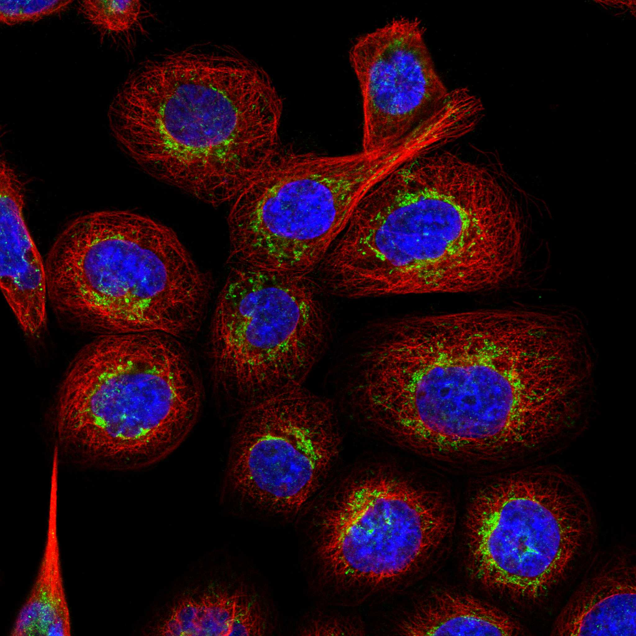
| What should an IF/ICC image show? | HPA reports enhanced mitochondrial ICC-IF localisation for both listed antibodies (HPA: subcellular ICC-IF and antibody validation). | Look for mitochondrial signal in the cell image; use the separate IF/ICC guide for its workflow. The ICC evidence supports localisation but supplies no paraffin IHC protocol (HPA: antibody validation). |
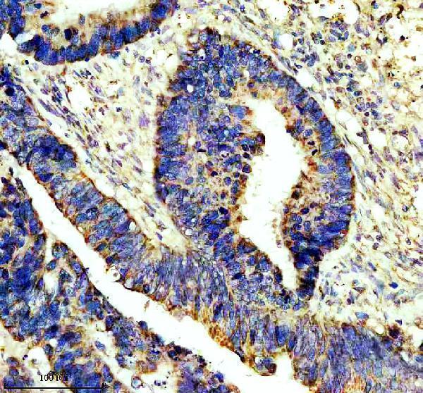
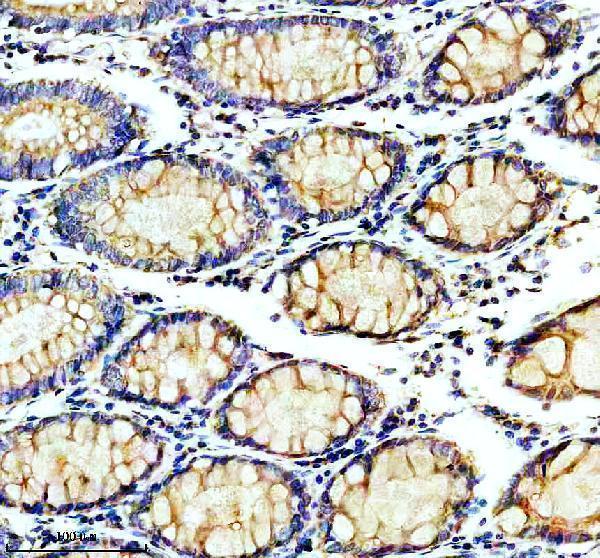
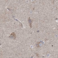


Comprehensive Human Protein Atlas IHC scoring per tissue (reliability: Approved — Medium consistency between antibody staining and RNA expression data.). Rows are taken directly from the HPA tissue chart — click any row's HPA link to view the source.
| Tissue | Cell type | Level | Evidence | Source |
|---|---|---|---|---|
| Adrenal gland | Glandular cells | High | Protein (IHC) | HPA → |
| Appendix | Glandular cells | High | Protein (IHC) | HPA → |
| Breast | Glandular cells | High | Protein (IHC) | HPA → |
| Bronchus | Respiratory epithelial cells | High | Protein (IHC) | HPA → |
| Cerebellum | Cells in granular layer | High | Protein (IHC) | HPA → |
| Tissue | Cell type | Level | Evidence | Source |
|---|---|---|---|---|
| None in HPA: AARS2 is detected in all 45 scored tissues. Use a no-primary (secondary-only) and an isotype control instead. | ||||
Use the catalog antibody’s paraffin-section IHC evidence to guide retrieval and detection, then assess AARS2 staining against its expected mitochondrial localisation.
A10160-2 has paraffin-section IHC images from human colon cancer and colon, and rat and mouse ovary (catalog IHC captions). No IF/ICC data are supplied (catalog applications; IF images).
A10160-2 is listed for IHC in human, mouse and rat (catalog applications; reactivity). Its IHC captions show paraffin sections of human colon cancer, human colon, rat ovary and mouse ovary (catalog IHC captions).
Which to pick: Choose A10160-2 for paraffin-section tissue IHC: it is a rabbit polyclonal antibody with an IHC application and images from those tissues (catalog host; dilution record; applications; IHC captions). The captions do not report a fixative (catalog IHC captions). No listed SKU has an IF/ICC application or image, so there is no evidence-backed IF/ICC choice; for cross-species IHC, A10160-2 has human, mouse and rat reactivity and corresponding tissue images (catalog applications; IF images; reactivity; IHC captions).