This website uses cookies to ensure you get the best experience on our website.


- Table of Contents
Plan chromogenic ABCA7 IHC on paraffin sections, using bone marrow hematopoietic cells as a strong reference (HPA tissue IHC). The guide covers fixation, staining interpretation and a 5 μg/mL starting concentration for the IHC-P catalog antibody (datasheet: A03163).

Expected localisation, validated protocols, controls and antibodies — the at-a-glance facts below, then the full design guide.
| Expected localisation | Cytoplasmic in marrow and immune cell subsets (HPA tissue IHC) | |
| Staining pattern | Cytoplasmic staining in marrow and immune cell subsets (HPA tissue IHC) | |
| Antigen retrieval | Citrate pH 6.0 HIER, 95–98 °C, 20 min (rule: cytoplasmic / membrane antigen) | |
| Positive control | Bone marrow+4 more · see all | |
| Negative control | Adipose tissue+4 more · see all |
| Fixation | Keep paraffin-section fixation consistent across samples. (standard IHC practice; not target-specific) | |
| Caveat | Presumed off-target staining may confound scoring (HPA tissue IHC) | |
| Regulation | Enriched in myelo-lymphatic tissues (UniProt) | |
| Isoform / epitope | Two isoforms; map extracellular vs cytoplasmic epitopes (UniProt) |
The catalog antibody’s IHC-P protocol is followed by two published ABCA7 protocols for paraffin sections (PMC6176408; PMC2976870).
| Sample | Tissue sections; selected-image fixative not specified (standard IHC workflow) |
| Fixation | Image fixative and duration unreported (datasheet A03163); verify before use. |
| Sectioning | 4–5 µm sections on charged slides (standard) |
| Deparaffinisation | Xylene, graded ethanol series to water (standard) |
| Antigen retrieval | Heat-induced epitope retrieval in citrate buffer, pH 6.0, 20 min at 95–98 °C (standard rule: cytoplasmic / membrane antigen) |
| Peroxidase block | 3% H2O2, 10 min, room temperature (standard) |
| Blocking | 10% normal serum of the secondary host, 30 min, room temperature (standard) |
| Primary antibody | Rabbit anti-ABCA7, 5 μg/mL (datasheet A03163) |
| Primary incubation | Overnight at 4 °C (standard) |
| Detection | HRP-polymer secondary, DAB chromogen 5–10 min (standard) |
| Counterstain | Hematoxylin, blue, dehydrate and mount (standard) |
| Expected result | ABCA7-positive staining in hematopoietic cells of bone marrow (HPA tissue IHC: High). HPA tissue profile: Cytoplasmic expression in bone marrow and in subsets of immune cells in several tissues. No signal in the no-primary control. |
ABCA7 is a multipass membrane protein that can also appear in intracellular compartments (UniProt Q8IZY2 topology and subcellular location). In paraffin-section IHC, expect cytoplasmic staining in bone marrow hematopoietic cells and subsets of immune cells; staining also occurs at ciliary rootlets in selected epithelia (HPA tissue IHC). HPA rates the tissue staining profile Enhanced but reports presumed off-target binding, so judge signal by cell type and compartment together (HPA tissue IHC).
| Strong cytoplasmic staining in bone marrow hematopoietic cells. | This matches a reported high-signal population and is a useful positive reference (HPA tissue IHC: High in bone marrow hematopoietic cells). Score the hematopoietic cells specifically; staining across every structure in the section would require a separate explanation (HPA tissue IHC; general IHC practice). |
| Strong staining concentrated at ciliary rootlets in fallopian tube or nasopharynx. | This matches the reported high-signal location in ciliated cells (HPA tissue IHC: High at ciliary rootlets). The restricted rootlet pattern matters as much as intensity: staining spread evenly through unrelated cells does not reproduce that observation (HPA tissue IHC; general IHC practice). |
| Predominantly nuclear signal, or uniform staining of every compartment. | A nuclear-dominant pattern does not match the reported membrane, Golgi and cytoplasmic locations (UniProt Q8IZY2 subcellular location; HPA subcellular). Treat it as suspect and check controls and detection background before assigning it to ABCA7 (general IHC practice). |
| Strong signal in a cell population recorded as not detected by HPA, such as adipocytes. | This conflicts with the reported IHC pattern for that population (HPA tissue IHC: adipocytes Not detected). Cross-reactivity is plausible because HPA notes presumed off-target binding; endogenous detection activity is another general chromogenic-IHC possibility (HPA tissue IHC; general IHC practice). |
| Diffuse color across tissue, or no staining in bone marrow hematopoietic cells. | Diffuse color obscures the compartment and cell-type pattern; an unstained reported high-signal population leaves the assay uninformative (HPA tissue IHC: bone marrow High; general IHC practice). Review background and positive controls before interpreting weaker or negative samples (general IHC practice). |
| Tissue and cell selection | Bone marrow hematopoietic cells and fallopian-tube or nasopharyngeal ciliary rootlets are reported High; placenta decidual cells and spleen red-pulp cells are Medium (HPA tissue IHC). Choose and score the named cell population, since a tissue-level label does not describe every cell in that section (HPA tissue IHC). |
| Topology and compartment | ABCA7 has 15 annotated transmembrane segments and extracellular regions at residues 43–549 and 1264–1537 (UniProt Q8IZY2 topology). Its membrane association supports compartment-aware interpretation, but topology alone cannot predict a chromogenic epitope's visibility in a paraffin section (UniProt Q8IZY2 topology; general IHC practice). |
| Cell state | Resting macrophages are reported to contain ABCA7 in the cytoplasm, probably Golgi and endosomes; stimulation with C1q or apoptotic cells shifts it toward ruffles and phagocytic cups (UniProt Q8IZY2 subcellular location). A change in distribution within macrophages can therefore be biologically plausible without establishing a change in total abundance (UniProt Q8IZY2 subcellular location). |
| Antibody evidence | HPA041564 has Enhanced IHC validation, while the HPA tissue profile reports medium staining-versus-RNA consistency and disregarded presumed off-target binding (HPA antibodies; HPA tissue IHC). Match the observed cell and compartment pattern before accepting isolated strong staining as specific (HPA tissue IHC; general IHC practice). |
| Isoforms and processing | UniProt records 2 isoforms and a full-length chain spanning residues 1–2146, with no annotated propeptide (UniProt Q8IZY2 isoforms and processing). Without an antibody epitope in the supplied evidence, this record cannot establish which isoform a stain detects (UniProt Q8IZY2 isoforms; general IHC practice). |
| IF/ICC Q&A: where should ABCA7 appear? | Mainly at the plasma membrane and Golgi apparatus, with additional cell-junction signal in the reported ICC-IF images (HPA subcellular). This is an IF/ICC localisation answer; interpret paraffin-section chromogenic staining against the tissue IHC cell patterns above (HPA subcellular; HPA tissue IHC). |
| Situation | Likely cause | Next action |
|---|---|---|
| No signal in bone marrow hematopoietic cells. | A reported High population has failed as a positive reference (HPA tissue IHC). The failure's specific technical cause is undetermined from these sources. | Check that the hematopoietic population is present, then review antigen retrieval, primary-antibody and detection steps against the assay controls (HPA tissue IHC; general IHC practice). |
| Signal is strong in adipocytes or lymph-node germinal-center cells. | Both populations are reported Not detected; presumed off-target binding is documented in the HPA profile (HPA tissue IHC). | Compare the same run with the reported positive cell patterns and a negative control; investigate detection background before scoring this signal as ABCA7 (HPA tissue IHC; general IHC practice). |
| Color appears broadly in tissue despite little cell-specific pattern. | Nonspecific background or endogenous chromogenic activity can obscure a restricted stain (general IHC practice). | Inspect the no-primary control, blocking and washes; where the detection chemistry requires it, address endogenous enzyme activity (general IHC practice). |
| A section looks negative although the tissue is listed as ABCA7 positive. | HPA positives are assigned to named populations: for example, spleen red-pulp cells are Medium, rather than every spleen cell (HPA tissue IHC). | Find the annotated population and score it separately; compare it with a reported High positive control before calling the assay negative (HPA tissue IHC; general IHC practice). |
| Macrophage staining shifts from cytoplasm toward the cell edge. | Macrophage state may alter localisation: resting cytoplasmic signal and stimulated ruffle or phagocytic-cup localisation are reported (UniProt Q8IZY2 subcellular location). | Check whether the edge signal follows cell structures and remains cell restricted; record compartment separately from staining intensity (UniProt Q8IZY2 subcellular location; general IHC practice). |
| Nuclear staining dominates while expected cells lack a clear cytoplasmic or membrane pattern. | Nuclear-dominant staining is unsupported by the supplied ABCA7 localisation annotations (UniProt Q8IZY2 subcellular location; HPA subcellular). | Review controls, detection background and scoring boundaries; withhold a positive ABCA7 call until a supported cell and compartment pattern is visible (general IHC practice). |
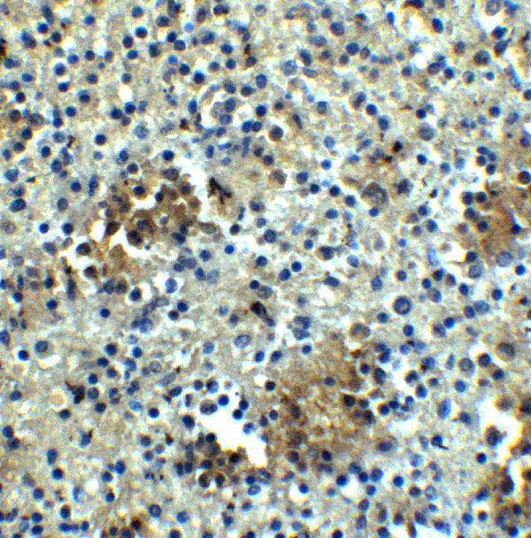
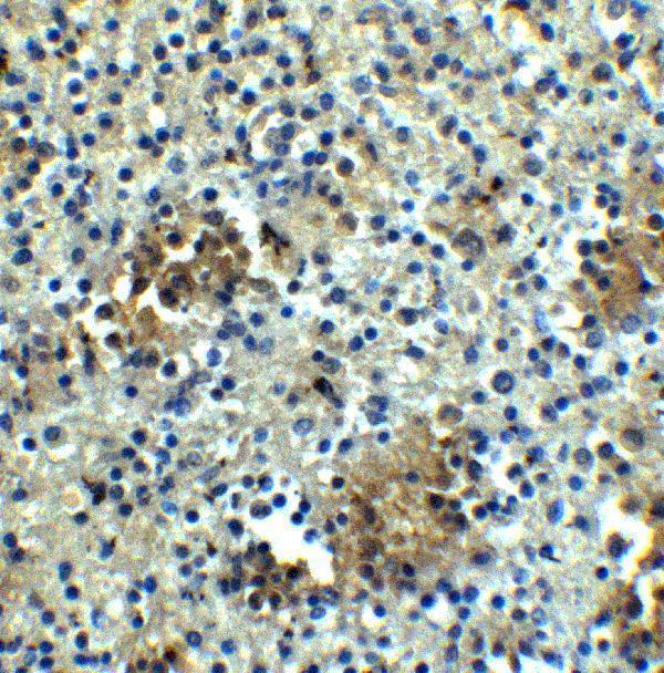
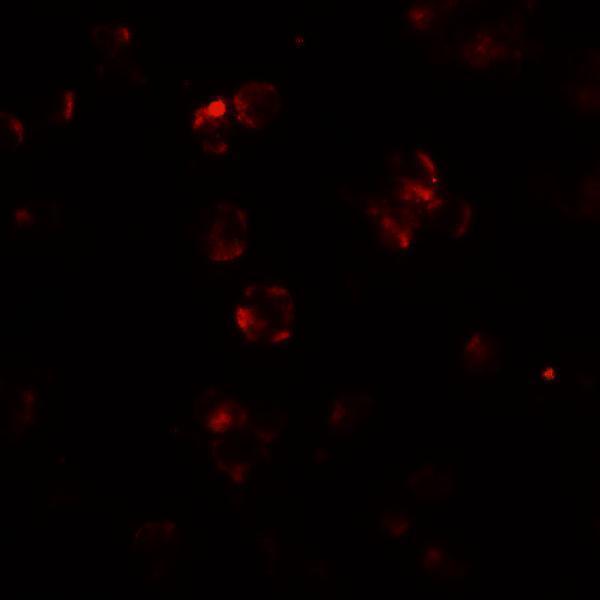
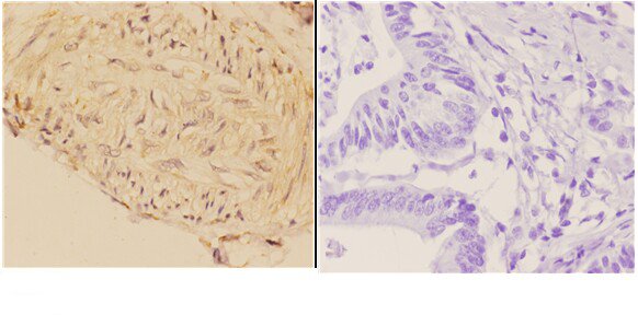
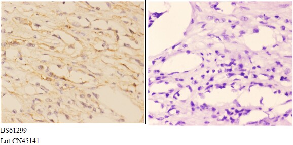


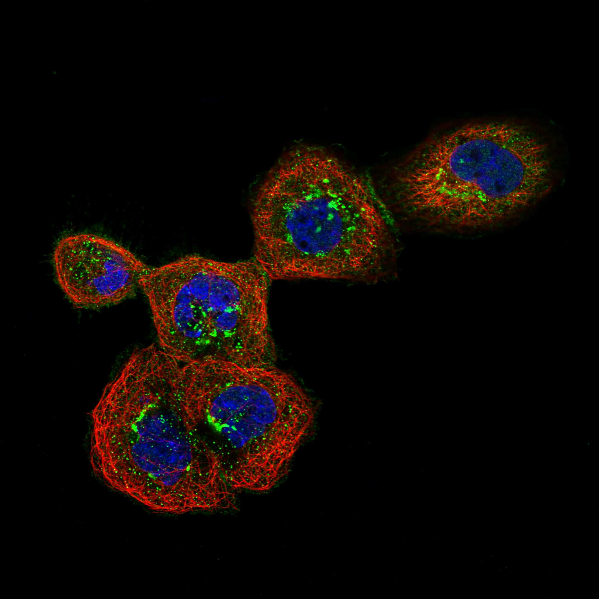
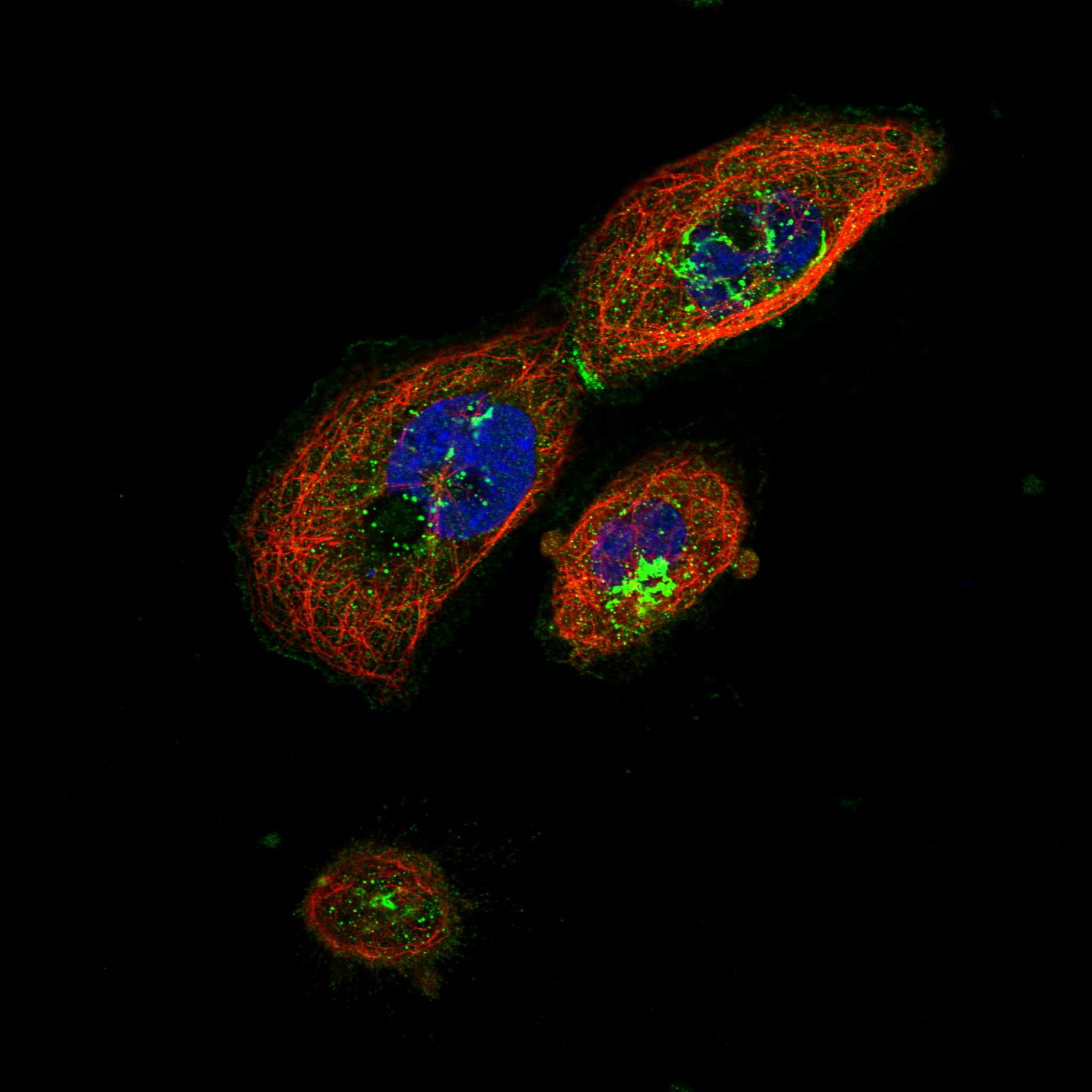
Comprehensive Human Protein Atlas IHC scoring per tissue (reliability: Enhanced — Medium consistency between antibody staining and RNA expression data. Presumed off target binding observed and disregarded.). Rows are taken directly from the HPA tissue chart — click any row's HPA link to view the source.
| Tissue | Cell type | Level | Evidence | Source |
|---|---|---|---|---|
| Bone marrow | Hematopoietic cells | High | Protein (IHC) | HPA → |
| Fallopian tube | Ciliated cells (ciliary rootlets) | High | Protein (IHC) | HPA → |
| Nasopharynx | Ciliated cells (ciliary rootlets) | High | Protein (IHC) | HPA → |
| Placenta | Decidual cells | Medium | Protein (IHC) | HPA → |
| Spleen | Cells in red pulp | Medium | Protein (IHC) | HPA → |
| Tissue | Cell type | Level | Evidence | Source |
|---|---|---|---|---|
| Adipose tissue | Adipocytes | Not detected | Protein (IHC) | HPA → |
| Breast | Adipocytes | Not detected | Protein (IHC) | HPA → |
| Caudate | Glial cells | Not detected | Protein (IHC) | HPA → |
| Cerebellum | Cells in granular layer | Not detected | Protein (IHC) | HPA → |
| Cerebral cortex | Endothelial cells | Not detected | Protein (IHC) | HPA → |
Troubleshoot ABCA7 staining in paraffin sections by checking retrieval, cell identity, compartment and controls before comparing staining intensity.
Two anti-ABCA7 antibodies have human tissue IHC images; A03163 also has IF images in 293 cells and human spleen, while A03163-2 lists human, mouse and rat reactivity (catalog images, applications and reactivity).
A03163 lists IHC-P, IF and ICC, with IHC shown in human spleen and IF shown in 293 cells and human spleen (catalog applications and image captions). A03163-2 lists IHC and shows membrane staining in paraffin-embedded human colon and kidney carcinoma at 1:50 (catalog applications and IHC image captions).
Which to pick: For paraffin-section IHC, choose A03163-2: its own images show human colon and kidney carcinoma sections, although the fixative is unreported (A03163-2 IHC image captions). For IF/ICC, choose A03163, which lists both applications and has IF images; its IHC image shows human spleen tissue without reporting the tissue fixative (A03163 applications and image captions). For mouse or rat samples, A03163-2 is the listed cross-species option, but its pictured IHC samples are human; both antibodies list rabbit host with no clone designation (catalog reactivity, host, clone and IHC image captions).