ABCF2 / ATP-binding cassette sub-family F member 2 · IHC design guide

Design Immunohistochemistry for ABCF2

Plan chromogenic IHC on paraffin sections using the cytoplasmic tissue pattern as a reference (HPA tissue IHC). This guide covers tissue controls, scoring, and the catalog antibody’s 1:50–1:200 IHC dilution range (datasheet).

Evidence assembled Oct 2026 · For research use; verify linked source records and product datasheet before use
Immunohistochemistry protocol sheet for ABCF2 (IHC for ABCF2): expected localisation Cytoplasm in most tissues (HPA tissue IHC), antibody A10873-1, validated IHC image, and IHC protocol steps
Printable ABCF2 IHC protocol sheet — expected localisation Cytoplasm in most tissues (HPA tissue IHC), antibody A10873-1, controls and protocol steps. Open the full ABCF2 IHC guide →

ABCF2 Immunohistochemistry Experimental Design Guide

Expected localisation, validated protocols, controls and antibodies — the at-a-glance facts below, then the full design guide.

Must know before staining
Expected localisation Cytoplasm in most tissues (HPA tissue IHC)
Staining pattern Cytoplasmic staining in cells across most tissues (HPA tissue IHC)
Antigen retrieval Citrate pH 6.0 HIER, 95–98 °C, 20 min (rule: cytoplasmic / membrane antigen)
Positive control ⓘ Adrenal gland+4 more · see all
Negative control ⓘ Adipose tissue+4 more · see all
Important caveats
Reasons your staining may differ from the expected pattern.
Fixation Keep formalin fixation consistent across sections. (standard IHC practice; not target-specific)
Caveat Staining may reflect proteins from more than one gene (HPA tissue IHC)
Regulation No expression regulation annotated (UniProt)
Isoform / epitope 2 isoforms; epitope differences are unknown (UniProt)
Section 1

Recommended ABCF2 IHC & IF Protocols

The catalog antibody’s IHC-P protocol (datasheet) is followed by published ABCF2 IHC methods for uterine, ovarian and liver cancer specimens (PMC2584961; PMC5509336; PMC6998385).

Recommended immunohistochemistry (IHC-P) protocol parameters
SampleParaffin-embedded mouse lung tissue; fixative not specified (datasheet A10873-1)
FixationImage fixative and duration unreported (datasheet A10873-1); verify before use.
Sectioning4–5 µm sections on charged slides (standard)
DeparaffinisationXylene, graded ethanol series to water (standard)
Antigen retrievalHeat-induced epitope retrieval in citrate buffer, pH 6.0, 20 min at 95–98 °C (standard rule: cytoplasmic / membrane antigen)
Peroxidase block3% H2O2, 10 min, room temperature (standard)
Blocking10% normal serum of the secondary host, 30 min, room temperature (standard)
Primary antibodyRabbit anti-ABCF2, 1:50-1:200 (datasheet A10873-1)
Primary incubationOvernight at 4 °C (standard)
DetectionHRP-polymer secondary, DAB chromogen 5–10 min (standard)
CounterstainHematoxylin, blue, dehydrate and mount (standard)
Expected resultABCF2-positive staining in glandular cells of adrenal gland (HPA tissue IHC: Medium). HPA tissue profile: Cytoplasmic expression in most tissues. No signal in the no-primary control.
💡Decision noteStart with citrate pH 6.0 retrieval at 95–98 °C for 20 min (page retrieval); heated citrate retrieval was also reported for ABCF2 staining (PMC6998385).
Section 2

What Is the Expected ABCF2 Staining Pattern?

In paraffin-section IHC, expect predominantly cytoplasmic ABCF2 staining across many tissues, including colon glandular cells, pancreatic exocrine glandular cells and cerebral cortex neurons (HPA: tissue IHC). HPA rates tissue IHC “Approved” but reports medium agreement with RNA data and cautions that the antibodies target proteins from more than one gene (HPA: tissue IHC). UniProt gives no subcellular annotation and reports no transmembrane segment (UniProt Q9UG63 topology).

What am I looking at on my slide?
Cytoplasmic chromogen in colon glandular cells or pancreatic exocrine glandular cells, at moderate intensity.This matches the reported compartment and cell populations; both examples are scored Medium (HPA: tissue IHC). Score the relevant cells and their cytoplasm rather than treating every stained area as equivalent (general IHC practice).
Predominantly nuclear or sharply membrane-like staining, with little cytoplasmic signal.That distribution differs from HPA’s cytoplasmic tissue profile (HPA: tissue IHC). Check counterstain boundaries, detection background and antibody controls before assigning it to ABCF2 (general IHC practice). UniProt supplies no compartment annotation that would establish an alternative location (UniProt Q9UG63).
Strong staining in a population reported as Not detected, such as adipocytes or cardiomyocytes.This conflicts with those specific HPA observations, but “Not detected” is not an absolute biological absence (HPA: tissue IHC). Consider cross-reactivity or endogenous chromogenic activity; HPA explicitly cautions that its antibodies target proteins from more than one gene (HPA: tissue IHC; general IHC practice).
Diffuse chromogen coats tissue, empty spaces or multiple compartments without clear cell boundaries.Such a pattern cannot establish the reported cytoplasmic localisation (HPA: tissue IHC). Review background in the negative detection control, blocking, washes and chromogen development before scoring cells (general IHC practice).
No visible cytoplasmic signal in an adequately represented colon glandular or pancreatic exocrine compartment.Both populations have Medium HPA staining, making them useful comparisons, though an individual section is not guaranteed to stain (HPA: tissue IHC). Check tissue identity, control staining and the IHC antibody workflow before calling the sample ABCF2-negative (general IHC practice).
💡Expected ABCF2 appearanceA plausible positive is Medium cytoplasmic staining in colon glandular or pancreatic exocrine glandular cells (HPA: tissue IHC); dominant nuclear staining or strong signal in HPA Not detected populations warrants control-based review (HPA: tissue IHC; general IHC practice).
How each factor affects the staining
Tissue and cell populationHPA reports cytoplasmic expression in most tissues and low tissue RNA specificity, yet staining varies by cell population: cortical neurons are Medium, whereas cerebellar granular-layer cells are Not detected (HPA: tissue IHC). Match the scored compartment to the sampled anatomy.
Strength of antibody evidenceThree listed antibodies have IHC “Approved” status, while the tissue profile reports medium consistency with RNA and cautions about targeting proteins from multiple genes (HPA: antibodies; HPA: tissue IHC). Treat a matching pattern as supportive, not uniquely identifying ABCF2.
Topology and protein formsUniProt reports no transmembrane segment, no signal peptide or propeptide, one chain spanning residues 1–623, and two isoforms (UniProt Q9UG63). These facts do not identify the antibody epitope or predict different IHC staining between isoforms.
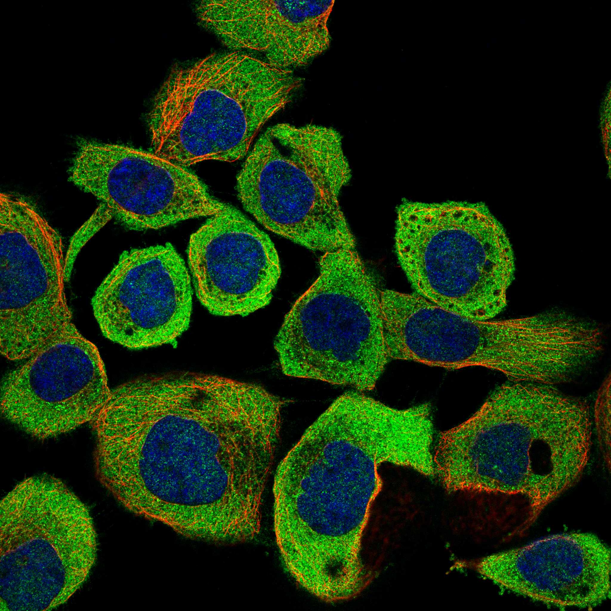
IF/ICC Q&A: Where should signal appear?HPA calls cytosol the main location, with uncertainty, and shows ICC-IF images from A-431, U-251MG and U2OS (HPA: subcellular). Its multi-gene antibody caution applies to that localisation; use the separate IF/ICC guide for assay design (HPA: subcellular).
Why is my staining missing, weak or wrong?
SituationLikely causeNext action
Known-positive tissue lacks cytoplasmic staining.Tissue representation, detection failure or an unsuitable run condition may explain the result (general IHC practice); target-specific fixation sensitivity is unreported in the supplied UniProt and HPA records.Confirm colon glandular or pancreatic exocrine cells are present, inspect the run’s positive control, and review the antibody’s IHC-P instructions for retrieval and dilution (HPA: tissue IHC; general IHC practice).
Most cells show uniform brown haze.Nonspecific detection or excessive chromogen development can obscure compartment boundaries (general IHC practice).Compare a negative detection control; then review blocking, washes, detection reagents and development time using the established IHC workflow (general IHC practice). Score only distinguishable cellular staining.
Signal is mainly nuclear or forms a crisp membrane rim.The appearance conflicts with the cytoplasmic tissue profile, although morphology or background may contribute (HPA: tissue IHC; general IHC practice).Inspect the counterstain and cell boundaries at higher magnification, compare controls, and repeat interpretation with the expected cytoplasmic compartment in view (general IHC practice).
Adipocytes or cardiomyocytes stain strongly.HPA reports these cells as Not detected; antibody cross-reactivity or endogenous detection activity is possible (HPA: tissue IHC; general IHC practice).Check negative detection controls and cellular localisation. Consider an independently validated antibody when specificity is central to the conclusion (general IHC practice; HPA: antibodies).
Weak signal appears in a tissue expected to stain.HPA intensity depends on the sampled population: hippocampal neurons are Low, while cerebral cortex neurons are Medium (HPA: tissue IHC). A weak field alone does not establish assay failure.Verify the exact cell population and compare a Medium HPA reference population processed in the same run before changing conditions (HPA: tissue IHC; general IHC practice).
IF/ICC localisation seems inconsistent with the IHC slide.HPA describes the ICC-IF cytosolic assignment as uncertain and cautions about antibodies targeting multiple genes (HPA: subcellular). Section morphology can also complicate compartment scoring (general IHC practice).Interpret each assay with its own controls and compare cytoplasmic versus cytosolic localisation cautiously; consult the separate IF/ICC guide for that assay (HPA: tissue IHC; HPA: subcellular; general IHC practice).

Sample controls for ABCF2 IHC & IF

🧪Run colon first: glandular cells should stain at the HPA Medium level (HPA: colon glandular cells, Medium); use cervix glandular cells as a comparison with Not detected staining (HPA: cervix glandular cells, Not detected). On the colon slide, adjacent cells without convincing specific staining can serve as internal negative references, but their ABCF2 status is not established by the supplied HPA row (HPA: colon glandular cells, Medium).
Positive control tissue: Adrenal gland (Glandular cells, HPA Medium)
Negative control tissue: Adipose tissue (HPA Not detected)
ICC-IF cell lines (HPA subcellular resource): HPA ICC-IF images show ABCF2 in A-431, U-251MG, U2OS, with annotated localisation: Cytosol (uncertain) (HPA subcellular).
Technical controls: Include a no-primary, secondary-only control and an isotype control matched to the primary antibody’s host species and clonality; use ABCF2 knockout material or a cognate-peptide block as a biological specificity control if available (standard IHC practice). For chromogenic colon IHC, quench endogenous peroxidase and check background before scoring glandular staining; assess tissue autofluorescence if using IF (standard IHC/IF practice).
⚠️Feasibility: A target-specific fixation window or fixation effect is unreported, and the selected 1:100 mouse-lung paraffin-section caption does not state a fixative (selected-SKU tissue-IHC caption: 1:100; fixative unreported). ABCF2-specific antigen-retrieval dependence is unreported, so retrieval conditions require empirical optimization; the supplied evidence does not establish whether frozen sections or IF would be easier (supplied target/application evidence: no matched comparison). In colon, endogenous peroxidase can complicate chromogenic interpretation, so evaluate it with the no-primary control (standard IHC practice).

HPA tissue IHC evidence for ABCF2

Comprehensive Human Protein Atlas IHC scoring per tissue (reliability: Approved — Medium consistency between antibody staining and RNA expression data. Caution, targets protein from more than one gene.). Rows are taken directly from the HPA tissue chart — click any row's HPA link to view the source.

Positive expression · recommended positive controls

TissueCell typeLevelEvidenceSource
Adrenal gland Glandular cells Medium Protein (IHC) HPA →
Caudate Neuronal cells Medium Protein (IHC) HPA →
Cerebral cortex Neuronal cells Medium Protein (IHC) HPA →
Colon Glandular cells Medium Protein (IHC) HPA →
Duodenum Glandular cells Medium Protein (IHC) HPA →

Undetected expression · recommended negative controls

TissueCell typeLevelEvidenceSource
Adipose tissue Adipocytes Not detected Protein (IHC) HPA →
Bone marrow Hematopoietic cells Not detected Protein (IHC) HPA →
Cerebellum Cells in granular layer Not detected Protein (IHC) HPA →
Cervix Glandular cells Not detected Protein (IHC) HPA →
Epididymis Glandular cells Not detected Protein (IHC) HPA →
Section 3

Advanced ABCF2 IHC Tips

Troubleshoot ABCF2 staining in paraffin sections using the reported mouse lung image, the page retrieval conditions, and cautious interpretation of tissue and subcellular evidence.

How should I adjust retrieval when ABCF2 staining is weak in paraffin sections?
Start with citrate buffer at pH 6.0, heated to 95–98 °C for 20 min (page antigen-retrieval rule). If staining is weak, check that sections were fully deparaffinised, the bath reached temperature, and slides remained covered by buffer throughout heating (standard IHC practice). Compare a small retrieval-time series while keeping section thickness, antibody dilution and detection conditions constant, because excessive heating can damage tissue morphology (standard IHC practice). Include a no-primary control and a section processed alongside the reported paraffin-embedded mouse lung example at 1:100, while recognising that its caption does not state a fixative (selected A10873-1 tissue-IHC caption).
Could fixation explain weak or uneven ABCF2 staining?
ABCF2-specific fixation sensitivity is unknown from the supplied evidence; the mouse lung caption identifies paraffin embedding but does not report a fixative (selected A10873-1 tissue-IHC caption). Record the actual fixative and fixation duration for each specimen, then compare sections processed under the same retrieval and staining conditions (standard IHC practice). If a fixation difference is suspected, test matched material with citrate pH 6.0 retrieval at 95–98 °C for 20 min before changing antibody concentration (page antigen-retrieval rule; standard IHC practice). Judge any improvement against morphology and no-primary staining, and avoid treating either HPA tissue patterns or ABCF2 topology as evidence of fixation sensitivity (standard IHC practice; UniProt Q9UG63 topology).
Which staining compartment should count as plausible ABCF2 signal?
Prioritise cytoplasmic staining when evaluating ABCF2 in chromogenic sections, since the tissue profile reports cytoplasmic expression in most tissues (HPA tissue IHC). A mainly cytosolic location is also reported, but that subcellular assignment is uncertain because the underlying antibodies target proteins from multiple genes (HPA subcellular). ABCF2 has no annotated transmembrane segment, so a crisp membrane-only rim needs independent validation before it is scored as target signal (UniProt Q9UG63 topology; standard IHC practice). Review the same cell population at higher magnification and compare with a no-primary slide to distinguish diffuse cytoplasmic staining from pigment, precipitate or tissue-edge deposition (standard IHC practice).
How can epitope uncertainty affect interpretation of ABCF2 IHC?
ABCF2 has 2 listed isoforms, while the supplied caption does not identify the catalog antibody’s epitope or establish isoform recognition (UniProt Q9UG63 isoforms; selected A10873-1 tissue-IHC caption). Its two annotated ABC transporter domains span residues 86–325 and 396–613, with reported modification sites including Thr218, Lys304 and Ser512 (UniProt Q9UG63 domains and modified residues). Check the antibody’s stated immunogen or epitope before claiming that staining represents both isoforms or a particular modification state (standard IHC practice). If that information remains unavailable, describe results as ABCF2 antibody staining, document the detection method, and avoid assigning a negative region to loss of one isoform (standard IHC practice).
How should I cross-check ABCF2 staining by multiplex IF?
For a separate IF experiment, pair ABCF2 with a validated marker for the cell population being assessed; lung macrophages are one low-staining population in the tissue profile (HPA tissue IHC; standard IF practice). Choose spectrally separated fluorophores after checking tissue autofluorescence in an unstained section, and include single-stain controls to assess channel bleed-through (standard IF practice). Because ABCF2 has no annotated transmembrane segment and its reported cytosolic location is uncertain, optimise permeabilisation for access to an intracellular epitope rather than assuming surface exposure (UniProt Q9UG63 topology; HPA subcellular; standard IF practice). Compare any apparent colocalisation with the IHC cell distribution, while treating the IF signal as a separate validation result (standard IF practice).
What should I check when ABCF2 chromogenic staining looks diffuse or granular?
First compare the stained section with a no-primary control processed through the same detection and counterstaining steps; shared colour points to background from the workflow (standard IHC practice). Check endogenous peroxidase blocking before chromogen development, especially when judging a low signal such as that reported for lung macrophages (standard IHC practice; HPA tissue IHC). The catalog image uses 1:100 on paraffin-embedded mouse lung, so use that documented dilution as a reference while titrating the antibody on your own sections (selected A10873-1 tissue-IHC caption; standard IHC practice). Reassess blocking, wash stringency and development time together with tissue morphology before accepting coarse granules or widespread haze as cytoplasmic ABCF2 (standard IHC practice; HPA tissue IHC).
How should I score ABCF2 IHC across tissue samples? ⚠ ANSWER MARKED FOR VERIFICATION
Define the cell population and tissue area before scoring, then use either an H-score from staining intensity and positive-cell fraction or the percentage of positive cells (standard IHC practice). For sparse populations, report positive cells per mm² of evaluable tissue and normalise counts to that measured area (standard IHC practice). Keep section processing, citrate pH 6.0 retrieval at 95–98 °C for 20 min, imaging and positivity thresholds consistent across samples (page antigen-retrieval rule; standard IHC practice). Report compartment and cell type alongside each score, because HPA describes cytoplasmic staining and its tissue antibody evidence has medium staining–RNA consistency with cross-gene caution (HPA tissue IHC).
How can I distinguish credible ABCF2 positivity from section artefacts?
Look for reproducible cytoplasmic signal in identifiable cells, consistent with the reported tissue pattern, and compare it with matched no-primary staining (HPA tissue IHC; standard IHC practice). A membrane-only outline is less persuasive because ABCF2 lacks an annotated transmembrane segment; a nuclear-only pattern also warrants independent checking against the reported mainly cytosolic, uncertain localisation (UniProt Q9UG63 topology; HPA subcellular). Exclude staining concentrated at section edges, damaged or necrotic areas, or sites showing endogenous enzyme activity before assigning a positive score (standard IHC practice). Interpret apparent positives cautiously across tissues because the HPA tissue antibody has medium staining–RNA consistency and may recognise proteins from more than one gene (HPA tissue IHC).
Boster reagents

Best ABCF2 / ATP-binding cassette sub-family F member 2 IHC Antibodies

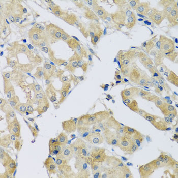
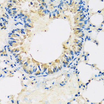
A10873-1 has IHC images of paraffin-embedded mouse lung and human stomach (catalog image captions). IF is listed, but no IF image is supplied (catalog applications; image list).

Real IHC data Immunohistochemistry of paraffin-embedded mouse lung using ABCF2 antibody at dilution of 1:100 (x40 lens).
Anti-ABCF2 Antibody
Cat # A10873-1

A10873-1 is shown in IHC on paraffin-embedded mouse lung and human stomach at 1:100 (catalog image captions). Its listed applications include IHC and IF, and its stated reactivity is human, mouse and rat (catalog applications; reactivity).

Which to pick: For tissue IHC, choose A10873-1: its images show paraffin-embedded mouse lung and human stomach; the fixative is unreported (catalog image captions). For IF, A10873-1 is listed at 1:50–1:200, but no IF image or ICC validation is supplied (catalog applications; dilution; image list). It is the only listed cross-species option, with stated human, mouse and rat reactivity; its host is rabbit and its clone is unspecified (catalog reactivity; host; clone).

Each figure is that product's own IHC / IF validation image from its datasheet.

References

  1. UniProt Consortium. UniProt entry Q9UG63 (ABCF2_HUMAN, ATP-binding cassette sub-family F member 2).
  2. Human Protein Atlas. ABCF2 tissue IHC expression (reliability: Approved).
  3. Human Protein Atlas. ABCF2 subcellular location (ICC-IF): Mainly localized to the cytosol. Caution: Based on antibodies targeting proteins from multiple genes..
  4. Human Protein Atlas. ABCF2 antibody validation summary (3 antibodies).
  5. Can ABCF2 protein expression predict the prognosis of uterine cancer?. British journal of cancer 2008 — PMC2584961.
  6. CircRNA_000864 Upregulates B-cell Translocation Gene 2 Expression and Represses Migration and Invasion in Pancreatic Cancer Cells by Binding to miR-361-3p. Frontiers in oncology 2020 — PMC7793745.
  7. ABCF2, an Nrf2 target gene, contributes to cisplatin resistance in ovarian cancer cells. Molecular carcinogenesis 2017 — PMC5509336.
  8. Circ-TCF4.85 silencing inhibits cancer progression through microRNA-486-5p-targeted inhibition of ABCF2 in hepatocellular carcinoma. Molecular oncology 2020 — PMC6998385.
  9. PubMed PMID:10944468 — UniProt-cited evidence.
  10. PubMed PMID:17974005 — UniProt-cited evidence.
  11. PubMed PMID:12853948 — UniProt-cited evidence.