This website uses cookies to ensure you get the best experience on our website.
- Table of Contents
1 Citations 6 Q&As
1 Citations
Facts about Abl interactor 1.
Involved in cytoskeletal reorganization and EGFR signaling. Together with EPS8 participates in transduction of signals from Ras to Rac.
Human | |
---|---|
Gene Name: | ABI1 |
Uniprot: | Q8IZP0 |
Entrez: | 10006 |
Belongs to: |
---|
ABI family |
Abelson interactor 1; ABI1; ABI-1; abl interactor 1; Abl-binding protein 4; ABLBP4; abl-interactor 1; Abl-interactor protein 1 long; E3B1; eps8 binding protein; Eps8 SH3 domain-binding protein; Eps8-binding protein; interactor protein AblBP4; nap1 binding protein; Nap1-binding protein; NAP1BP; spectrin SH3 domain binding protein 1; Spectrin SH3 domain-binding protein 1; SSH3BP; SSH3BP1
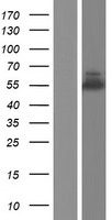
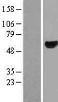
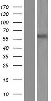
Mass (kDA):
55.081 kDA
Human | |
---|---|
Location: | 10p12.1 |
Sequence: | 10; NC_000010.11 (26746596..26861087, complement) |
Widely expressed, with highest expression in brain.
Cytoplasm. Nucleus. Cell projection, lamellipodium. Cell projection, filopodium. Cell projection, growth cone. Cell junction, synapse, postsynaptic density. Cytoplasm, cytoskeleton. Localized to protruding lamellipodia and filopodia tips. Also localized to neuronal growth cones and synaptosomes. May shuttle from the postsynaptic densities to the nucleus (By similarity).
The ABI1 Marker can be used for a variety of different research applications. Scientists can submit their results for species-specific samples and applications and receive product credits. The marker can be used by scientists of all types worldwide. The best uses of the ABI1 are listed below. Let's take a closer look at them. Let's start with the most common application.
In the field of biotechnology, the SSH3BP1/ABI1 PolycloNal Antibody is a highly specific and highly cited primary antibody. Boster has been manufacturing primary antibodies for over 25 years, and its selection includes both monoclonal and polyclonal antibody types. The Boster Bio Anti-SSH3BP1/ABI1 Polyclonal Antibody is available as a 100ul Liquid solution.
The company's Anti-SSH3BP1/ABI ELISA Kits are highly sensitive, with sensitivities as low as picograms. Each kit is tested against a panel of over 250 different tissue samples and validated for specificity and high affinity. Additionally, Boster offers product credits to the first reviewers of its products. This means that researchers around the world can get credits when they review the Boster Anti-SSH3BP1/ABI1 Polyclonal Antibody.
ELISA is an important stage in the development of antibodies. The first test-bleed after animal immunization is titrated with an ELISA assay. Then, spleen cells from the best responders undergo hybridoma fusion. The supernatant is then screened in a limiting-dilution subcloning technique until all wells are growing at the same rate. Finally, selected antibodies are subjected to further application testing.
The Boster Bio ABI1 ELISA kit is a sandwich ELISA that measures the levels of Canine CD117 in samples. The kit is highly sensitive at 12pg/ml and comes with removable strips. It is highly specific, so the results of the test will be specific to this particular animal. It is also designed to be precise with samples from this species. The kit contains a sample preparation guide and data analysis tips, which will make your experiments more accurate.
The Picokine(tm) platform enhances ELISA sensitivity to the picogram level. These kits have been validated on a variety of samples. Images and validation methods are available on request. Picoband saves time in IHC by avoiding the need to perform the additional steps required for secondary antibodies. The PicoKine(tm) platform is supported by insights into immunogen design. This enables high-sensitivity ELISA kits with unsurpassed sensitivity and reproducibility.
The gene phi31 is involved in the regulation of the phage's sensitivity to the phage defense mechanism AbiA. The phi31 phage encodes an open reading frame (ORF245) and a 118-bp fragment of DNA. These regions are responsible for the sensitivity of phi31 to AbiA. The phi31 phage has a mutation (G to A) within an inverted repeat.
The ABI1 gene encodes a protein that is expressed at the leading edge of cancer cells and has high expression in tumours with infiltrating growth and budding. Interestingly, Abi1 IHC staining scores are significantly higher at the invasive margin than the tumour center. Abi1 (pY435) is more nuclearly expressed and phosphorylated in tumours at the invasive margin than in the centre.
Abi1 is expressed in human gastric cancer and is involved in the progression of the disease. It is also found in breast cancer cell lines, which may indicate its prognostic and progression characteristics. In addition, ABI1 expression is downregulated in prostate3 and gastric4 cancers, suggesting that ABI1 has a tumor suppressor role. While the ABI1 gene is still under research, it does appear to be a promising tool for gastric cancer diagnosis.
In addition to its IHC utility, Abi1 has many potential applications in prostate cancer research. By disrupting Abi1, cellular proliferation is enhanced and cellular adhesion markers are downregulated. Both of these processes contribute to prostate cancer progression. Further, this protein may contribute to fibrosis. In addition, it may be involved in regulating cellular adhesion, which is a hallmark of certain types of cancer.
Abi1 is a member of the WAVE complex and is thought to play a role in prostate cancer progression. Its loss may lead to tumor progression and the mechanism of tumor suppression is not yet clear. To explore the role of Abi1 in prostate cancer progression, the researchers used CRISPR-based gene editing and retroviral expression to manipulate the level of Abi1 in prostate organoid tumor cell lines. The results demonstrated that Abi1 loss interferes with CHD1 cell attachment to fibronectin surfaces.
The applications of the ABI1 marker are diverse, including brain tissue imaging, gene therapy, and drug discovery. Abi-1 is a neuron-specific marker. In neurons, it is expressed on the dendrites, spines, and synapses. It colocalizes with ProSAP2/Shank3 and Bassoon in synapses. Although not found in the cytoskeletal PSD matrix, Abi-1 colocalizes with ProSAP2/Shank3 and Bassoon.
Researchers have also studied the effects of ABI1 on tumor development and progression. ABI1 loss is associated with increased WAVE3 expression, which may promote tumor invasion. However, the exact role of ABI1 in tumor progression remains unclear. Although ABI1 binds to several other proteins, its function is unclear. This research demonstrates that ABI1 is a functional marker for the epithelial cell.
The ABI1 protein is an adaptor protein involved in cytoskeletal reorganization and EGFR signaling. Activation of ABI1 inhibits EGF-induced activation of Erks, which negatively regulate cell growth and transformation. In addition to its role in tumor growth, ABI1 is also important in controlling dendritic outgrowth and determining the shape of synaptic contacts.
ABI1 has been linked to biochemical recurrence, metastasis, and death in prostate cancer. Furthermore, a study on RWPE-1 cell line revealed that low expression of ABI1 is associated with highly invasive prostate cancer. In addition, the loss of the ABI1 gene results in the loss of cell-cell adhesion markers, activating the EMT and non-canonical WNT signaling pathways. However, reexpression of ABI1 rescues this phenotype.
ABI1 is a marker for the Armadillo repeat domain, also known as the ARM domain. This domain interacts with OST1 and PUB12/13 proteins. ABA signaling activates these proteins, which phosphorylate downstream targets, including the transcriptional factors SLAC1 and PUB13. This interaction is specific to the ARM domain of PUB12/13.
The ABA signalling pathway in Arabidopsis has been successfully replicated in vitro. The ABA-activated OST1/SnRK2.6 is one of the outputs of ABA signalling. ABI1 preferentially interacts with PYLs, and the mutant Pub12/13 depletes ABI1 and decreases ABA signalling in plants. These two PP2Cs are part of the ABA signalling pathway and function in different tissues.
The integrins of the WAVE complex have important roles in prostate cancer progression. Abi1 interacts with WAVE complex proteins, which regulate the dynamics of actin at cell-cell junctions. Loss of ABI1 results in a reduced ability of epithelial cells to adhere to each other. Loss of this protein may also contribute to tumor suppression. In a recent study, researchers found that mice lacking the ABI1 gene showed decreased cell-cell adhesion and increased tumor proliferative activity.
The ABI1 gene is a versatile protein with many roles. It regulates actin cytoskeleton dynamics and binds to ENA/VASP. It may also act as an adapter in a complex of proteins. It is associated with a number of different functions in the body, including regulating the ABL1/c-Abl phosphorylation of ENAH and recruiting WASF1 to lamellipodia.
The ABI1 marker is a potential therapeutic target for the treatment of cancer. Its interaction with the a4 tail may mediate specific functions associated with a4-dependent processes, such as tumorigenesis. The role of the ABI1 marker in this process is still poorly understood. We are seeking to understand more about this gene and its function in cancer. To do this, we must first understand how it interacts with the a4 tail.
PMID: 9010225 by Biesova Z., et al. Isolation and characterization of e3B1, an eps8 binding protein that regulates cell growth.
PMID: 9593709 by Ziemnicka-Kotula D., et al. Identification of a candidate human spectrin Src homology 3 domain- binding protein suggests a general mechanism of association of tyrosine kinases with the spectrin-based membrane skeleton.
*More publications can be found for each product on its corresponding product page