This website uses cookies to ensure you get the best experience on our website.


- Table of Contents
Plan ABI2 chromogenic IHC in paraffin sections using the A04302 protocol at 2 μg/ml (datasheet: A04302). Use strong neuronal and Purkinje cell staining as tissue reference points, while checking antibody specificity because HPA reports medium consistency with RNA data (HPA tissue IHC).

Expected localisation, validated protocols, controls and antibodies — the at-a-glance facts below, then the full design guide.
| Expected localisation | Expected: cytoplasm, nucleus, projections, junctions (UniProt) | |
| Staining pattern | Strong staining in CNS neurons and Purkinje cells (HPA tissue IHC) | |
| Antigen retrieval | EDTA pH 8.0 HIER, heat-mediated (datasheet A04302) | |
| Positive control | Adrenal gland+4 more · see all | |
| Negative control | Adipose tissue+4 more · see all |
| Fixation | Keep fixation consistent across sections (standard IHC practice; not target-specific) | |
| Caveat | Isoform 1, unlike isoform 3, reaches protrusion tips (UniProt) | |
| Regulation | CNS enriched; intensity varies by tissue (HPA tissue IHC) | |
| Isoform / epitope | 4 isoforms; epitope coverage is unspecified (UniProt) |
The catalog antibody protocol is followed by published ABI2 IHC methods for mouse femur, paired human TNBC samples, and HCC specimens (PMC12690970; PMC11212232; PMC9825985).
| Sample | Paraffin-embedded mouse brain tissue; fixative not specified (datasheet A04302) |
| Fixation | Image fixative and duration unreported (datasheet A04302); verify before use. |
| Sectioning | 4–5 µm sections on charged slides (standard) |
| Deparaffinisation | Xylene, graded ethanol series to water (standard) |
| Antigen retrieval | Heat retrieval: EDTA pH 8.0 (datasheet A04302); 20 min, 95–100 °C (standard) |
| Peroxidase block | 3% H2O2, 10 min, room temperature (standard) |
| Blocking | 10% goat serum (datasheet A04302) |
| Primary antibody | Rabbit anti-ABI2, 2-5 μg/ml (datasheet A04302) |
| Primary incubation | Overnight at 4 °C (datasheet A04302) |
| Detection | HRP-conjugated secondary, DAB chromogen (datasheet A04302) |
| Counterstain | Hematoxylin, blue, dehydrate and mount (standard) |
| Expected result | ABI2-positive staining in glandular cells of adrenal gland (HPA tissue IHC: High). HPA tissue profile: Expression in most tissues, most abundant in CNS. No signal in the no-primary control. |
ABI2 is a nonmembrane protein associated with the cytoplasm, nucleus, cell projections and adherens junctions (UniProt Q9NYB9 topology and localization). In paraffin IHC, expect staining chiefly in neuronal cells, Purkinje cells and adrenal glandular cells, which HPA scores High (HPA tissue IHC). HPA describes expression across most tissues, with greatest abundance in the CNS; its tissue profile has Enhanced reliability but awaits external verification (HPA tissue IHC).
| Strong staining in cerebellar Purkinje cells, cortical neurons or adrenal glandular cells. | These are HPA High cell populations and useful positive references (HPA tissue IHC). Assess staining in the identified cells, not by whole-section darkness (general IHC practice). |
| Cell-associated cytoplasmic or nuclear staining, with possible signal near cell edges. | Cytoplasm, nucleus, projections and adherens junctions are reported locations (UniProt Q9NYB9 localization). Fine projection-tip patterns may be difficult to resolve in chromogenic sections (general IHC practice). |
| Predominant staining of a compartment inconsistent with the expected cellular pattern, such as a crisp plasma-membrane rim throughout the section. | ABI2 has no transmembrane segment (UniProt Q9NYB9 topology). Recheck morphology and controls before interpreting a pervasive membrane-rim pattern as ABI2 (general IHC practice). |
| Strong staining in adipocytes, cardiomyocytes or prostate glandular cells, especially when positive controls also look atypical. | HPA reports these cell populations as Not detected by tissue IHC (HPA tissue IHC). Consider antibody cross-reactivity or endogenous chromogen activity, and inspect controls (general IHC practice). |
| Uniform haze across cells and stroma, or no staining in an HPA High reference tissue. | Haze lacks the reported cell-selective pattern (HPA tissue IHC); absent reference staining leaves the run uninterpretable. Check detection background or assay performance before scoring ABI2 (general IHC practice). |
| Tissue and cell selection | HPA scores Purkinje cells, cortical neurons and adrenal glandular cells High, but breast glandular and bronchial ciliated cells Medium (HPA tissue IHC). Compare the named cell population within each tissue rather than imposing one intensity threshold across tissues. |
| Subcellular interpretation | UniProt lists cytoplasm, nucleus, cytoskeleton, projections and adherens junctions (UniProt Q9NYB9 localization). A paraffin-section pattern may emphasize a subset; judge localization alongside cell identity and the positive control (general IHC practice). |
| Isoform and resolution | Four isoforms are listed; isoform 1, unlike isoform 3, reaches protruding lamellipodia and filopodia tips (UniProt Q9NYB9 isoforms and localization). A lack of visible tip staining in IHC does not alone exclude ABI2 (general IHC practice). |
| Topology and processing | ABI2 lacks a transmembrane segment, signal peptide and propeptide; the listed chain spans residues 1–513 (UniProt Q9NYB9 topology and processing). The supplied record gives no antibody epitope, so it cannot predict epitope-specific retrieval or isoform detection. |
| Antibody evidence | The listed antibody HPA070567 has IHC Enhanced validation, meaning its pattern was reproduced by independent antibodies or orthogonal data (HPA antibodies). The tissue profile still notes medium RNA–staining consistency and pending external verification (HPA tissue IHC). |
| IF/ICC: where should ABI2 appear? | HPA reports mainly supported nucleoplasmic localization, additional supported cytosol and uncertain vesicles; the listed antibody has ICC Supported status (HPA subcellular; HPA antibodies). Use that as an IF interpretation reference, not an IHC compartment requirement. |
| Situation | Likely cause | Next action |
|---|---|---|
| No signal in Purkinje cells, cortical neurons or adrenal glandular cells. | These populations are HPA High, so absent staining may reflect an unsuccessful IHC run or unsuitable assay conditions (HPA tissue IHC; general IHC practice). | Verify tissue identity and the positive control, then review the catalog antibody's IHC-P instructions for retrieval and dilution; no ABI2-specific retrieval condition is supplied here (general IHC practice). |
| A supposedly negative section shows widespread brown precipitate. | Endogenous detection activity, nonspecific antibody binding or residual reagent can produce background in chromogenic IHC (general IHC practice). | Inspect a no-primary control, review detection-system blocking and washes, and compare staining with the expected HPA cell distribution (general IHC practice; HPA tissue IHC). |
| Adipocytes, cardiomyocytes or prostate glandular cells stain more strongly than the reference cells. | HPA reports these cell populations as Not detected, while the selected neuronal and adrenal populations are High (HPA tissue IHC). | Confirm cell identity and compare controls; if the discordant pattern persists, treat specificity as unresolved rather than assigning ABI2 positivity (general IHC practice). |
| Only faint staining appears in breast or bronchus. | HPA scores breast glandular cells and bronchial ciliated cell bodies Medium, below its High reference populations (HPA tissue IHC). | Run an HPA High reference tissue alongside them and score the specified cells separately; faint signal alone does not establish assay failure (HPA tissue IHC; general IHC practice). |
| Signal appears mainly nuclear, raising concern because ABI2 regulates actin. | Nuclear localization is listed by UniProt, and HPA IF reports supported nucleoplasmic localization (UniProt Q9NYB9 localization; HPA subcellular). | Check the nuclear counterstain, cell identity and controls before rejecting nuclear signal; do not require visible projection tips in paraffin IHC (general IHC practice). |
| The positive control stains, but the test section has diffuse haze that obscures cells. | The HPA reference pattern identifies particular stained cell populations; uniform haze cannot be scored as that pattern (HPA tissue IHC; general IHC practice). | Review no-primary background, blocking, detection and washes; adjust conditions using the catalog IHC-P instructions, then rescore only clearly identifiable cells (general IHC practice). |


Comprehensive Human Protein Atlas IHC scoring per tissue (reliability: Enhanced — Medium consistency between antibody staining and RNA expression data. Pending external verification.). Rows are taken directly from the HPA tissue chart — click any row's HPA link to view the source.
| Tissue | Cell type | Level | Evidence | Source |
|---|---|---|---|---|
| Adrenal gland | Glandular cells | High | Protein (IHC) | HPA → |
| Cerebellum | Purkinje cells | High | Protein (IHC) | HPA → |
| Cerebral cortex | Neuronal cells | High | Protein (IHC) | HPA → |
| Breast | Glandular cells | Medium | Protein (IHC) | HPA → |
| Bronchus | Ciliated cells (cell body) | Medium | Protein (IHC) | HPA → |
| Tissue | Cell type | Level | Evidence | Source |
|---|---|---|---|---|
| Adipose tissue | Adipocytes | Not detected | Protein (IHC) | HPA → |
| Bone marrow | Hematopoietic cells | Not detected | Protein (IHC) | HPA → |
| Epididymis | Glandular cells | Not detected | Protein (IHC) | HPA → |
| Heart muscle | Cardiomyocytes | Not detected | Protein (IHC) | HPA → |
| Liver | Cholangiocytes | Not detected | Protein (IHC) | HPA → |
Use the A04302 paraffin-section result as a starting point, then judge ABI2 staining by compartment, cell type, controls, and section quality.
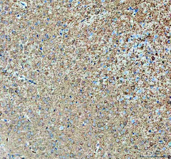
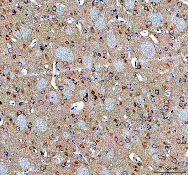
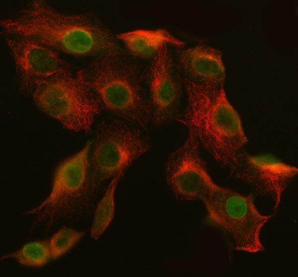
Real ABI2 IHC images show paraffin sections of human, mouse, and rat brain (A04302 IHC captions). IF/ICC is listed, but the supplied IF caption identifies a different target (catalog IF caption).
A04302 is the only SKU with a rendered card and its own IHC figure, showing ABI2 staining in a mouse brain paraffin section (A04302 card caption). Additional A04302 IHC captions describe rat and human brain paraffin sections; its applications also list IF/ICC (A04302 IHC captions; catalog applications).
Which to pick: Choose A04302 for paraffin-section IHC: its own brain captions document staining across human, mouse, and rat, with 2 μg/ml primary antibody after EDTA pH 8.0 retrieval; the fixative is unreported (A04302 IHC captions). For IF/ICC, PA2068 lists both applications and 5 μg/ml for human immunocytochemistry/immunofluorescence, but has no supplied IF image (PA2068 catalog applications and dilution; IF image alts). For cross-species IHC, A04302 has the strongest pictured support across the three species; M04302 lists IHC and human, mouse, and rat reactivity but has no supplied IHC figure (A04302 IHC captions; M04302 catalog applications, reactivity, and IHC image alts).