This website uses cookies to ensure you get the best experience on our website.


- Table of Contents
Plan chromogenic ACAD11 IHC in paraffin sections around the granular cytoplasmic staining reported in brain, kidney and liver (HPA tissue IHC). This guide covers fixation consistency, the catalog antibody’s 1:100–1:300 IHC dilution (datasheet), and interpretation of cell-specific staining (HPA tissue IHC).

Expected localisation, validated protocols, controls and antibodies — the at-a-glance facts below, then the full design guide.
| Expected localisation | Tissue: granular cytoplasm (HPA tissue IHC); annotated: peroxisome and mitochondrial membrane (UniProt) | |
| Staining pattern | Granular cytoplasm in hepatocytes and renal tubules (HPA tissue IHC) | |
| Antigen retrieval | Citrate pH 6.0 HIER, 95–98 °C, 20 min (rule: cytoplasmic / membrane antigen) | |
| Positive control | Appendix+4 more · see all | |
| Negative control | Adipose tissue+4 more · see all |
| Fixation | Keep paraffin-section fixation consistent across samples. (standard IHC practice; not target-specific) | |
| Caveat | Cerebellar staining favors granular-layer processes (HPA tissue IHC) | |
| Regulation | Tissue-dependent staining intensity (HPA tissue IHC) | |
| Isoform / epitope | 3 isoforms; antibody epitope coverage is unspecified (UniProt; datasheet) |
The catalog antibody’s IHC-P protocol is accompanied by a published ACAD11 IHC protocol for human cerebellum and muscle (PMC3073726).
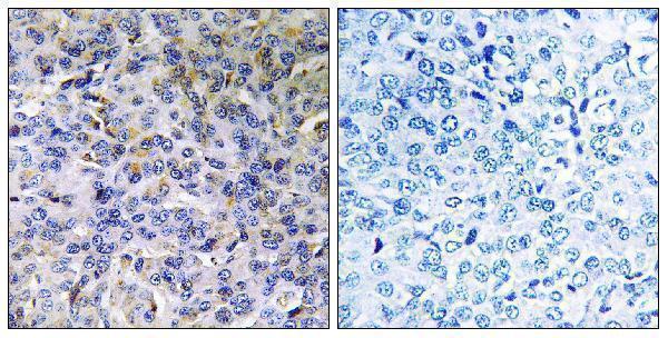
| Sample | Paraffin-embedded human breast carcinoma tissue; fixative not specified (datasheet A10875-1) |
| Fixation | Image fixative and duration unreported (datasheet A10875-1); verify before use. |
| Sectioning | 4–5 µm sections on charged slides (standard) |
| Deparaffinisation | Xylene, graded ethanol series to water (standard) |
| Antigen retrieval | Heat-induced epitope retrieval in citrate buffer, pH 6.0, 20 min at 95–98 °C (standard rule: cytoplasmic / membrane antigen) |
| Peroxidase block | 3% H2O2, 10 min, room temperature (standard) |
| Blocking | 10% normal serum of the secondary host, 30 min, room temperature (standard) |
| Primary antibody | Rabbit anti-ACAD11, 1:100-1:300 (datasheet A10875-1) |
| Primary incubation | Overnight at 4 °C (standard) |
| Detection | HRP-polymer secondary, DAB chromogen 5–10 min (standard) |
| Counterstain | Hematoxylin, blue, dehydrate and mount (standard) |
| Expected result | ACAD11-positive staining in enterocytes - Microvilli of appendix (HPA tissue IHC: High). HPA tissue profile: Granular cytoplasmic expression in selected tissues, most abundant in brain, kidney and liver. No signal in the no-primary control. |
ACAD11 is reported at peroxisomes and, in some settings, at the mitochondrial membrane; it has no transmembrane segment (UniProt Q709F0). In paraffin-section IHC, expect granular cytoplasmic staining, especially in cerebellar granular-layer processes, kidney proximal tubule cells and hepatocytes (HPA tissue IHC). HPA rates its IHC evidence Enhanced, with medium consistency between staining and RNA data (HPA tissue IHC).
| Granular cytoplasmic staining is strong in cerebellar granular-layer processes and moderate in kidney proximal tubule cell bodies or hepatocytes. | This matches the reported cell-level pattern: High in cerebellar processes and Medium in those kidney and liver cells (HPA tissue IHC). Score each cell population and its compartment; intensity need not match across tissues. |
| Signal is predominantly nuclear or forms a continuous cell-surface outline instead of cytoplasmic granules. | That distribution differs from the reported granular cytoplasmic IHC pattern (HPA tissue IHC). UniProt reports no transmembrane segment, although membrane association occurs in some settings (UniProt Q709F0). Check specificity before calling it ACAD11. |
| Strong staining appears in adipocytes or bronchial respiratory epithelial cells. | HPA reports ACAD11 as Not detected in those cell populations (HPA tissue IHC). Consider nonspecific antibody binding or endogenous detection activity; compare a matched control. An HPA negative is a reference observation, not proof that every specimen must be negative. |
| Brown signal spreads across cells or tissue without a discernible granular cytoplasmic pattern. | Diffuse signal cannot be confidently assigned to ACAD11's reported pattern (HPA tissue IHC). As a general IHC check, inspect the no-primary control and assess blocking, antibody concentration and chromogen development before scoring. |
| A cerebellar granular layer, kidney proximal tubule region or hepatocyte region has no detectable signal. | Each is a reported positive cell population, although kidney and liver are rated Medium rather than High (HPA tissue IHC). Confirm that the intended cells are present, then check the staining run and a positive control before interpreting absence as biological. |
| Which tissue gives the clearest reference pattern? | Cerebellar granular-layer processes are rated High; proximal tubule cell bodies and hepatocytes are rated Medium (HPA tissue IHC). Use the named cell population when judging a section, rather than its tissue-wide color. |
| How far can organelle identity be inferred? | UniProt lists peroxisomes and mitochondrial membrane association, with observations differing by tissue and cell context (UniProt Q709F0). Granular cytoplasmic chromogen alone does not distinguish those organelles; organelle assignment needs separate evidence. |
| How strong is the IHC pattern evidence? | HPA reports Enhanced IHC validation for HPA048317 and describes medium consistency between antibody staining and RNA expression (HPA antibodies; HPA tissue IHC). Treat the mapped pattern as a reference while retaining the stated consistency limit. |
| Do isoforms or processing define a different staining pattern? | UniProt lists 3 isoforms and one annotated chain spanning residues 1–780 (UniProt Q709F0). The supplied evidence does not establish which isoforms the antibody detects, so staining cannot identify an isoform. |
| Could chromogenic detection create a misleading positive? | Endogenous enzyme activity can contribute color in routine chromogenic IHC (general IHC practice). A matched no-primary control helps identify detection-derived signal; this is a general workflow check, not an ACAD11-specific HPA finding. |
| IF/ICC Q&A: is a cultured-cell pattern established? | No HPA ICC-IF images or main subcellular location are available for ACAD11, and the listed antibody has no ICC validation entry (HPA subcellular; HPA antibodies). The tissue IHC pattern therefore does not establish an expected IF/ICC image. |
| Situation | Likely cause | Next action |
|---|---|---|
| No signal appears in a section containing the reported positive cells. | A staining-run problem is possible; kidney and liver positives are only Medium on the HPA scale (HPA tissue IHC). | Verify the relevant cells on the section and inspect a concurrent positive control. As general IHC practice, review retrieval, primary incubation and detection if that control also fails; no ACAD11-specific retrieval effect is established. |
| The positive control stains, but the test section lacks visible granules. | The sampled cells may differ from the HPA-positive populations, or the test specimen may genuinely differ (HPA tissue IHC). | Confirm cell identity and morphology before calling a negative. Compare like cell populations and report the control result; avoid treating whole-tissue absence as proof of absent ACAD11. |
| Nuclei or uninterrupted cell borders dominate the signal. | This conflicts with HPA's granular cytoplasmic pattern; UniProt reports no transmembrane segment (HPA tissue IHC; UniProt Q709F0). | Check the no-primary control and review antibody concentration and detection as general IHC practice. Score ACAD11 only where the cell-level pattern is supported. |
| Unexpected strong color appears in adipocytes or bronchial respiratory epithelium. | Those cells are listed as Not detected by HPA; nonspecific binding or detection background is possible (HPA tissue IHC; general IHC practice). | Compare matched controls and examine whether color follows the expected granular cytoplasmic distribution. Record the discrepancy rather than reclassifying those cells as established positives. |
| Diffuse brown background obscures tissue boundaries. | Excess antibody, incomplete blocking or prolonged chromogen development can raise background in routine IHC (general IHC practice). | Inspect the no-primary control and adjust one general staining variable at a time. Reassess whether cytoplasmic granules remain distinct in a reported positive cell population (HPA tissue IHC). |
| Kidney or liver looks weaker than cerebellum. | That ordering is compatible with HPA's High rating for cerebellar processes and Medium ratings for proximal tubule cell bodies and hepatocytes (HPA tissue IHC). | Score the specified cells and compartment within each tissue. Investigate a run problem only if the expected pattern or a concurrent positive control is also lost. |


Comprehensive Human Protein Atlas IHC scoring per tissue (reliability: Enhanced — Medium consistency between antibody staining and RNA expression data.). Rows are taken directly from the HPA tissue chart — click any row's HPA link to view the source.
| Tissue | Cell type | Level | Evidence | Source |
|---|---|---|---|---|
| Appendix | Enterocytes - Microvilli | High | Protein (IHC) | HPA → |
| Cerebellum | Processes in granular layer | High | Protein (IHC) | HPA → |
| Cerebral cortex | Neuropil | Medium | Protein (IHC) | HPA → |
| Colon | Enterocytes - Microvilli | Medium | Protein (IHC) | HPA → |
| Heart muscle | Cardiomyocytes | Medium | Protein (IHC) | HPA → |
| Tissue | Cell type | Level | Evidence | Source |
|---|---|---|---|---|
| Adipose tissue | Adipocytes | Not detected | Protein (IHC) | HPA → |
| Adrenal gland | Glandular cells | Not detected | Protein (IHC) | HPA → |
| Bone marrow | Hematopoietic cells | Not detected | Protein (IHC) | HPA → |
| Breast | Adipocytes | Not detected | Protein (IHC) | HPA → |
| Bronchus | Respiratory epithelial cells | Not detected | Protein (IHC) | HPA → |
Troubleshoot ACAD11 staining in paraffin sections by checking retrieval, cellular pattern, controls, and scoring before interpreting chromogenic signal.
A10875-1 has an IHC image of paraffin-embedded human breast carcinoma (IHC image caption). IF is listed without an IF image (catalog: applications; IF image alts). Reactivity includes Human, Mouse, and Rat (catalog: reactivity).
A10875-1 has an IHC image of paraffin-embedded human breast carcinoma with a peptide-blocked comparator (IHC image caption). It lists IHC and IF applications and Human, Mouse, and Rat reactivity; no IF image is supplied (catalog: applications, reactivity, IF image alts).
Which to pick: Choose A10875-1 for tissue IHC because its image shows staining in a paraffin-embedded human breast carcinoma section; the fixative is unreported (IHC image caption). For IF, A10875-1 lists that application, but no IF image or ICC application is supplied (catalog: applications, IF image alts). It is also the listed cross-species option for Human, Mouse, and Rat, though the supplied IHC image shows only human tissue (catalog: reactivity; IHC image caption).