ACKR3 / Atypical chemokine receptor 3 · IHC design guide

Design Immunohistochemistry for ACKR3

Plan ACKR3 chromogenic IHC in paraffin sections with the IHC-validated antibody at 2–5 µg/ml (datasheet A02656-2). Assess variable cytoplasmic staining in trophoblasts and immune cells, bearing in mind the low consistency between antibody staining and RNA expression (HPA tissue IHC).

Evidence assembled Oct 2026 · For research use; verify linked source records and product datasheet before use
Immunohistochemistry protocol sheet for ACKR3 (IHC for ACKR3): expected localisation Observed cytoplasmic (HPA tissue IHC); membrane/endosomes (UniProt), antibody A02656-2, validated IHC image, and IHC protocol steps
Printable ACKR3 IHC protocol sheet — expected localisation Observed cytoplasmic (HPA tissue IHC); membrane/endosomes (UniProt), antibody A02656-2, controls and protocol steps. Open the full ACKR3 IHC guide →

ACKR3 Immunohistochemistry Experimental Design Guide

Expected localisation, validated protocols, controls and antibodies — the at-a-glance facts below, then the full design guide.

Must know before staining
Expected localisation Observed cytoplasmic (HPA tissue IHC); membrane/endosomes (UniProt)
Staining pattern Variable cytoplasmic staining in trophoblasts and immune cells (HPA tissue IHC)
Antigen retrieval EDTA pH 8.0 HIER, heat-mediated (datasheet A02656-2)
Positive control ⓘ Appendix+4 more · see all
Negative control ⓘ Adrenal gland+4 more · see all
Important caveats
Reasons your staining may differ from the expected pattern.
Fixation Keep fixation consistent across paraffin sections (standard IHC practice; not target-specific)
Caveat Antibody staining and RNA show low consistency (HPA tissue IHC)
Regulation Ligand induces internalization (UniProt)
Isoform / epitope No isoforms; check extracellular vs cytoplasmic epitope (UniProt)
Section 1

Recommended ACKR3 IHC & IF Protocols

Compare the catalog antibody’s EDTA pH 8.0 IHC-P protocol (datasheet A02656-2) with three published ACKR3 chromogenic IHC protocols (PMC9046756; PMC10523940; PMC6274818).

Recommended immunohistochemistry (IHC-P) protocol parameters
SampleParaffin-embedded human breast cancer tissue; fixative not specified (datasheet A02656-2)
FixationImage fixative and duration unreported (datasheet A02656-2); verify before use.
Sectioning4–5 µm sections on charged slides (standard)
DeparaffinisationXylene, graded ethanol series to water (standard)
Antigen retrievalHeat retrieval: EDTA pH 8.0 (datasheet A02656-2); 20 min, 95–100 °C (standard)
Peroxidase block3% H2O2, 10 min, room temperature (standard)
Blocking10% goat serum (datasheet A02656-2)
Primary antibodyRabbit anti-ACKR3, 2-5 µg/ml (datasheet A02656-2)
Primary incubationOvernight at 4 °C (datasheet A02656-2)
DetectionHRP-conjugated secondary, DAB chromogen (datasheet A02656-2)
CounterstainHematoxylin, blue, dehydrate and mount (standard)
Expected resultACKR3-positive staining in lymphoid tissue of appendix (HPA tissue IHC: Medium). HPA tissue profile: Cytoplasmic expression at variable levels in several different cell types, including trophoblasts and immune cells. No signal in the no-primary control.
💡Decision noteStart with heat-mediated EDTA pH 8.0 retrieval (datasheet A02656-2); a published ACKR3 IHC protocol also uses EDTA pH 8.0 (PMC10523940).
Section 2

What Is the Expected ACKR3 Staining Pattern?

ACKR3 is a 7-pass receptor at the cell membrane and in endosomes, with predominant localization in endocytic vesicles (UniProt P25106 topology and subcellular location). In tissue IHC, expect variable cytoplasmic staining in several cell types, including trophoblasts and immune cells (HPA tissue IHC). HPA rates its tissue IHC evidence Approved but reports low consistency between antibody staining and RNA expression (HPA tissue IHC).

What am I looking at on my slide?
Granular cytoplasmic staining in placental trophoblasts or kidney tubular cells, with possible membrane accentuation.This fits HPA's medium staining in those cells and UniProt's predominantly vesicular, recycling receptor (HPA tissue IHC; UniProt P25106 subcellular location). Judge the pattern within identifiable cells; a uniformly filled field gives less evidence of receptor localization (standard IHC practice).
Strong, isolated nuclear staining without a cell-associated cytoplasmic or membrane pattern.A nuclear-only pattern is discordant with the annotated membrane and endosomal locations and the HPA cytoplasmic tissue profile (UniProt P25106 subcellular location; HPA tissue IHC). Treat it as suspect and inspect controls before scoring it as ACKR3 (standard IHC practice).
Prominent staining in a cell population reported as not detected, such as adrenal glandular cells.HPA reports no staining in that specific population, so unexpected signal warrants checking cell identity, antibody specificity and endogenous detection activity (HPA tissue IHC; standard IHC practice). A single discordant section cannot establish expression because HPA reports low staining–RNA consistency (HPA tissue IHC).
Diffuse color across cells and surrounding tissue, with little difference between expected positive and negative areas.This does not resolve the variable, cell-associated cytoplasmic pattern described by HPA (HPA tissue IHC). Background from detection reagents, inadequate blocking or excessive antibody concentration is possible; compare a matched no-primary control and review detection conditions (standard IHC practice).
No staining in placental trophoblasts or kidney tubular cells.Both populations have medium HPA IHC staining, so absence of signal calls for checking section quality, antibody performance, retrieval and detection with an appropriate positive control (HPA tissue IHC; standard IHC practice). HPA's Approved rating and low RNA concordance limit how decisively one negative result can be read (HPA tissue IHC).
💡Expected ACKR3 appearanceCall a section positive when identifiable cells show variable, cell-associated cytoplasmic or vesicular staining, potentially with membrane signal; placental trophoblasts and kidney tubular cells are medium HPA examples, while isolated nuclear staining or uniform tissue-wide color is suspect (HPA tissue IHC; UniProt P25106 subcellular location; standard IHC practice).
How each factor affects the staining
Receptor topology and epitope locationACKR3 has 7 transmembrane segments, extracellular and cytoplasmic regions, and annotated glycosylation sites at residues 13, 22 and 39 (UniProt P25106 topology and glycosylation). Epitope location may matter when interpreting antibody access, but no antibody epitope or ACKR3-specific retrieval response is supplied.
Internalization and recyclingUniProt places ACKR3 predominantly in endocytic vesicles and describes ligand-induced internalization followed by receptor recycling (UniProt P25106 subcellular location). Cytoplasmic puncta can therefore fit receptor biology; membrane-only staining is not required for a plausible positive pattern (UniProt P25106 subcellular location).
Tissue pattern and confidenceHPA reports medium staining in placental trophoblasts, kidney tubular cells and appendix lymphoid tissue, but no detection in adrenal glandular cells or bone marrow hematopoietic cells (HPA tissue IHC). These observations are cell-specific; the Approved rating carries a low staining–RNA consistency caveat (HPA tissue IHC).
Antibody validationHPA lists IHC as Approved for HPA049718 and ICC as Supported for HPA032003 (HPA antibodies). Those are different application records, so the ICC result does not by itself validate staining in paraffin sections (HPA antibodies; standard IHC practice).
Q: What should IF/ICC show?A: Vesicles are the approved main location and plasma membrane is supported; HPA lists ICC-IF images in HeLa and U2OS cells (HPA subcellular ICC-IF). This answers the localization question without making an IF/ICC protocol recommendation.
Why is my staining missing, weak or wrong?
SituationLikely causeNext action
A known-positive tissue has no visible signal.The assay may have insufficient signal, or the chosen section may lack the expected cell population (standard IHC practice).Confirm placental trophoblasts or kidney tubular cells are present, then review positive-control performance, retrieval, antibody dilution and detection steps (HPA tissue IHC; standard IHC practice). No ACKR3-specific retrieval condition is supplied.
Adrenal glandular cells stain as strongly as the chosen positive control.This conflicts with HPA's not-detected result for those cells; nonspecific binding or endogenous detection activity is possible (HPA tissue IHC; standard IHC practice).Compare matched no-primary and detection controls, then reassess blocking and cell identity before assigning the signal to ACKR3 (standard IHC practice).
Only nuclei are colored.A nuclear-only pattern is inconsistent with ACKR3's annotated membrane and endosomal locations (UniProt P25106 subcellular location).Check counterstain and detection controls, and seek cell-associated cytoplasmic or membrane signal in an HPA-positive population before scoring (HPA tissue IHC; standard IHC practice).
The whole section has diffuse color.Poorly resolved background can obscure the variable cytoplasmic pattern reported by HPA (HPA tissue IHC; standard IHC practice).Inspect a no-primary control and review blocking, antibody dilution, washes and chromogen development (standard IHC practice). Interpret only staining that resolves to identifiable cells.
Staining appears only at cell borders, without obvious puncta.Membrane staining is biologically plausible, although UniProt describes predominant vesicular localization (UniProt P25106 subcellular location).Check that the border signal follows identifiable cells and compare it with HPA-positive tissue and detection controls; do not reject it solely for lacking puncta (HPA tissue IHC; UniProt P25106 subcellular location; standard IHC practice).
Endothelial or brain staining differs from the expected tissue profile.UniProt reports ACKR3 in some brain endothelial cells, while HPA reports no detection in cerebral cortex endothelial cells by tissue IHC (UniProt P25106 tissue specificity; HPA tissue IHC).Record the exact tissue and cell population, compare matched controls, and report the source disagreement alongside the observed pattern rather than forcing a positive or negative call (standard IHC practice).

Sample controls for ACKR3 IHC & IF

🧪Run kidney first and score staining in tubular cells (HPA: Kidney, cells in tubules, Medium). Use adrenal gland glandular cells as the negative tissue (HPA: Adrenal gland, glandular cells, Not detected); on the kidney slide, compare tubules with adjacent non-tubular cells, treating them as an internal negative only if they remain at background level on the no-primary control.
Positive control tissue: Appendix (Lymphoid tissue, HPA Medium)
Negative control tissue: Adrenal gland (HPA Not detected)
ICC-IF cell lines (HPA subcellular resource): HPA ICC-IF images show ACKR3 in HeLa, U2OS, with annotated localisation: Vesicles (approved), Plasma membrane (supported) (HPA subcellular).
Technical controls: Include no-primary (secondary-only) and host-species- and isotype-matched rabbit IgG controls, and confirm specificity with ACKR3-knockout material or peptide competition if the immunizing peptide is available (selected-SKU caption: rabbit primary; standard IHC controls). Quench endogenous peroxidase and check renal tubules for background before interpreting DAB staining (selected-SKU caption: HRP/DAB; standard kidney IHC practice).
⚠️Feasibility: A target-specific fixation window or fixation effect is unreported, and the selected-SKU paraffin-section caption does not state the fixative (selected-SKU caption: fixative not stated). The demonstrated IHC procedure used heat retrieval in EDTA at pH 8.0, so check retrieval performance for the chosen specimens (selected-SKU caption: EDTA heat retrieval); the supplied evidence does not establish whether frozen sections or IF are easier. IF/ICC images exist in HeLa and U2OS, with plasma-membrane and vesicular localization, but they do not establish tissue IF performance (HPA subcellular: HeLa and U2OS ICC-IF; vesicles and plasma membrane).

HPA tissue IHC evidence for ACKR3

Comprehensive Human Protein Atlas IHC scoring per tissue (reliability: Approved — Low consistency between antibody staining and RNA expression data.). Rows are taken directly from the HPA tissue chart — click any row's HPA link to view the source.

Positive expression · recommended positive controls

TissueCell typeLevelEvidenceSource
Appendix Lymphoid tissue Medium Protein (IHC) HPA →
Bronchus Ciliated cells (ciliary rootlets) Medium Protein (IHC) HPA →
Fallopian tube Ciliated cells (cell body) Medium Protein (IHC) HPA →
Kidney Cells in tubules Medium Protein (IHC) HPA →
Pancreas Exocrine glandular cells Medium Protein (IHC) HPA →

Undetected expression · recommended negative controls

TissueCell typeLevelEvidenceSource
Adrenal gland Glandular cells Not detected Protein (IHC) HPA →
Bone marrow Hematopoietic cells Not detected Protein (IHC) HPA →
Caudate Glial cells Not detected Protein (IHC) HPA →
Cerebellum Cells in granular layer Not detected Protein (IHC) HPA →
Cerebral cortex Endothelial cells Not detected Protein (IHC) HPA →
Section 3

Advanced ACKR3 IHC Tips

Troubleshoot ACKR3 staining in paraffin section chromogenic IHC using the catalog antibody’s tissue example, receptor biology, and reported staining patterns.

What retrieval should I try first when ACKR3 staining is weak?
Start with heat-mediated retrieval in EDTA pH 8.0 for paraffin sections stained with A02656-2 (datasheet A02656-2). The documented breast cancer section used this buffer before overnight primary incubation at 4°C (caption A02656-2). Keep heating and cooling conditions consistent across comparison slides, and record them because the caption gives no retrieval duration (caption A02656-2; standard IHC practice). If staining remains weak, test a different retrieval condition on matched sections while retaining the documented condition as a reference (standard IHC practice). Compare signal in the intended cells with tissue damage and background before adopting the change (standard IHC practice).
Could fixation explain weak or uneven ACKR3 staining?
The A02656-2 tissue example identifies a paraffin-embedded section but does not state its fixative (caption A02656-2). Target-specific fixation sensitivity is therefore unknown; the reported tissue patterns do not establish which fixation conditions preserve this antibody’s epitope (caption A02656-2; HPA: tissue IHC). For a controlled comparison, stain sections with documented processing histories together, using the same EDTA pH 8.0 retrieval and detection conditions (datasheet A02656-2; standard IHC practice). Examine morphology alongside signal, because poorly preserved tissue can make cell boundaries and staining distribution difficult to assess (standard IHC practice). Record any fixation differences before attributing an intensity change to ACKR3 abundance (standard IHC practice).
Should ACKR3 appear at the membrane or inside cells?
Assess both membranous and cytoplasmic punctate staining: ACKR3 is reported at the cell membrane and in early and recycling endosomes (UniProt P25106 subcellular). Predominant vesicular localisation and ligand-associated internalisation make a cytoplasmic pattern biologically plausible (UniProt P25106 subcellular). HPA also reports variable cytoplasmic tissue staining and identifies vesicles and plasma membrane in its subcellular assessment (HPA: tissue IHC; HPA: subcellular). At 20×–40× review magnification, compare the pattern with cell outlines and intact tissue morphology before assigning compartments (standard IHC practice). Record membrane and cytoplasmic signal separately, since a single combined score can conceal a change in distribution (standard IHC practice).
How can epitope position affect ACKR3 staining in sections?
Confirm the antibody’s immunogen or mapped epitope before interpreting a negative section, because its position is not supplied here (caption A02656-2). ACKR3 has 7 transmembrane segments, with extracellular residues 1–40 and a cytoplasmic tail at 320–362 (UniProt P25106 topology). Extracellular residues 13, 22 and 39 are annotated glycosylation sites, and tail residues 347, 350 and 355 include annotated phosphoserines (UniProt P25106 modifications). These features identify possible epitope contexts; they do not establish how this antibody responds to retrieval or processing (UniProt P25106 topology; caption A02656-2). No isoforms are listed in the supplied record, so avoid assigning staining differences to a specific splice isoform (UniProt P25106 isoforms).
How should I investigate ACKR3 localisation with multiplex IF?
On the separate IF/ICC guide, pair ACKR3 with a marker for the cell population under study and assess whether their signals occupy the same cells (standard IF practice). HPA reports ACKR3 signal in trophoblasts and immune cells in tissue, with vesicles and plasma membrane in its subcellular assessment (HPA: tissue IHC; HPA: subcellular). Choose spectrally separated fluorophores and compare an unstained section to identify tissue autofluorescence before assigning faint signal (standard IF practice). If the antibody epitope is intracellular, evaluate permeabilisation; if extracellular, first assess access without it, since this antibody’s epitope is unspecified (UniProt P25106 topology; caption A02656-2; standard IF practice). Keep the EDTA pH 8.0 condition as IHC evidence only when planning that separate assay (datasheet A02656-2).
What should I check when DAB background obscures ACKR3?
The documented paraffin section used 10% goat serum, 2 μg/ml primary antibody overnight at 4°C, and DAB detection (caption A02656-2). Check whether background follows the tissue, section edges, or the whole slide, and compare a primary-omission control processed with the same detection reagents (standard IHC practice). Include a peroxidase-blocking step and verify wash quality when using HRP and DAB; these are general chromogenic IHC controls (standard IHC practice). If diffuse staining persists, titrate the primary around the documented concentration while keeping retrieval and development consistent (caption A02656-2; standard IHC practice). Interpret faint diffuse colour cautiously when it lacks discernible cellular localisation (UniProt P25106 subcellular; standard IHC practice).
How should I score ACKR3 across paraffin sections? ⚠ ANSWER MARKED FOR VERIFICATION
Define the cell population and analysis region before scoring, since HPA reports variable cytoplasmic staining across cell types (HPA: tissue IHC; standard IHC practice). For a cell-based readout, record the percentage of positive cells and intensity from 0–3, then calculate an H-score from 0–300 using the percentage at each intensity (standard IHC practice). Score membrane and cytoplasmic compartments separately when the research question concerns receptor distribution (UniProt P25106 subcellular; standard IHC practice). For dispersed positive cells, report counts per mm² of evaluable tissue and identify the denominator used (standard IHC practice). Apply identical thresholds, exclusion rules, and imaging conditions across sections before comparing results (standard IHC practice).
How do I distinguish credible ACKR3 staining from artefact?
A credible pattern should resolve to intact cells and may include membrane or vesicular cytoplasmic signal (UniProt P25106 subcellular; HPA: subcellular). Check whether stained cells fit the sampled tissue: the catalog example shows breast cancer staining, while HPA reports medium staining in kidney tubules and placental trophoblasts (caption A02656-2; HPA: tissue IHC). Treat staining restricted to section edges, necrotic regions, or a primary-omission control as suspect, and investigate endogenous peroxidase when DAB appears without primary antibody (standard IHC practice). Avoid calling isolated nuclear colour ACKR3 localisation without independent validation, given the reported membrane and endosomal distribution (UniProt P25106 subcellular). HPA labels its tissue antibody approved but notes low consistency with RNA data, so corroborate unexpected patterns independently (HPA: tissue IHC).
Boster reagents

Best ACKR3 / Atypical chemokine receptor 3 IHC Antibodies

Anti-ACKR3 antibodies have IHC data from human paraffin cancer sections (A02656-2 image captions) and IF data from COS7 cells (A30784 image caption).

Real IHC data IHC analysis of GPCR RDC1/CXCR-7/ACKR3 using anti-GPCR RDC1/CXCR-7/ACKR3 antibody (A02656-2). GPCR RDC1/CXCR-7/ACKR3 was detected in a paraffin-embedded section of human breast cancer tissue. Heat mediated antigen retrieval was performed in EDTA buffer (pH 8.0, epitope retrieval solution). The tissue section was blocked with 10% goat serum. The tissue section was then incubated with 2 μg/ml rabbit anti-GPCR RDC1/CXCR-7/ACKR3 Antibody (A02656-2) overnight at 4°C. Peroxidase Conjugated Goat Anti-rabbit IgG was used as secondary antibody and incubated for 30 minutes at 37°C. The tissue section was developed using HRP Conjugated Rabbit IgG Super Vision Assay Kit (Catalog # SV0002) with DAB as the chromogen.
Anti-GPCR RDC1/CXCR-7/ACKR3 Antibody ®
Cat # A02656-2
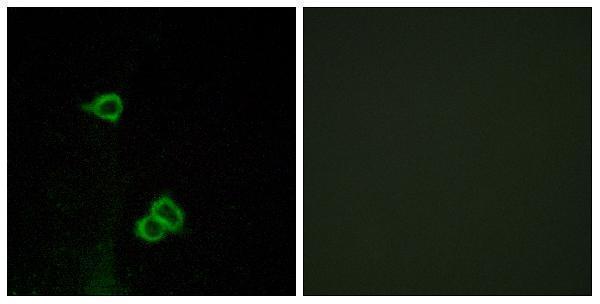
Real IF data Immunofluorescence analysis of COS7 cells, using CXCR7 Antibody. The picture on the right is blocked with the synthesized peptide.
Anti-CXCR7 ACKR3 Antibody
Cat # A30784

A02656-2 is listed for human IHC, with images from paraffin sections of breast, colon, endometrial, and urothelial cancers (catalog applications, reactivity, and image captions). A30784 is listed for IF/ICC with human, monkey, mouse, and rat reactivity, and its IF image shows COS7 cells (catalog applications, reactivity, and image caption).

Which to pick: Choose A02656-2 for tissue IHC: its images show paraffin sections, while the fixative is unreported (A02656-2 image captions). Choose A30784 for IF/ICC; its IF image shows COS7 cells, and its listed reactivity covers human, monkey, mouse, and rat for cross-species planning (A30784 applications, image caption, and reactivity). Both antibodies are listed as polyclonal (catalog dilution notes).

Each figure is that product's own IHC / IF validation image from its datasheet.

References

  1. UniProt Consortium. UniProt entry P25106 (ACKR3_HUMAN, Atypical chemokine receptor 3).
  2. Human Protein Atlas. ACKR3 tissue IHC expression (reliability: Approved).
  3. Human Protein Atlas. ACKR3 subcellular location (ICC-IF): Localized to the plasma membrane and vesicles..
  4. Human Protein Atlas. ACKR3 antibody validation summary (2 antibodies).
  5. CXCR4/ACKR3/CXCL12 axis in the lymphatic metastasis of vulvar squamous cell carcinoma. Journal of clinical pathology 2022 — PMC9046756.
  6. Circulating hsa_circ_0072309, acting via the miR-100/ACKR3 pathway, maybe a potential biomarker for the diagnosis, prognosis, and treatment of brain metastasis from non-small-cell lung cancer. Cancer medicine 2023 — PMC10523940.
  7. Breast Cancer: An Examination of the Potential of ACKR3 to Modify the Response of CXCR4 to CXCL12. International journal of molecular sciences 2018 — PMC6274818.
  8. Constructing a chemokine-based model and identifying CCL17 as a core biomarker associated with immune infiltrates in thyroid cancer. Translational cancer research 2025 — PMC12554474.
  9. PubMed PMID:1675791 — UniProt-cited evidence.
  10. PubMed PMID:14702039 — UniProt-cited evidence.
  11. PubMed PMID:15815621 — UniProt-cited evidence.