This website uses cookies to ensure you get the best experience on our website.


- Table of Contents
Plan chromogenic ACOT13 IHC in paraffin sections using liver hepatocytes or kidney tubule cells as positive tissue references (HPA tissue IHC). The catalog antibody starts at 2.5 μg/mL (datasheet); assess granular cytoplasmic staining against a negative control (HPA tissue IHC; standard IHC practice).

Expected localisation, validated protocols, controls and antibodies — the at-a-glance facts below, then the full design guide.
| Expected localisation | Granular cytoplasm in tissue sections (HPA tissue IHC) | |
| Staining pattern | Granular cytoplasm in hepatocytes and kidney tubule cells (HPA tissue IHC) | |
| Antigen retrieval | Tris-EDTA pH 9.0 HIER, 95–98 °C, 20 min (rule: nuclear antigen) | |
| Positive control | Gallbladder+4 more · see all | |
| Negative control | Adipose tissue+4 more · see all |
| Fixation | Matched tissue-IHC evidence does not establish the fixation claim. Validate the specimen-specific method before use. (selected-SKU IHC image A07288-1) | |
| Caveat | Antibody–RNA consistency is medium (HPA tissue IHC) | |
| Regulation | No regulatory stimulus specified (UniProt record) | |
| Isoform / epitope | 2 isoforms; N-terminal processing may affect N-terminal epitopes (UniProt) |
Compare the catalog antibody’s IHC-P protocol with the published rat caudal intervertebral disc protocol (PMC12983563 methods).
| Sample | Tissue sections; selected-image fixative not specified (standard IHC workflow) |
| Fixation | Image fixative and duration unreported (datasheet A07288-1); verify before use. |
| Sectioning | 4–5 µm sections on charged slides (standard) |
| Deparaffinisation | Xylene, graded ethanol series to water (standard) |
| Antigen retrieval | Heat-induced epitope retrieval in Tris-EDTA buffer, pH 9.0, 20 min at 95–98 °C (standard rule: nuclear antigen) |
| Peroxidase block | 3% H2O2, 10 min, room temperature (standard) |
| Blocking | 10% normal serum of the secondary host, 30 min, room temperature (standard) |
| Primary antibody | Rabbit anti-ACOT13, 2.5 μg/mL (datasheet A07288-1) |
| Primary incubation | Overnight at 4 °C (standard) |
| Detection | HRP-polymer secondary, DAB chromogen 5–10 min (standard) |
| Counterstain | Hematoxylin, blue, dehydrate and mount (standard) |
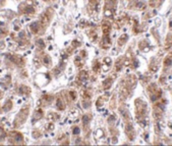
| Expected result | ACOT13-positive staining in glandular cells of gallbladder (HPA tissue IHC: High). HPA tissue profile: General cytoplasmic expression with a granular pattern. No signal in the no-primary control. |
ACOT13 should show chiefly granular cytoplasmic staining in tissue sections (HPA tissue IHC: general cytoplasmic expression with a granular pattern). Strong examples include hepatocytes, kidney tubule cells and gallbladder glandular cells (HPA tissue IHC: High). UniProt also lists cytosol, mitochondria, nucleus and mitotic spindle, and reports no transmembrane segment (UniProt Q9NPJ3). Interpret compartment differences cautiously: HPA rates tissue staining Enhanced, with medium consistency against RNA data (HPA tissue IHC: reliability).
| Granular cytoplasmic staining in hepatocytes or kidney tubule cells, with clear cell outlines. | This matches the reported tissue pattern and High staining in those cells (HPA tissue IHC). Compare the named cell population and its cytoplasmic distribution; intensity elsewhere need not match it (HPA tissue IHC: tissue-specific levels). |
| Staining appears exclusively nuclear, or as a continuous rim around most cells. | Neither is the reported general tissue IHC pattern (HPA tissue IHC: granular cytoplasm). UniProt does list nuclear ACOT13 during interphase, so nuclear signal alone cannot establish an artefact (UniProt Q9NPJ3). Check morphology and controls before assigning localisation (general IHC practice). |
| Strong staining dominates adipocytes, smooth muscle cells or oral squamous epithelial cells. | Those populations are listed as Not detected in the HPA tissue examples (HPA tissue IHC). Unexpected signal raises cross-reactivity or detection-background questions; it does not establish either cause by itself. Review cell identity and control sections (general IHC practice). |
| Color spreads across extracellular areas or the whole section without discernible cellular boundaries. | That appearance cannot be scored as the reported granular cytoplasmic pattern (HPA tissue IHC). Excess detection background or endogenous activity can produce broad color in chromogenic IHC; inspect a negative reagent control and section pigment (general IHC practice). |
| Hepatocytes remain unstained in an otherwise readable section. | Hepatocytes are a High HPA tissue example (HPA tissue IHC), so absent signal calls for checking that the positive control and detection run worked. It does not, on its own, show that the specimen lacks ACOT13 (general IHC practice). |
| Tissue and cell selection | Hepatocytes, kidney tubule cells, gallbladder glandular cells, ovary follicle cells, parathyroid glandular cells and late spermatids are High examples (HPA tissue IHC). Score the specified cells within each tissue; HPA levels are observations, not a guarantee for every section. |
| Compartment evidence | Tissue IHC is generally granular and cytoplasmic (HPA tissue IHC). UniProt additionally lists nucleus and mitotic spindle, including interphase nuclear localisation and mitotic tubulin colocalisation (UniProt Q9NPJ3). These annotations do not make nuclear-only tissue staining the expected IHC result. |
| Antibody validation | HPA019881 and HPA057134 each have Enhanced IHC status (HPA antibodies). HPA describes Enhanced as reproduction by independent antibodies or orthogonal data; the tissue summary still reports medium consistency with RNA expression (HPA tissue IHC). Interpret a new reagent against its own controls. |
| Isoforms and processing | UniProt lists 2 isoforms and chains beginning at residue 1 or 2, including an N-terminally processed form (UniProt Q9NPJ3). No antibody epitope is supplied here, so these facts cannot predict whether the catalog antibody stains the forms differently. |
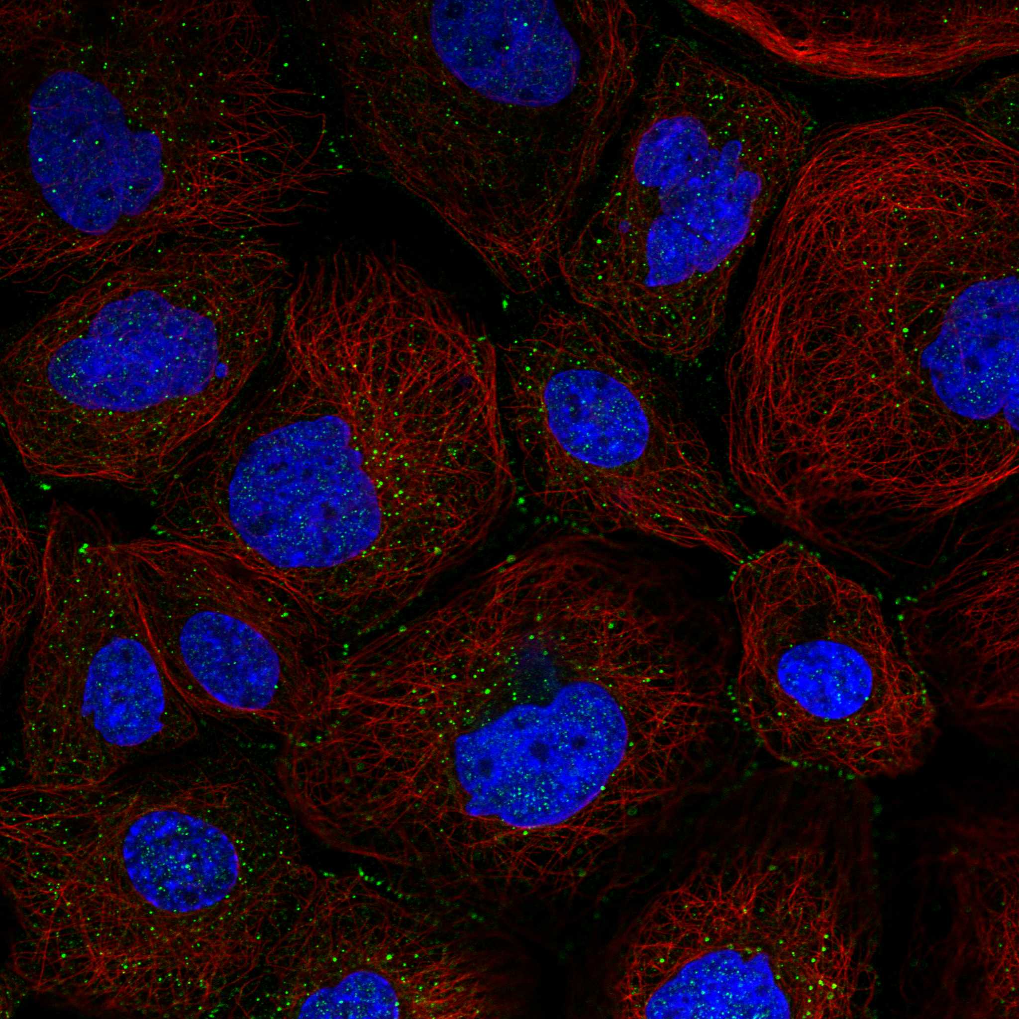
| IF/ICC Q&A | What if IF shows junctional signal? HPA ICC-IF reports mainly cell junction localisation and marks it Approved, with images in A-431 and U-251MG (HPA subcellular). That separate observation does not replace the granular cytoplasmic expectation for tissue IHC (HPA tissue IHC). |
| Situation | Likely cause | Next action |
|---|---|---|
| No color in the ACOT13 positive control. | A failed detection run or unsuitable staining conditions are possible (general IHC practice); HPA reports High hepatocyte and kidney tubule staining (HPA tissue IHC). | Confirm tissue identity, section integrity, reagent sequence and detection-control performance; review the catalog antibody's IHC-P instructions before changing conditions (general IHC practice). |
| Positive cells show faint, hard-to-score color. | HPA levels vary by cell population; cerebral cortical neurons and lung macrophages are Low examples (HPA tissue IHC). | Score a High example such as hepatocytes beside the test section, then assess cell-specific color and background on the same run (HPA tissue IHC; general IHC practice). |
| Color covers tissue and empty areas alike. | Detection background or endogenous chromogenic activity may obscure cellular staining (general IHC practice). | Inspect a control without primary antibody and any native pigment; check blocking, washes and detection timing using the established IHC workflow (general IHC practice). |
| Unexpected strong color appears in HPA Not detected cells. | Cross-reactivity, endogenous activity or mistaken cell identification are possibilities; HPA lists adipocytes and smooth muscle cells as Not detected examples (HPA tissue IHC; general IHC practice). | Verify cell morphology and compare control staining. Treat the result as unresolved until its cellular pattern and background are accounted for (general IHC practice). |
| A section appears predominantly nuclear. | UniProt permits nuclear localisation during interphase, while HPA describes general tissue IHC as granular cytoplasmic (UniProt Q9NPJ3; HPA tissue IHC). | Check whether cytoplasmic granules are also present and whether nuclear color persists in the negative reagent control; avoid assigning specificity from compartment alone (general IHC practice). |
| IF/ICC shows junctional signal while tissue IHC looks cytoplasmic. | The assays have different reported observations: junctions in HPA ICC-IF and granular cytoplasm in HPA tissue IHC (HPA subcellular; HPA tissue IHC). | Interpret each image against its own assay and control evidence. Use the dedicated IF/ICC guide for that experiment; do not use the ICC-IF location as a tissue IHC scoring rule (HPA subcellular; HPA tissue IHC). |

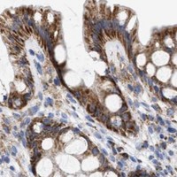
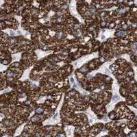


Comprehensive Human Protein Atlas IHC scoring per tissue (reliability: Enhanced — Medium consistency between antibody staining and RNA expression data.). Rows are taken directly from the HPA tissue chart — click any row's HPA link to view the source.
| Tissue | Cell type | Level | Evidence | Source |
|---|---|---|---|---|
| Gallbladder | Glandular cells | High | Protein (IHC) | HPA → |
| Kidney | Cells in tubules | High | Protein (IHC) | HPA → |
| Liver | Hepatocytes | High | Protein (IHC) | HPA → |
| Ovary | Follicle cells | High | Protein (IHC) | HPA → |
| Parathyroid gland | Glandular cells | High | Protein (IHC) | HPA → |
| Tissue | Cell type | Level | Evidence | Source |
|---|---|---|---|---|
| Adipose tissue | Adipocytes | Not detected | Protein (IHC) | HPA → |
| Oral mucosa | Squamous epithelial cells | Not detected | Protein (IHC) | HPA → |
| Smooth muscle | Smooth muscle cells | Not detected | Protein (IHC) | HPA → |
| Soft tissue | Fibroblasts | Not detected | Protein (IHC) | HPA → |
| Spleen | Cells in red pulp | Not detected | Protein (IHC) | HPA → |
Troubleshoot ACOT13 staining in paraffin sections by checking retrieval, cell type, subcellular pattern and controls before interpreting chromogenic signal.
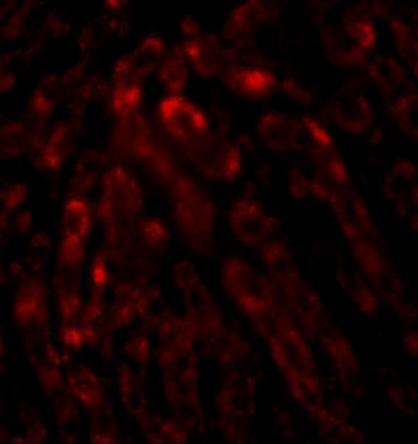
A07288-1 has human liver IHC and IF images (catalog image captions) and lists human, mouse, and rat reactivity (catalog: reactivity).
A07288-1 is listed for IHC-P and IF (catalog: applications). Its images show human liver IHC at 2.5 μg/mL and IF at 20 μg/mL (catalog image captions).
Which to pick: For paraffin-section tissue IHC, choose A07288-1: it lists IHC-P and shows human liver staining at 2.5 μg/mL; the caption does not report the fixative (catalog: applications; IHC image caption). For fluorescence, A07288-1 lists IF and shows human liver staining at 20 μg/mL; ICC validation is not listed (catalog: applications; IF image caption). For cross-species work, A07288-1 lists human, mouse, and rat reactivity and has a rabbit host, but the supplied images show human tissue only (catalog: reactivity, host, image captions).