ACOT2 / Acyl-coenzyme A thioesterase 2, mitochondrial · IHC design guide

Design Immunohistochemistry for ACOT2

Plan chromogenic IHC on paraffin sections with the catalog antibody at 1:100–1:300 (datasheet: IHC dilution). Compare staining with the cytoplasmic, mitochondrial tissue pattern (HPA tissue IHC), using highly stained kidney collecting ducts as a reference (HPA tissue IHC).

Evidence assembled Oct 2026 · For research use; verify linked source records and product datasheet before use
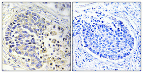
Immunohistochemistry protocol sheet for ACOT2 (IHC for ACOT2): expected localisation Cytoplasmic, with a mitochondrial pattern (HPA tissue IHC), antibody A10261-1, validated IHC image, and IHC protocol steps
Printable ACOT2 IHC protocol sheet — expected localisation Cytoplasmic, with a mitochondrial pattern (HPA tissue IHC), antibody A10261-1, controls and protocol steps. Open the full ACOT2 IHC guide →

ACOT2 Immunohistochemistry Experimental Design Guide

Expected localisation, validated protocols, controls and antibodies — the at-a-glance facts below, then the full design guide.

Must know before staining
Expected localisation Cytoplasmic, with a mitochondrial pattern (HPA tissue IHC)
Staining pattern Kidney collecting ducts: cytoplasmic, mitochondrial pattern (HPA tissue IHC)
Antigen retrieval Citrate pH 6.0 HIER, 95–98 °C, 20 min (rule: cytoplasmic / membrane antigen)
Positive control ⓘ Appendix+4 more · see all
Negative control ⓘ Adipose tissue
Important caveats
Reasons your staining may differ from the expected pattern.
Fixation Keep fixation consistent across sections (standard IHC practice; not target-specific)
Caveat Antibody staining may reflect proteins from more than one gene (HPA tissue IHC)
Regulation No staining-linked regulator reported (UniProt)
Isoform / epitope 2 isoforms; epitope impact is unreported (UniProt)
Section 1

Recommended ACOT2 IHC & IF Protocols

Compare the catalog antibody’s IHC-P protocol with the published mouse brain IHC protocol (PMC11005155).

Recommended immunohistochemistry (IHC-P) protocol parameters
SampleParaffin-embedded human lung cancer tissue; fixative not specified (datasheet A10261-1)
FixationImage fixative and duration unreported (datasheet A10261-1); verify before use.
Sectioning4–5 µm sections on charged slides (standard)
DeparaffinisationXylene, graded ethanol series to water (standard)
Antigen retrievalHeat-induced epitope retrieval in citrate buffer, pH 6.0, 20 min at 95–98 °C (standard rule: cytoplasmic / membrane antigen)
Peroxidase block3% H2O2, 10 min, room temperature (standard)
Blocking10% normal serum of the secondary host, 30 min, room temperature (standard)
Primary antibodyRabbit anti-ACOT2, 1:100-1:300 (datasheet A10261-1)
Primary incubationOvernight at 4 °C (standard)
DetectionHRP-polymer secondary, DAB chromogen 5–10 min (standard)
CounterstainHematoxylin, blue, dehydrate and mount (standard)
Expected resultACOT2-positive staining in endocrine cells of appendix (HPA tissue IHC: High). HPA tissue profile: Cytoplasmic expression wit a mitochondrial pattern in most tissues. No signal in the no-primary control.
💡Decision noteStart with citrate pH 6.0 HIER at 95–98 °C for 20 min (page antigen-retrieval setting).
Section 2

What Is the Expected ACOT2 Staining Pattern?

ACOT2 is mitochondrial and has no annotated transmembrane segment (UniProt P49753: subcellular location and topology). In paraffin section IHC, expect cytoplasmic staining with a mitochondrial pattern in many tissues, including strong staining in kidney collecting ducts and bronchial respiratory epithelial cells (HPA tissue IHC: High). HPA rates the tissue profile Enhanced, while warning that its antibodies may also target proteins from more than one gene (HPA tissue IHC: reliability description).

What am I looking at on my slide?
Granular cytoplasmic staining in kidney collecting ducts or bronchial respiratory epithelial cells.This fits the reported mitochondrial pattern and High staining in those cell types (HPA tissue IHC: profile; kidney and bronchus). Judge the pattern within the named cells; whole-section intensity alone cannot show whether the expected cells are positive (general IHC practice).
Predominantly nuclear or crisp plasma-membrane staining, without a cytoplasmic mitochondrial pattern.That compartment conflicts with mitochondrial localisation (UniProt P49753: subcellular location; HPA subcellular: mitochondria). Treat it as suspect and compare the same run with a known-positive tissue before interpreting it as ACOT2 (general IHC practice).
Strong staining in adipocytes, especially when expected positive cells stain weakly.HPA reports adipocytes as Not detected in adipose tissue (HPA tissue IHC: adipose tissue). Unexpected staining could reflect cross-reactivity or endogenous detection activity; the image alone cannot distinguish those causes (general IHC practice).
A uniform diffuse tint across cells, extracellular space, and the section background.This lacks the reported cytoplasmic mitochondrial pattern (HPA tissue IHC: profile). Check reagent background and washing with appropriate controls before scoring cellular positivity (general IHC practice).
No staining in kidney collecting ducts or bronchial respiratory epithelial cells.Those cells are reported High by HPA (HPA tissue IHC: kidney and bronchus). A blank known-positive section makes a technical failure plausible; assess controls and the staining run before calling a test specimen negative (general IHC practice).
💡Expected ACOT2 appearanceCall a result positive when named cells show clear cytoplasmic staining with a mitochondrial pattern, especially the High-staining kidney collecting ducts or bronchial respiratory epithelium (HPA tissue IHC); isolated nuclear staining or strong adipocyte staining is suspect (UniProt P49753: mitochondrion; HPA tissue IHC: adipocytes Not detected).
How each factor affects the staining
Cell type within the sectionCompare corresponding cells, not entire organs: HPA calls kidney collecting ducts High but adipocytes in adipose tissue Not detected (HPA tissue IHC).
Strength of tissue evidenceHPA rates the tissue IHC profile Enhanced but notes medium agreement with RNA and possible targeting of more than one gene (HPA tissue IHC: reliability description). Interpret unexpected staining cautiously.
Antibody validationHPA043705 is Supported for IHC; HPA060170 is Enhanced for IHC (HPA antibodies). Record which antibody generated the image when comparing patterns.
Isoforms and epitopeUniProt lists two ACOT2 isoforms but supplies no epitope location here (UniProt P49753: isoforms). Do not infer isoform-specific IHC staining from this record.
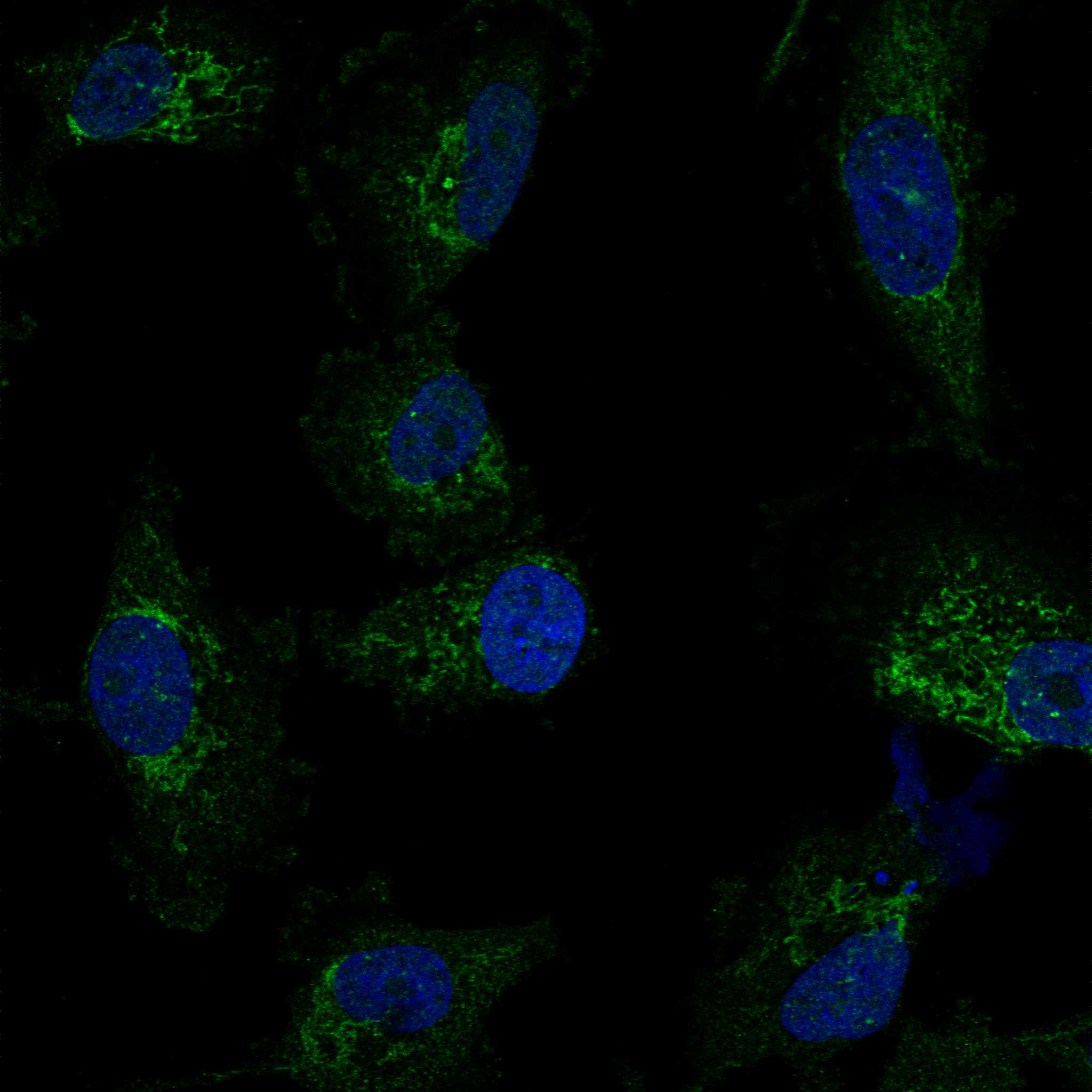
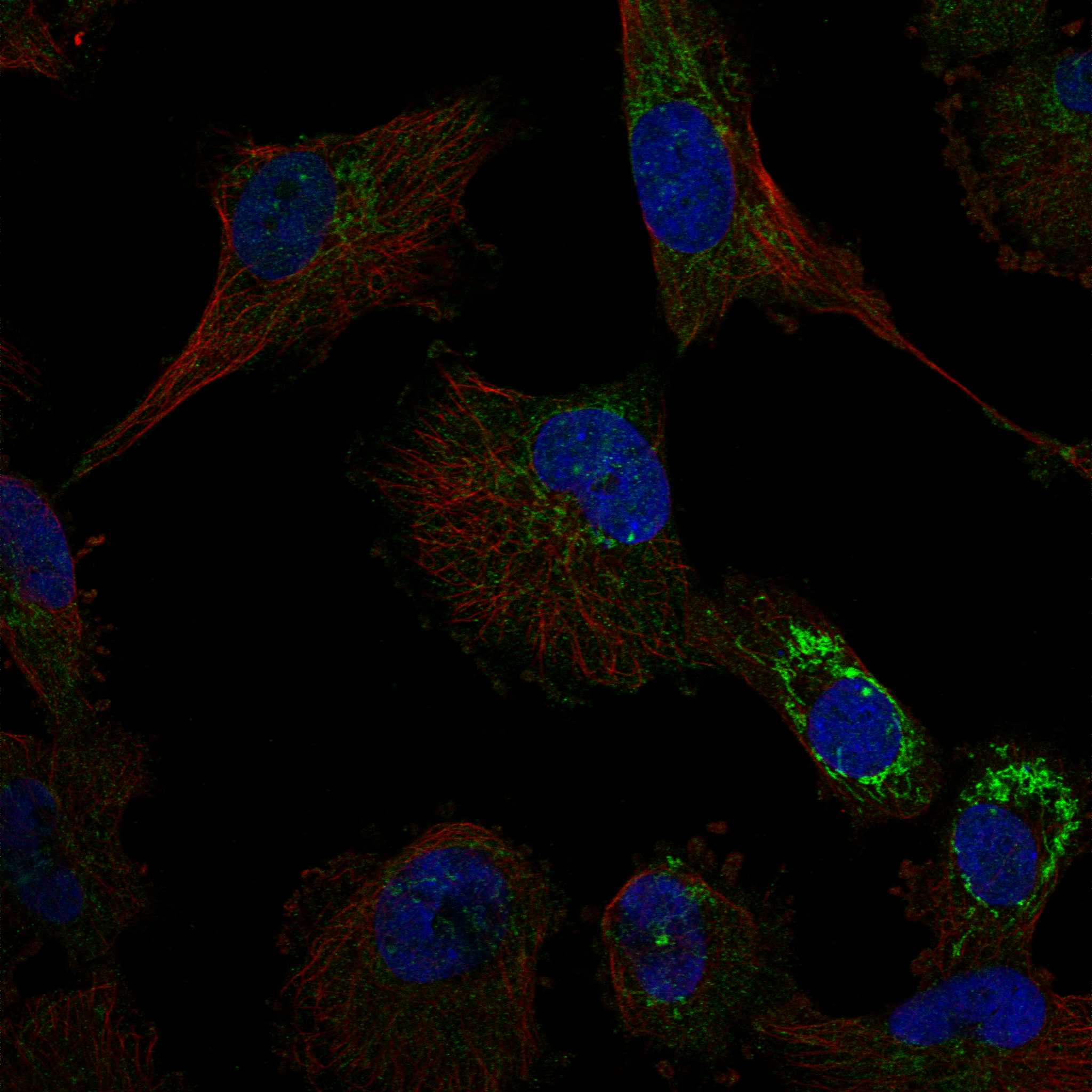
IF/ICC question: what location should fluorescence show?Mitochondrial localisation is approved in HPA ICC-IF images from U-251MG and U2OS; HPA cautions that the antibodies may target proteins from multiple genes (HPA subcellular).
Why is my staining missing, weak or wrong?
SituationLikely causeNext action
Known-positive collecting ducts or respiratory epithelium are blank.The run may have failed; these cells are reported High (HPA tissue IHC: kidney and bronchus).Check that control tissue stained, then review retrieval, primary-antibody dilution, detection reagents, and counterstain for this IHC run (general IHC practice).
Staining is mainly nuclear or outlines the cell surface.The distribution disagrees with mitochondrial localisation (UniProt P49753; HPA subcellular).Compare with a known-positive section and a control omitting primary antibody; review staining specificity before scoring (general IHC practice).
Adipocytes stain strongly.HPA reports adipocytes in adipose tissue as Not detected; cross-reactivity or detection background is possible (HPA tissue IHC; general IHC practice).Check a control omitting primary antibody and compare adipocytes with expected positive cells in the same run (general IHC practice).
The entire section has a diffuse brown haze.Non-specific reagent binding, incomplete washing, or endogenous chromogenic detection activity may contribute (general IHC practice).Inspect the control omitting primary antibody, review blocking and washes, and address endogenous enzyme activity if the detection system requires it (general IHC practice).
Many unexpected cell types stain as strongly as the expected cells.HPA warns that antibodies may target proteins from more than one gene; widespread signal alone cannot establish ACOT2 specificity (HPA tissue IHC: reliability description).Compare cell-level patterns with HPA and, when available, staining from an independently validated antibody (HPA tissue IHC: profile; general IHC practice).
A weak test section is being called ACOT2-negative.Some listed cells have Low staining, while HPA reports a cytoplasmic mitochondrial pattern in most tissues (HPA tissue IHC).First confirm an expected High-staining control in the same run, then score the relevant cell type and localisation in the test section (HPA tissue IHC; general IHC practice).

Sample controls for ACOT2 IHC & IF

🧪Run kidney first; collecting ducts should stain (HPA: Kidney collecting ducts, High). Run adipose tissue as the negative tissue; adipocytes are not detected (HPA: Adipose tissue adipocytes, Not detected). On the kidney slide, unstained neighboring cells can show background staining, but the supplied rows do not establish a specific kidney cell type as negative (HPA: Kidney collecting ducts, High).
Positive control tissue: Appendix (Endocrine cells, HPA High)
Negative control tissue: Adipose tissue (HPA Not detected)
ICC-IF cell lines (HPA subcellular resource): HPA ICC-IF images show ACOT2 in U-251MG, U2OS, with annotated localisation: Mitochondria (approved) (HPA subcellular).
Technical controls: Include a no-primary (secondary-only) control, a host-species/class-matched isotype control for a monoclonal antibody or matched normal host serum for a polyclonal antibody, and ACOT2 knockout tissue or a cognate-peptide block if available (standard IHC practice). For kidney chromogenic IHC, quench endogenous peroxidase and check endogenous biotin if using avidin–biotin detection (standard IHC practice).
⚠️Feasibility: A target-specific fixation window or fixation effect is unreported, and the selected A10261-1 paraffin-section caption does not state a fixative (caption: fixative not stated). Antigen-retrieval dependence is unreported; optimize retrieval empirically for IHC-P (standard IHC practice). The supplied evidence does not establish whether frozen sections or IF/ICC are easier; in kidney, endogenous biotin can complicate avidin–biotin detection (standard IHC practice).

HPA tissue IHC evidence for ACOT2

Comprehensive Human Protein Atlas IHC scoring per tissue (reliability: Enhanced — Medium consistency between antibody staining and RNA expression data. Caution, targets protein from more than one gene.). Rows are taken directly from the HPA tissue chart — click any row's HPA link to view the source.

Positive expression · recommended positive controls

TissueCell typeLevelEvidenceSource
Appendix Endocrine cells High Protein (IHC) HPA →
Bronchus Respiratory epithelial cells High Protein (IHC) HPA →
Caudate Glial cells High Protein (IHC) HPA →
Duodenum Endocrine cells High Protein (IHC) HPA →
Kidney Collecting ducts High Protein (IHC) HPA →

Undetected expression · recommended negative controls

TissueCell typeLevelEvidenceSource
Adipose tissue Adipocytes Not detected Protein (IHC) HPA →
Section 3

Advanced ACOT2 IHC Tips

Troubleshooting ACOT2 staining in paraffin sections requires attention to mitochondrial localisation, cell identity, and antibody specificity (UniProt P49753; HPA tissue IHC).

Which retrieval conditions should I start with for ACOT2 chromogenic IHC?
Start with citrate buffer at pH 6.0 for heat induced antigen retrieval at 95–98 °C for 20 minutes (page IHC retrieval protocol). Let slides cool in buffer, then keep wash conditions consistent across test sections so retrieval is the main variable (standard IHC practice). The selected A10261-1 image shows staining in paraffin embedded human lung cancer at 1:100, but its caption does not report retrieval conditions (A10261-1 IHC caption). If staining remains weak, test another retrieval condition on adjacent sections while holding antibody dilution and detection constant (standard IHC practice). Judge improvement by cytoplasmic staining with a mitochondrial pattern in intact cells, while accounting for the reported antibody specificity caution (HPA tissue IHC).
Could fixation explain weak or uneven ACOT2 staining?
ACOT2 specific sensitivity to fixation is unknown from the supplied evidence, so do not assign weak staining to a particular fixative or fixation duration (A10261-1 IHC caption: fixative unreported). The selected image documents paraffin embedded human lung cancer and a 1:100 antibody dilution, without identifying the fixative (A10261-1 IHC caption). Compare sections with documented processing histories, using the same citrate pH 6.0 retrieval and detection run before changing several variables (page IHC retrieval protocol; standard IHC practice). Record section thickness, processing history and tissue preservation when assessing uneven staining (standard IHC practice). Neither mitochondrial localisation nor the annotated modified residues establish a fixation effect (UniProt P49753).
Where should convincing ACOT2 staining appear in tissue sections?
Look for cytoplasmic staining with a mitochondrial pattern in intact cells, consistent with the reported tissue profile and mitochondrial localisation (HPA tissue IHC; UniProt P49753). ACOT2 has no annotated transmembrane segment, so a crisp plasma membrane outline alone would not match its recorded topology (UniProt P49753 topology). Compare stained cells with their neighbours within the same section; collecting ducts in kidney and respiratory epithelial cells in bronchus are reported as high staining examples (HPA tissue IHC). Review morphology before calling coarse pigment or diffuse staining mitochondrial (standard IHC practice). Because the HPA antibody evidence may include proteins from more than one gene, location alone cannot establish ACOT2 identity (HPA tissue IHC).
How do isoforms and epitope uncertainty affect an ACOT2 IHC result?
ACOT2 has two recorded isoforms, but the supplied antibody evidence does not map the recognized epitope to either one (UniProt P49753; A10261-1 IHC caption). Treat staining as evidence from the antibody assay rather than an isoform specific measurement until epitope coverage is documented (standard IHC interpretation). UniProt lists modified lysines at positions 104 and 470, without evidence here that either modification changes staining (UniProt P49753 modified residues). Compare any epitope information obtained later with the isoform sequences before making isoform claims (standard antibody validation practice). The HPA warning about recognition of proteins from multiple genes adds a separate specificity concern (HPA tissue IHC).
How should I assess ACOT2 by multiplex IF alongside this IHC guide?
Treat IF conditions as a separate assay: the selected A10261-1 caption documents paraffin section IHC, while HPA reports mitochondrial ICC/IF localisation with an antibody specificity caution (A10261-1 IHC caption; HPA subcellular). For a bronchus study, pair ACOT2 with an epithelial marker to identify the respiratory epithelial cells reported as high staining (HPA tissue IHC; standard multiplex IF practice). Choose spectrally separated fluorophores after checking tissue autofluorescence in unstained controls, and place the weaker signal in the cleaner channel (standard IF practice). Because the antibody epitope is unspecified and ACOT2 lacks an annotated transmembrane segment, optimise permeabilisation for access to its mitochondrial epitope rather than assuming a membrane facing side (UniProt P49753 topology; standard IF practice).
What causes diffuse or granular background in ACOT2 chromogenic IHC?
First inspect a no primary control and the tissue edge; signal there can reveal detection background or an edge effect (standard IHC practice). Use a peroxidase block before DAB detection, then check whether endogenous pigment or enzyme activity persists without primary antibody (standard chromogenic IHC practice). Titrate the antibody around the caption's 1:100 condition and compare matched sections, since that image alone does not define an optimum for every specimen (A10261-1 IHC caption; standard IHC practice). Preserve the same citrate pH 6.0 retrieval during the dilution comparison (page IHC retrieval protocol). Require interpretable cytoplasmic mitochondrial pattern and cell morphology before treating granular colour as ACOT2 staining (HPA tissue IHC; standard IHC interpretation).
How should I score ACOT2 staining across tissue sections? ⚠ ANSWER MARKED FOR VERIFICATION
Define cell populations and compartments before scoring, because HPA reports a cytoplasmic mitochondrial pattern across tissues and high staining in specific cell types (HPA tissue IHC). For a defined population, record the percentage of positive cells and intensity, or calculate an H-score using the same thresholds for every slide (standard IHC quantification practice). If comparing fields with different cellularity, report positive cells per mm² and normalise to viable tissue area or the relevant cell population (standard IHC quantification practice). Exclude necrosis, folds and section edges by prespecified morphology rules (standard IHC practice). Report the antibody specificity limitation alongside scores, since HPA cautions that its staining may include proteins from more than one gene (HPA tissue IHC).
When is an apparent ACOT2 positive cell likely to be an artefact?
A convincing result combines intact cell morphology with cytoplasmic mitochondrial pattern, consistent with the recorded localisation (HPA tissue IHC; UniProt P49753). Be cautious with isolated nuclear or plasma membrane staining: neither is the reported main location, and ACOT2 has no annotated transmembrane segment (HPA subcellular; UniProt P49753 topology). Check whether the stained cell type fits the tissue context; HPA reports high signal in kidney collecting ducts but no detection in adipocytes from adipose tissue (HPA tissue IHC). Reject staining confined to cut edges, necrosis or a no primary control as assay artefact (standard IHC practice). Even plausible staining needs independent specificity support because HPA warns its antibodies may recognize proteins from multiple genes (HPA tissue IHC).
Boster reagents

Best ACOT2 / Acyl-coenzyme A thioesterase 2, mitochondrial IHC Antibodies

A10261-1 has an IHC figure from paraffin-embedded human lung cancer (IHC image caption). IF is listed as an application, but no IF figure is supplied (catalog applications; image inventory).

Real IHC data Immunohistochemistry validation of ACOT2 using Anti-ACOT2 Antibody (A10261-1). Immunohistochemical analysis of paraffin-embedded human lung cancer. Antibody was diluted at 1:100 (4°C
Anti-ACOT2 Antibody
Cat # A10261-1

A10261-1 will render with its IHC image of paraffin-embedded human lung cancer (IHC image caption). The catalog lists IHC and IF applications and human, mouse and rat reactivity for A10261-1; it supplies no IF image (catalog applications; catalog reactivity; image inventory).

Which to pick: For tissue IHC, choose A10261-1: its image documents paraffin-embedded human lung cancer at 1:100, while the fixative is unreported (IHC image caption). For IF, A10261-1 has a listed dilution of 1:200–1:1000, but ICC validation and an IF image are unreported (catalog applications; catalog IF dilution; image inventory). For mouse or rat samples, A10261-1 lists reactivity with both species, although its IHC image shows human tissue only; clonality is unreported (catalog reactivity; IHC image caption; catalog clone field).

Each figure is that product's own IHC / IF validation image from its datasheet.