This website uses cookies to ensure you get the best experience on our website.


- Table of Contents
Plan ACOT8 chromogenic IHC in paraffin sections with a starting antibody dilution of 1:100–1:300 (datasheet A09392-1). Use liver hepatocytes or kidney tubules as high-staining reference cells (HPA tissue IHC), and assess cytoplasmic staining that may appear granular (HPA tissue IHC).

Expected localisation, validated protocols, controls and antibodies — the at-a-glance facts below, then the full design guide.
| Expected localisation | Observed cytoplasm; peroxisome matrix expected (HPA tissue IHC; UniProt) | |
| Staining pattern | Cytoplasmic staining, sometimes granular, across tissues (HPA tissue IHC) | |
| Antigen retrieval | Tris-EDTA pH 8.0 HIER, heat-mediated (datasheet A09392-1) | |
| Positive control | Adrenal gland+4 more · see all | |
| Negative control | None in HPA (detected in all 44 tissues); use no-primary + isotype controls |
| Fixation | Keep paraffin-section fixation consistent (standard IHC practice; not target-specific); Matched tissue-IHC evidence does not establish the fixation claim. Validate the specimen-specific method before use. (selected-SKU IHC image A09392-1) | |
| Caveat | Cytosolic signal may accompany peroxisomal signal (UniProt) | |
| Regulation | Ubiquitous expression (UniProt) | |
| Isoform / epitope | No annotated isoforms or signal/propeptide; epitope unknown (UniProt) |
The catalog antibody’s IHC-P protocol (datasheet: A09392-1) is supplemented by one published ACOT8 protocol for breast cancer tissue (PMC11346483).
| Sample | Paraffin-embedded Human brain tissue; fixative not specified (datasheet A09392-1) |
| Fixation | Image fixative and duration unreported (datasheet A09392-1); verify before use. |
| Sectioning | 4–5 µm sections on charged slides (standard) |
| Deparaffinisation | Xylene, graded ethanol series to water (standard) |
| Antigen retrieval | Heat retrieval: Tris-EDTA pH 8.0 (datasheet A09392-1); 20 min, 95–100 °C (standard) |
| Peroxidase block | 3% H2O2, 10 min, room temperature (standard) |
| Blocking | 10% normal serum of the secondary host, 30 min, room temperature (standard) |
| Primary antibody | Rabbit anti-ACOT8, 1:100-1:300 (datasheet A09392-1) |
| Primary incubation | Overnight at 4 °C (standard) |
| Detection | HRP-polymer secondary, DAB chromogen 5–10 min (standard) |
| Counterstain | Hematoxylin, blue, dehydrate and mount (standard) |
| Expected result | ACOT8-positive staining in glandular cells of adrenal gland (HPA tissue IHC: High). HPA tissue profile: Ubiquitous cytoplasmic expression, sometimes in a granular pattern. No signal in the no-primary control. |
ACOT8 is predominantly in the peroxisome matrix, although cytosolic localization cannot be excluded; it has no transmembrane segment (UniProt O14734: localization and topology). In paraffin-section IHC, expect cytoplasmic staining that may look granular, especially in hepatocytes, kidney tubular cells, and several glandular cell populations (HPA: tissue IHC). HPA rates the tissue staining Approved, with medium consistency between staining and RNA expression (HPA: tissue IHC reliability).
| Cytoplasmic staining, sometimes granular, is strong in hepatocytes or kidney tubular cells. | This fits the reported IHC pattern: both cell populations are rated High, and the overall profile is ubiquitous cytoplasmic expression that can be granular (HPA: tissue IHC). Granules are compatible with the predominantly peroxisomal localization, but chromogenic appearance alone does not establish peroxisome identity (UniProt O14734: localization; general IHC interpretation). |
| The nucleus is the dominant stained compartment while cytoplasm is faint or unstained. | A predominantly nuclear result does not match the reported cytoplasmic tissue pattern or peroxisome-matrix localization (HPA: tissue IHC; UniProt O14734: localization). Treat it as a possible staining artefact and compare with an appropriate control and the expected positive cells before assigning biological meaning (general IHC practice). |
| Hematopoietic cells dominate the signal while nearby expected positive cells stain weakly. | HPA rates bone-marrow hematopoietic cells Low, while hepatocytes and kidney tubular cells are High (HPA: tissue IHC). A reversal of that expected pattern warrants checks for cross-reactivity or endogenous detection activity; Low means reduced reported staining, not proven absence of ACOT8 (HPA: tissue IHC; general IHC practice). |
| Color covers many cells or the section broadly, obscuring cytoplasmic boundaries. | Diffuse background prevents a reliable call about ACOT8 localization or cell-specific intensity (general IHC interpretation). Inspect a matched negative control for nonspecific antibody binding or detection background, then reassess whether discrete cytoplasmic signal remains in expected positive cells (general IHC practice; HPA: tissue IHC). |
| A liver or kidney section shows no convincing cytoplasmic signal. | Hepatocytes and kidney tubular cells are reported High, so a blank result in either is unexpected under a working assay (HPA: tissue IHC). First check section quality, reagent performance, retrieval conditions, and detection controls as general IHC variables; the supplied sources do not establish ACOT8-specific fixation or retrieval sensitivity (general IHC practice). |
| Compartment and morphology | The peroxisome matrix is the predominant UniProt location, with cytosol not excluded; HPA describes ubiquitous cytoplasmic IHC that can be granular (UniProt O14734: localization; HPA: tissue IHC). Neither source makes every cytoplasmic granule a confirmed peroxisome. |
| Choice of comparison cells | HPA rates hepatocytes, kidney tubular cells, Purkinje cells, and listed glandular populations High; squamous epithelial cells of the vagina, smooth muscle cells, and bone-marrow hematopoietic cells are Low (HPA: tissue IHC). These are relative staining references, not absolute positive and negative controls. |
| Strength of tissue evidence | The HPA tissue profile is Approved with medium consistency between antibody staining and RNA expression; CAB010261 is IHC Approved (HPA: tissue IHC reliability; HPA: antibodies). Interpret an unusual pattern with controls rather than treating the profile as a guarantee for every section (general IHC practice). |
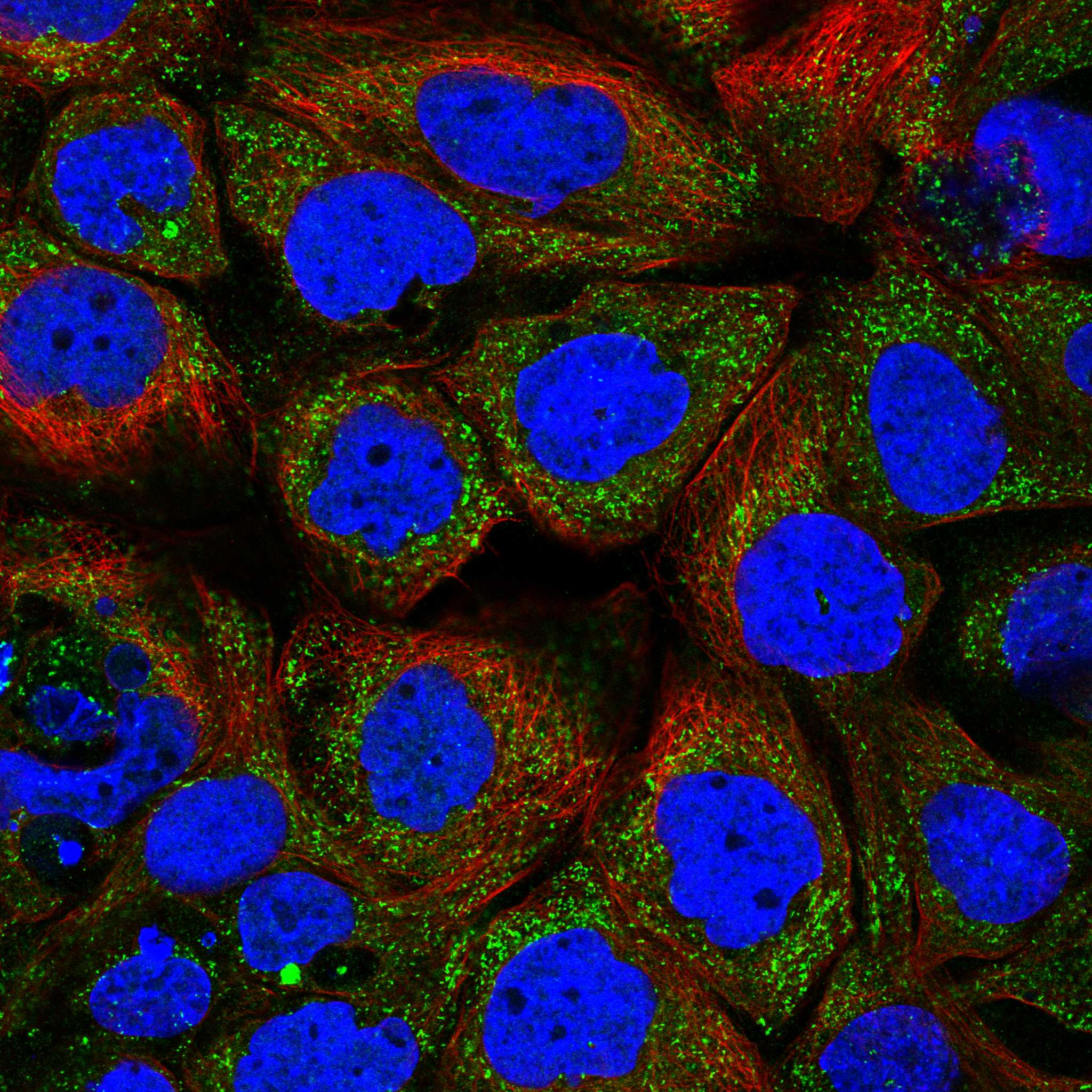
| What should IF/ICC show? | HPA reports mainly vesicular localization, with additional mitochondrial localization, in ICC-IF; its vesicle location is Supported and mitochondrial location Approved (HPA: subcellular ICC-IF). UniProt places ACOT8 predominantly in the peroxisome matrix and does not exclude cytosol (UniProt O14734: localization). Resolve a compartment claim with suitable markers rather than appearance alone (general IF practice). |
| Situation | Likely cause | Next action |
|---|---|---|
| No staining in liver hepatocytes or kidney tubular cells. | Both are High in the HPA tissue profile, so assay failure or unsuitable staining conditions are possible (HPA: tissue IHC; general IHC practice). | Check the section and detection controls, then review retrieval and antibody incubation under the chosen general IHC workflow; no ACOT8-specific fixation or retrieval effect is supplied (general IHC practice). |
| Strong signal appears mainly in nuclei. | This conflicts with the reported cytoplasmic IHC pattern and peroxisome-matrix localization; artefact is possible (HPA: tissue IHC; UniProt O14734: localization). | Compare expected positive cells with a matched negative control, then reassess the detection signal and staining specificity (general IHC practice). |
| Color appears without a clear cellular pattern. | Nonspecific background or endogenous detection activity can obscure specific chromogenic staining (general IHC practice). | Check a control lacking primary antibody and review blocking, washing, and detection controls before interpreting tissue distribution (general IHC practice). |
| Bone-marrow hematopoietic cells stain more strongly than the expected positive cells. | HPA rates these cells Low, so a strong reversal raises concern about background or cross-reactivity; Low does not mean absent (HPA: tissue IHC; general IHC interpretation). | Compare the same run with a High-rated tissue and a matched negative control; assess the cellular distribution before scoring ACOT8 (HPA: tissue IHC; general IHC practice). |
| Only weak, diffuse cytoplasm is visible in a High-rated tissue. | HPA permits cytoplasmic staining without obvious granules, but weak signal can also reflect a technical limitation (HPA: tissue IHC; general IHC practice). | Judge signal against background and an expected positive control; do not require visible puncta as the sole criterion for positivity (HPA: tissue IHC; general IHC interpretation). |
| A granular pattern is interpreted as definitive peroxisomal staining. | Granularity is reported in tissue IHC, but morphology alone cannot identify the organelle; ICC-IF also reports vesicles and mitochondria (HPA: tissue IHC; HPA: subcellular ICC-IF). | Describe the IHC result as granular cytoplasmic staining. Reserve organelle assignment for an appropriately controlled localization experiment (general IHC/IF interpretation). |
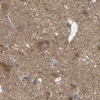


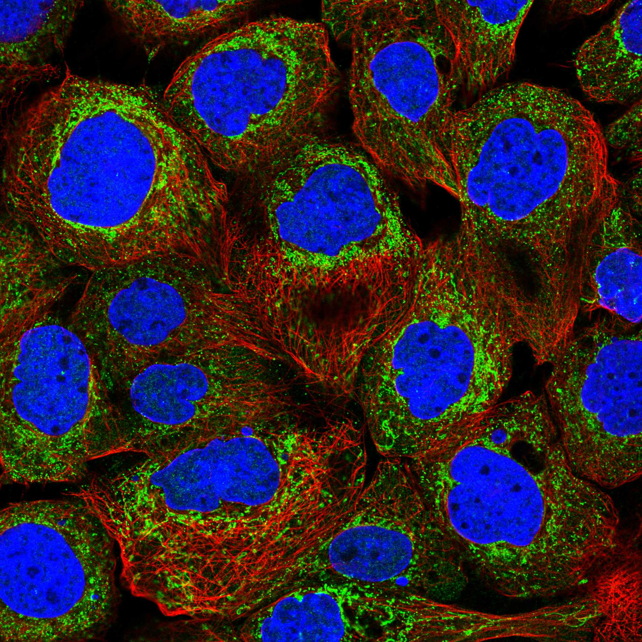
Comprehensive Human Protein Atlas IHC scoring per tissue (reliability: Approved — Medium consistency between antibody staining and RNA expression data.). Rows are taken directly from the HPA tissue chart — click any row's HPA link to view the source.
| Tissue | Cell type | Level | Evidence | Source |
|---|---|---|---|---|
| Adrenal gland | Glandular cells | High | Protein (IHC) | HPA → |
| Cerebellum | Purkinje cells | High | Protein (IHC) | HPA → |
| Colon | Glandular cells | High | Protein (IHC) | HPA → |
| Duodenum | Glandular cells | High | Protein (IHC) | HPA → |
| Epididymis | Glandular cells | High | Protein (IHC) | HPA → |
| Tissue | Cell type | Level | Evidence | Source |
|---|---|---|---|---|
| None in HPA: ACOT8 is detected in all 44 scored tissues. Use a no-primary (secondary-only) and an isotype control instead. | ||||
Use the catalog antibody’s paraffin-section evidence alongside ACOT8 localisation and tissue-expression data when troubleshooting chromogenic IHC.
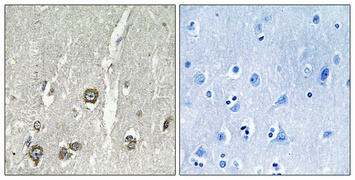
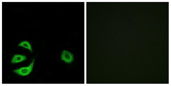
A09392-1 has IHC data from paraffin-embedded human brain (IHC image caption) and IF data from A549 cells (IF image caption). Its listed reactivity covers human, mouse and rat (catalog reactivity).
A09392-1 is shown staining paraffin-embedded human brain by IHC (IHC image caption). A09392-1 is also shown in IF images of A549 cells, including a peptide-blocked comparison (IF image caption).
Which to pick: Choose A09392-1 for paraffin-section tissue IHC: its own human-brain image used 1:100 antibody overnight at 4°C with Tris-EDTA pH 8.0 retrieval; the fixative is unreported (IHC image caption). For IF/ICC, A09392-1 is the listed option because the rabbit polyclonal antibody lists IF and has an A549-cell IF image (catalog host and dilution data; catalog applications; IF image caption). For mouse or rat work, A09392-1 lists reactivity with both species, while the supplied IHC and IF images show human brain and A549 cells, respectively (catalog reactivity; IHC image caption; IF image caption).