This website uses cookies to ensure you get the best experience on our website.


- Table of Contents
Plan chromogenic ACSL6 IHC in paraffin sections using seminal vesicle glandular cells or late spermatids as positive controls (HPA tissue IHC). This guide covers fixation consistency, antibody titration and scoring of cytoplasmic staining (standard IHC practice; HPA tissue IHC).

Expected localisation, validated protocols, controls and antibodies — the at-a-glance facts below, then the full design guide.
| Expected localisation | Cytoplasm observed; organelle membranes annotated (HPA tissue IHC; UniProt) | |
| Staining pattern | Cytoplasmic signal in glandular cells and late spermatids (HPA tissue IHC) | |
| Antigen retrieval | Citrate pH 6.0 HIER, 95–98 °C, 20 min (rule: cytoplasmic / membrane antigen) | |
| Positive control | Seminal vesicle+4 more · see all | |
| Negative control | Bone marrow+4 more · see all |
| Fixation | Keep fixation consistent across paraffin sections. (standard IHC practice; not target-specific) | |
| Caveat | Medium consistency between staining and RNA data (HPA tissue IHC) | |
| Regulation | Staining regulation unreported (UniProt) | |
| Isoform / epitope | 9 isoforms; cytoplasmic region aa 46–697 (UniProt) |
The catalog antibody’s IHC-P protocol is accompanied by published ACSL6 staining details for liver cancer specimens (PMC11409972) and paraffin sections (PMC11759416).
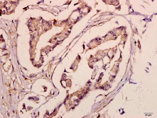
| Sample | Paraffin-embedded human breast carcinoma tissue; fixative not specified (datasheet A07813-2) |
| Fixation | Image fixative and duration unreported (datasheet A07813-2); verify before use. |
| Sectioning | 4–5 µm sections on charged slides (standard) |
| Deparaffinisation | Xylene, graded ethanol series to water (standard) |
| Antigen retrieval | Heat-induced epitope retrieval in citrate buffer, pH 6.0, 20 min at 95–98 °C (standard rule: cytoplasmic / membrane antigen) |
| Peroxidase block | 3% H2O2, 10 min, room temperature (standard) |
| Blocking | 10% normal serum of the secondary host, 30 min, room temperature (standard) |
| Primary antibody | Rabbit anti-ACSL6, 1:50-1:200 (datasheet A07813-2) |
| Primary incubation | Overnight at 4 °C (standard) |
| Detection | HRP-polymer secondary, DAB chromogen 5–10 min (standard) |
| Counterstain | Hematoxylin, blue, dehydrate and mount (standard) |
| Expected result | ACSL6-positive staining in glandular cells of seminal vesicle (HPA tissue IHC: High). HPA tissue profile: Cytoplasmic expression mainly in testis, seminal vesicle, small intestines and brain. No signal in the no-primary control. |
ACSL6 is a membrane-associated enzyme with a cytoplasmic domain; UniProt places it at mitochondrial outer, peroxisomal, microsomal and endoplasmic reticulum membranes (UniProt Q9UKU0 topology and subcellular location). In tissue IHC, expect cytoplasmic staining, especially in seminal vesicle and small-intestine glandular cells and testicular late spermatids (HPA tissue IHC: High). HPA rates its tissue staining Enhanced, with medium agreement between staining and RNA data; external verification remains pending (HPA tissue IHC).
| Cytoplasmic staining in seminal vesicle or small-intestine glandular cells, or testicular late spermatids. | This matches HPA's strongest reported tissue IHC pattern (HPA tissue IHC: High in these cells). Assess the named cells against neighboring cells on the same section; a uniformly stained field gives less confidence in cell-specific signal (standard IHC interpretation). |
| Predominantly nuclear staining with little cytoplasmic signal. | This conflicts with the cytoplasmic tissue profile and membrane-associated UniProt locations (HPA tissue IHC; UniProt Q9UKU0 subcellular location). Treat an isolated nuclear pattern as suspect and compare it with a known-positive tissue and a no-primary control (standard IHC practice). |
| Strong signal in a cell type HPA reports as undetected, such as breast adipocytes or bronchial respiratory epithelium. | That pattern is discordant with the supplied tissue observations (HPA tissue IHC: Not detected in those cells). Cross-reactivity or endogenous detection activity are possibilities, not diagnoses; compare a no-primary control and repeat with an independently validated antibody (standard IHC practice). |
| Diffuse color across cells, stroma and section edges, obscuring cell boundaries. | The result cannot establish the cell-restricted cytoplasmic pattern reported by HPA (HPA tissue IHC). Uneven detection or background may be involved; inspect control slides and staining distribution before scoring ACSL6 expression (standard IHC interpretation). |
| No visible signal in a well-preserved seminal vesicle or small-intestine section. | This disagrees with High glandular-cell staining reported for those tissues (HPA tissue IHC). First verify that the expected glands are present; then check the IHC-validated antibody, detection reagents and run controls before interpreting the tissue as negative (standard IHC practice). |
| Membrane topology | UniProt annotates one transmembrane segment at residues 25–45 and a cytoplasmic region at 46–697 (UniProt Q9UKU0 topology). This supports a cytoplasmic, membrane-associated interpretation but does not resolve individual organelles by chromogenic tissue IHC (standard IHC interpretation). |
| Tissue and cell selection | HPA reports High staining in seminal vesicle and small-intestine glandular cells and testicular late spermatids; cerebellar Purkinje cells and cortical glia are Medium (HPA tissue IHC). Select the specific cell population when judging a control, since an organ-wide score could hide a restricted pattern (standard IHC interpretation). |
| Bone marrow evidence differs by method | UniProt reports expression in erythrocyte precursors and bone marrow, while HPA tissue IHC lists bone-marrow hematopoietic cells as Not detected (UniProt Q9UKU0 tissue specificity; HPA tissue IHC). Do not treat marrow IHC as an assured positive control from these sources alone. |
| Antibody validation and isoforms | HPA lists antibody HPA040470 as IHC Enhanced and ICC Uncertain (HPA antibodies). UniProt lists 9 isoforms (UniProt Q9UKU0 isoforms); the supplied record gives no antibody epitope, so isoform coverage cannot be inferred. |
| IF/ICC: what appearance should I expect? | HPA's ICC-IF summary places ACSL6 mainly in vesicles, with additional cytosol localization marked uncertain; its main vesicle location is approved (HPA subcellular ICC-IF). This is an IF/ICC observation, not a required vesicle pattern for paraffin-section chromogenic IHC (HPA subcellular ICC-IF; HPA tissue IHC). |
| Fixation and retrieval evidence | Target-specific fixation effects are not established by the supplied assay evidence. Verify with a matched IHC source before attributing a result to fixation. |
| Situation | Likely cause | Next action |
|---|---|---|
| Strong nuclear staining dominates a nominal positive tissue. | The compartment conflicts with HPA's cytoplasmic tissue profile and UniProt's membrane locations (HPA tissue IHC; UniProt Q9UKU0 subcellular location). | Compare a known-positive section and no-primary control; score ACSL6 only where the expected cell-associated cytoplasmic pattern is supported (standard IHC practice). |
| A known-positive glandular tissue has no signal. | Absent staining conflicts with HPA's High glandular-cell observations, but does not identify which assay step failed (HPA tissue IHC). | Confirm glandular cells are present, inspect run controls and detection reagents, then optimize retrieval or antibody dilution using the IHC-validated antibody (standard IHC practice). |
| Brown color appears broadly, including stroma or slide edges. | Diffuse deposition can obscure HPA's cell-specific cytoplasmic pattern (HPA tissue IHC; standard IHC interpretation). | Review no-primary and reagent controls; check blocking, washing and chromogen development before assigning positive cells (standard IHC practice). |
| An HPA-undetected cell population stains strongly. | Cross-reactivity or endogenous chromogenic detection activity may explain discordant color; the HPA observation alone cannot distinguish them (HPA tissue IHC; standard IHC practice). | Compare the no-primary control, assess endogenous detection activity and seek an independent antibody result before reporting unexpected expression (standard IHC practice). |
| Bone marrow is negative despite an expectation from the UniProt tissue note. | The sources describe different evidence: UniProt reports expression in marrow-associated precursors, whereas HPA detects no staining in sampled hematopoietic cells (UniProt Q9UKU0 tissue specificity; HPA tissue IHC). | Use an HPA High tissue as the IHC positive control; describe a marrow result by its observed cell type and assay rather than treating the sources as equivalent (HPA tissue IHC; standard IHC interpretation). |
| An IF/ICC vesicle pattern seems different from broad cytoplasmic tissue IHC. | HPA reports approved vesicle localization in ICC-IF but a cytoplasmic tissue IHC profile; the assays resolve different features (HPA subcellular ICC-IF; HPA tissue IHC). | Interpret each assay against its own HPA reference and validation status; HPA lists ICC as Uncertain for HPA040470 (HPA antibodies; HPA subcellular ICC-IF). |


Comprehensive Human Protein Atlas IHC scoring per tissue (reliability: Enhanced — Medium consistency between antibody staining and RNA expression data. Pending external verification.). Rows are taken directly from the HPA tissue chart — click any row's HPA link to view the source.
| Tissue | Cell type | Level | Evidence | Source |
|---|---|---|---|---|
| Seminal vesicle | Glandular cells | High | Protein (IHC) | HPA → |
| Small intestine | Glandular cells | High | Protein (IHC) | HPA → |
| Testis | Elongated or late spermatids | High | Protein (IHC) | HPA → |
| Adrenal gland | Glandular cells | Medium | Protein (IHC) | HPA → |
| Cerebellum | Purkinje cells | Medium | Protein (IHC) | HPA → |
| Tissue | Cell type | Level | Evidence | Source |
|---|---|---|---|---|
| Bone marrow | Hematopoietic cells | Not detected | Protein (IHC) | HPA → |
| Breast | Adipocytes | Not detected | Protein (IHC) | HPA → |
| Bronchus | Respiratory epithelial cells | Not detected | Protein (IHC) | HPA → |
| Endometrium | Cells in endometrial stroma | Not detected | Protein (IHC) | HPA → |
| Epididymis | Glandular cells | Not detected | Protein (IHC) | HPA → |
Troubleshoot ACSL6 chromogenic IHC in paraffin sections using the selected antibody caption, expression references, and standard section controls.
Anti-ACSL6 antibodies list IHC and IF applications across human, mouse and rat (catalog applications/reactivity); the supplied image documents IHC in a human paraffin section (catalog IHC image caption).
The rendered card is A07813-2, listed for IHC in human, mouse and rat (catalog applications/reactivity). Its own figure shows paraffin-embedded human breast carcinoma at 1:50 (catalog IHC image caption).
Which to pick: For tissue IHC, choose A07813-2: its own figure documents a human paraffin section, and the fixative is unreported (catalog IHC image caption). For IF/ICC, consider the polyclonal A07813-1 because IF is listed; ICC-specific testing and an IF image are unreported (catalog dilution raw; catalog applications; catalog image alts). Both SKUs list human, mouse and rat reactivity, but the supplied tissue image documents human tissue only (catalog reactivity; catalog IHC image caption).