This website uses cookies to ensure you get the best experience on our website.


- Table of Contents
Plan chromogenic ACVR1C IHC on paraffin sections with the catalog antibody's 1:100–1:300 dilution range (datasheet). Compare staining with the cytoplasmic tissue pattern while accounting for uncertain reliability and presumed off-target binding (HPA tissue IHC).

Expected localisation, validated protocols, controls and antibodies — the at-a-glance facts below, then the full design guide.
| Expected localisation | Observed cytoplasmic staining (HPA tissue IHC); membrane localization (UniProt) | |
| Staining pattern | Cytoplasmic staining in glandular cells across several tissues (HPA tissue IHC) | |
| Antigen retrieval | Citrate pH 6.0 HIER, 95–98 °C, 20 min (rule: cytoplasmic / membrane antigen) | |
| Positive control | Adrenal gland+4 more · see all | |
| Negative control | None in HPA (detected in all 45 tissues); use no-primary + isotype controls |
| Fixation | Keep fixation consistent across sections. (standard IHC practice; not target-specific) | |
| Caveat | Presumed off-target staining; reliability uncertain (HPA tissue IHC) | |
| Regulation | Placental expression throughout pregnancy (UniProt) | |
| Isoform / epitope | 4 isoforms; check epitope against extracellular and cytoplasmic regions (UniProt) |
The catalog antibody’s IHC-P protocol is accompanied by a published ACVR1C protocol for colorectal cancer tissue (PMC13371181).
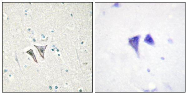
| Sample | Paraffin-embedded human brain tissue; fixative not specified (datasheet A30526) |
| Fixation | Image fixative and duration unreported (datasheet A30526); verify before use. |
| Sectioning | 4–5 µm sections on charged slides (standard) |
| Deparaffinisation | Xylene, graded ethanol series to water (standard) |
| Antigen retrieval | Heat-induced epitope retrieval in citrate buffer, pH 6.0, 20 min at 95–98 °C (standard rule: cytoplasmic / membrane antigen) |
| Peroxidase block | 3% H2O2, 10 min, room temperature (standard) |
| Blocking | 10% normal serum of the secondary host, 30 min, room temperature (standard) |
| Primary antibody | Rabbit anti-ACVR1C, 1:100 - 1:300 (datasheet A30526) |
| Primary incubation | Overnight at 4 °C (standard) |
| Detection | HRP-polymer secondary, DAB chromogen 5–10 min (standard) |
| Counterstain | Hematoxylin, blue, dehydrate and mount (standard) |
| Expected result | ACVR1C-positive staining in glandular cells of adrenal gland (HPA tissue IHC: High). HPA tissue profile: General cytoplasmic expression in several different tissue types. No signal in the no-primary control. |
ACVR1C is a single-pass membrane receptor with an extracellular region and cytoplasmic kinase domain (UniProt Q8NER5 topology). In paraffin IHC, HPA reports general cytoplasmic staining across several tissues, including high staining in colon glandular cells, cerebral cortical neurons and lung macrophages (HPA tissue IHC). Interpret these observations cautiously: HPA rates tissue staining Uncertain because antibody staining and RNA expression show low consistency, and presumed off-target binding was disregarded (HPA reliability).
| Cell-associated cytoplasmic or peripheral staining in colon glandular cells, cerebral cortical neurons, or lung macrophages. | These cell types have High tissue IHC staining reported by HPA (HPA tissue IHC). A peripheral component fits the single-pass membrane topology (UniProt Q8NER5 topology), while cytoplasmic staining matches HPA's observed profile (HPA tissue IHC). Neither appearance alone establishes antibody specificity: both assessed HPA antibodies have Uncertain IHC validation (HPA antibodies HPA007982, HPA011933). |
| Predominantly nuclear staining, with little staining at the cell periphery or in the cytoplasm. | A nuclear-dominant pattern conflicts with the membrane topology and HPA's general cytoplasmic tissue profile (UniProt Q8NER5 topology; HPA tissue IHC). Treat it as suspect and compare it with the counterstain and controls (general IHC practice). Cytoplasmic staining by itself cannot resolve the discrepancy between predicted membrane location and observed tissue IHC. |
| Strong staining in a different cell population while the HPA-reported cell population is faint. | For example, strong lung epithelial staining with faint macrophages would differ from HPA's High macrophage observation (HPA tissue IHC). Consider cross-reactivity or endogenous chromogen activity, and check cell identity and detection controls (general IHC practice). HPA's Uncertain reliability makes an unexpected pattern a reason to validate, rather than immediate proof of either true expression or artefact (HPA reliability). |
| Uniform haze over tissue and spaces between cells, obscuring cell boundaries. | This is difficult to score as cell-associated ACVR1C staining. Assess nonspecific binding, incomplete washing and endogenous detection activity with appropriate controls (general IHC practice). HPA describes cytoplasmic expression in several tissue types, but that observation does not validate diffuse slide-wide background (HPA tissue IHC). |
| No detectable staining in a section selected for an HPA-reported High cell population. | First check tissue identity, the presence of the expected cells, antibody and detection performance, and the retrieval conditions (general IHC practice). Colon glandular cells and lung macrophages are reported High examples, but HPA assigns the tissue IHC evidence Uncertain reliability; an unstained section therefore needs technical review and independent biological interpretation (HPA tissue IHC; HPA reliability). |
| Receptor topology | The extracellular region spans residues 22–113, the transmembrane segment 114–134, and the cytoplasmic region 135–493 (UniProt Q8NER5 topology). This supports a membrane-associated expectation, but HPA tissue IHC reports a general cytoplasmic appearance (HPA tissue IHC). |
| Antibody validation | Both listed HPA antibodies, HPA007982 and HPA011933, have Uncertain IHC status; HPA reports low consistency between staining and RNA data and disregarded presumed off-target binding (HPA antibodies; HPA reliability). Use cell-specific controls before treating staining intensity as confirmed ACVR1C abundance (general IHC practice). |
| Choice of comparison tissue | HPA reports High staining in several cell populations, including adrenal and breast glandular cells, kidney glomerular cells, and lung macrophages; it lists low staining in ovarian stroma and skin fibroblasts (HPA tissue IHC). Low is not an established absence, and no negative tissue is supplied (HPA tissue IHC). |
| Processing and epitope | UniProt lists a signal peptide at residues 1–20 and the mature chain at 21–493 (UniProt Q8NER5 processing). Antibody epitope location is not supplied, so the effect of processing on either listed antibody cannot be predicted. |
| IF/ICC: what location should be expected? | A membrane-associated signal is consistent with UniProt topology and HPA's subcellular summary of Membrane (UniProt Q8NER5 topology; HPA subcellular). HPA supplies no ICC-IF image cell lines or main-location assignment, so this section cannot establish a cell-specific IF pattern (HPA subcellular). |
| Situation | Likely cause | Next action |
|---|---|---|
| No signal in an HPA-reported High population. | Missing expected cells, a detection failure, or retrieval conditions could explain the result (general IHC practice); HPA's High call is itself Uncertain (HPA tissue IHC; HPA reliability). | Confirm the cell population and run appropriate positive and detection controls; review the antibody instructions and optimise retrieval as a general paraffin IHC step (general IHC practice). |
| Nuclear signal dominates. | The compartment differs from UniProt's membrane topology and HPA's observed cytoplasmic profile (UniProt Q8NER5 topology; HPA tissue IHC). | Check the counterstain and negative controls, then assess whether a second independently validated approach reproduces the cell-specific pattern (general IHC practice). |
| Strong signal appears mainly in an unexpected cell type. | Cross-reactivity or endogenous detection activity is possible (general IHC practice), especially given HPA's Uncertain IHC validation (HPA reliability). | Verify cell identity on the section; compare HPA-reported cell populations and use reagent and detection controls before scoring the signal (HPA tissue IHC; general IHC practice). |
| Diffuse brown haze masks cellular detail. | Nonspecific reagent binding, inadequate washing, or endogenous peroxidase activity can produce background in chromogenic IHC (general IHC practice). | Inspect reagent controls and review blocking, washing, antibody concentration and endogenous peroxidase control conditions (general IHC practice). |
| A low-staining comparison tissue is strongly positive. | HPA lists ovarian stroma and skin fibroblasts as Low, not negative; unexpected intensity may also reflect nonspecific staining (HPA tissue IHC; general IHC practice). | Check the cell population, staining distribution and controls; do not use a Low entry as an absolute negative standard (HPA tissue IHC; general IHC practice). |
| Only a sharp membrane rim stains, with little cytoplasmic signal. | A rim is topology-consistent, but HPA describes general cytoplasmic tissue IHC and rates reliability Uncertain (UniProt Q8NER5 topology; HPA tissue IHC; HPA reliability). | Record the compartment and cell type separately, then seek independent specificity evidence before calling the rim confirmed ACVR1C (general IHC practice). |


Comprehensive Human Protein Atlas IHC scoring per tissue (reliability: Uncertain — Low consistency between antibody staining and RNA expression data. Presumed off target binding observed and disregarded.). Rows are taken directly from the HPA tissue chart — click any row's HPA link to view the source.
| Tissue | Cell type | Level | Evidence | Source |
|---|---|---|---|---|
| Adrenal gland | Glandular cells | High | Protein (IHC) | HPA → |
| Breast | Glandular cells | High | Protein (IHC) | HPA → |
| Cerebral cortex | Neuronal cells | High | Protein (IHC) | HPA → |
| Colon | Glandular cells | High | Protein (IHC) | HPA → |
| Duodenum | Glandular cells | High | Protein (IHC) | HPA → |
| Tissue | Cell type | Level | Evidence | Source |
|---|---|---|---|---|
| None in HPA: ACVR1C is detected in all 45 scored tissues. Use a no-primary (secondary-only) and an isotype control instead. | ||||
Troubleshoot ACVR1C staining in paraffin sections by checking retrieval, cell context, membrane topology and the reliability of the observed pattern.
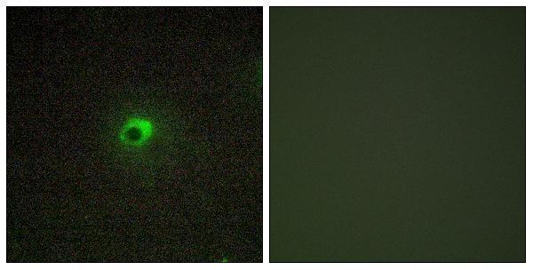
The anti-ACVR1C catalog antibody has IHC data from paraffin-embedded human brain tissue and IF data from COS7 cells (A30526 image captions). Listed reactivity covers human, mouse, and rat (catalog reactivity).
A30526 is listed for IHC and IF/ICC, with IHC shown in paraffin-embedded human brain tissue and IF shown in COS7 cells (catalog applications; A30526 image captions). Both images include a peptide-blocked comparison (A30526 image captions).
Which to pick: Choose A30526 for paraffin-section tissue IHC; its own image shows human brain tissue, and the listed IHC dilution is 1:100–1:300 (A30526 IHC image caption; datasheet: IHC 1:100–1:300). For IF/ICC, A30526 is listed for both applications, with IF shown in COS7 cells and a listed ICC/IF dilution of 1:200–1:1000 (catalog applications; A30526 IF image caption; datasheet: ICC/IF 1:200–1:1000). For cross-species work, A30526 is a rabbit polyclonal listed as reactive with human, mouse, and rat; the fixative used for its paraffin-section IHC image is unreported (catalog host, clonality, and reactivity; A30526 IHC image caption).