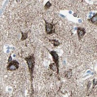
ACVRL1 / Activin receptor type-1-like · IHC design guide

Design Immunohistochemistry for ACVRL1

Plan chromogenic ACVRL1 IHC on paraffin sections using the catalog antibody at 2–5 μg/ml (datasheet: A01468-2). Compare gallbladder glandular cells reported as high with adipose tissue adipocytes reported as undetected, while accounting for the reported low concordance between staining and RNA expression (HPA tissue IHC).

Evidence assembled Oct 2026 · For research use; verify linked source records and product datasheet before use
Immunohistochemistry protocol sheet for ACVRL1 (IHC for ACVRL1): expected localisation Cytoplasmic tissue staining (HPA tissue IHC); cell membrane (UniProt), antibody A01468-2, validated IHC image, and IHC protocol steps
Printable ACVRL1 IHC protocol sheet — expected localisation Cytoplasmic tissue staining (HPA tissue IHC); cell membrane (UniProt), antibody A01468-2, controls and protocol steps. Open the full ACVRL1 IHC guide →

ACVRL1 Immunohistochemistry Experimental Design Guide

Expected localisation, validated protocols, controls and antibodies — the at-a-glance facts below, then the full design guide.

Must know before staining
Expected localisation Cytoplasmic tissue staining (HPA tissue IHC); cell membrane (UniProt)
Staining pattern Cytoplasmic staining in glandular, neuronal and tubular cells (HPA tissue IHC)
Antigen retrieval EDTA pH 8.0 HIER, heat-mediated (datasheet A01468-2)
Positive control ⓘ Caudate+4 more · see all
Negative control ⓘ Adipose tissue+4 more · see all
Important caveats
Reasons your staining may differ from the expected pattern.
Fixation Keep fixation consistent across sections (standard IHC practice; not target-specific)
Caveat Staining has low concordance with RNA expression (HPA tissue IHC)
Regulation Expression regulation not annotated (UniProt)
Isoform / epitope No isoforms listed; epitope side matters across the membrane (UniProt)
Section 1

Recommended ACVRL1 IHC & IF Protocols

The catalog antibody’s IHC-P protocol uses EDTA pH 8.0 heat retrieval (datasheet A01468-2). A published human hippocampus protocol provides a comparison (PMC5988976).

Recommended immunohistochemistry (IHC-P) protocol parameters
SampleParaffin-embedded human breast cancer tissue; fixative not specified (datasheet A01468-2)
FixationImage fixative and duration unreported (datasheet A01468-2); verify before use.
Sectioning4–5 µm sections on charged slides (standard)
DeparaffinisationXylene, graded ethanol series to water (standard)
Antigen retrievalHeat retrieval: EDTA pH 8.0 (datasheet A01468-2); 20 min, 95–100 °C (standard)
Peroxidase block3% H2O2, 10 min, room temperature (standard)
Blocking10% goat serum (datasheet A01468-2)
Primary antibodyRabbit anti-ACVRL1, 2-5μg/ml (datasheet A01468-2)
Primary incubationOvernight at 4 °C (datasheet A01468-2)
DetectionStreptavidin-biotin complex (SABC), DAB chromogen (datasheet A01468-2)
CounterstainHematoxylin, blue, dehydrate and mount (standard)
Expected resultACVRL1-positive staining in neuronal cells of caudate (HPA tissue IHC: High). HPA tissue profile: Cytoplasmic expression in several tissue types. No signal in the no-primary control.
💡Decision noteStart with EDTA pH 8.0 heat retrieval (datasheet A01468-2); the published hippocampus excerpt does not specify a retrieval method (PMC5988976).
Section 2

What Is the Expected ACVRL1 Staining Pattern?

ACVRL1 is a cell-membrane receptor with an extracellular segment and a cytoplasmic kinase region (UniProt P37023 topology). In paraffin-section IHC, HPA reports strong staining in selected neuronal, glandular, and kidney tubular cells, but describes the overall tissue pattern as cytoplasmic (HPA tissue IHC). HPA rates its IHC antibody Approved while noting low consistency between staining and RNA expression (HPA tissue IHC); assess both cell identity and compartment cautiously.

What am I looking at on my slide?
Membrane-associated staining in an expected cell population, with some cytoplasmic staining.This is compatible with UniProt's membrane assignment and HPA's cytoplasmic IHC profile; neither source establishes that staining must be exclusively membranous (UniProt P37023; HPA tissue IHC). Compare signal with background in the same section before calling cells positive (general IHC practice).
Strong staining in duodenal or small-intestinal glandular cells, or kidney tubular cells.These cell populations are listed as High by HPA and provide useful observed patterns for slide comparison (HPA tissue IHC). High describes the reported staining level, not a required intensity for every specimen or run; HPA also reports low staining–RNA consistency (HPA tissue IHC).
Strong, isolated nuclear staining without a convincing membrane or cytoplasmic pattern.A nuclear-only pattern does not match UniProt's cell-membrane location or HPA's cytoplasmic tissue profile (UniProt P37023; HPA tissue IHC). Treat it as suspect staining and inspect control slides and morphology before assigning it to ACVRL1 (general IHC practice).
Strong signal in bronchial respiratory epithelium or adipocytes.HPA reports ACVRL1 as Not detected in these listed cell populations (HPA tissue IHC). Recheck cell identity and controls; unexpected signal could reflect nonspecific antibody binding or endogenous detection activity, but the image alone cannot distinguish those causes (general IHC practice).
No signal in glandular cells of an otherwise well-preserved duodenal section.HPA lists these cells as High, so an absent result warrants a technical check (HPA tissue IHC). It does not alone prove assay failure: sample variation and HPA's low staining–RNA consistency limit how strongly one reference image predicts a new section (HPA tissue IHC).
💡Expected ACVRL1 appearanceCall a positive result when discrete chromogenic staining is above local background in HPA-listed High cells, such as duodenal glandular or kidney tubular cells; membrane-associated signal fits UniProt topology, while cytoplasmic signal fits HPA IHC, and uniform background or nuclear-only staining is suspect (HPA tissue IHC; UniProt P37023; general IHC practice).
How each factor affects the staining
Compartment and epitope locationUniProt places residues 22–118 outside the cell, 119–141 across the membrane, and 142–503 in the cytoplasm (UniProt P37023 topology). The supplied sources do not locate the IHC antibody's epitope, so topology cannot predict its retrieval requirement or staining intensity.
Tissue-pattern confidenceHPA calls the tissue IHC reliability Approved but reports low consistency with RNA expression; its summary says cytoplasmic expression in several tissue types (HPA tissue IHC). Use the listed cells as comparison points, not as a universal map of positive tissues.
Antibody validationThe listed rabbit polyclonal antibody HPA007041 has IHC status Approved; no IHC Enhanced designation is supplied (HPA antibodies). An Approved result supports using its images as a reference, but the supplied validation does not independently settle every unexpected cell or compartment.
Antigen retrieval in paraffin IHCThe supplied UniProt and HPA records give no ACVRL1-specific retrieval condition or fixation sensitivity. Retrieval is a general paraffin-IHC workflow variable; assess it against control staining under the chosen assay conditions (general IHC practice).
Can IF/ICC show the same location?HPA's ICC-IF summary says Membrane, but gives no main-location entry or cell-line images (HPA subcellular). That limited summary is consistent with UniProt's membrane assignment; it does not establish an IF/ICC staining pattern for a particular preparation (UniProt P37023).
Why is my staining missing, weak or wrong?
SituationLikely causeNext action
The expected High cell population appears negative.Possible assay failure or specimen variation; the HPA reference is not a guarantee for each section (HPA tissue IHC; general IHC practice).Check tissue preservation and assay controls, then review retrieval and primary-antibody conditions within the validated IHC workflow (general IHC practice).
Chromogen covers many structures evenly, obscuring cell boundaries.Diffuse background can arise from blocking, washing, or detection conditions (general IHC practice); it does not resemble a discrete cell pattern (HPA tissue IHC).Inspect the negative control and reduce background through the assay's blocking, washing, and detection controls before scoring ACVRL1 (general IHC practice).
Only nuclei are strongly stained.The dominant compartment conflicts with UniProt's membrane assignment and HPA's cytoplasmic IHC summary (UniProt P37023; HPA tissue IHC).Check counterstain versus chromogen and compare the negative control; score nuclear-only signal as unresolved until a plausible specific pattern appears (general IHC practice).
Bronchial respiratory epithelial cells stain strongly.HPA lists this cell population as Not detected, making the result discordant with its tissue reference (HPA tissue IHC).Confirm the cell identification and compare negative and detection controls; investigate nonspecific binding or endogenous activity before calling it ACVRL1 (general IHC practice).
Cytoplasmic staining appears, but a crisp membrane rim does not.HPA reports cytoplasmic tissue staining even though UniProt assigns ACVRL1 to the cell membrane (HPA tissue IHC; UniProt P37023).Assess whether staining is cell-restricted and above background in an HPA-listed population; do not reject it solely for lacking a membrane rim (HPA tissue IHC; general IHC practice).
Endothelial staining is weak while nearby glandular cells are stronger.HPA lists colon endothelial cells as Low, whereas several glandular cell populations are High (HPA tissue IHC). These ratings concern specific tissues and cells, not all vessels or glands.Record tissue and cell type separately, compare with the appropriate HPA entry, and avoid applying one tissue's intensity rating across organs (HPA tissue IHC).

Sample controls for ACVRL1 IHC & IF

🧪Run duodenum first and score glandular cells, which HPA rates High for ACVRL1 (HPA: High in duodenum glandular cells). Use cervix glandular cells as the tissue negative (HPA: Not detected in cervix glandular cells); on the duodenum slide, cells outside the glandular compartment should provide a background comparator, but their ACVRL1 status is not established by the supplied HPA row.
Positive control tissue: Caudate (Neuronal cells, HPA High)
Negative control tissue: Adipose tissue (HPA Not detected)
ICC-IF cell lines (HPA subcellular resource): HPA carries no ICC-IF cell line for ACVRL1; derive a cell-line control from the positive tissue's cell type (Neuronal cells) and confirm it by RNA or western blot first.
Technical controls: Include no-primary and secondary-only controls, plus nonimmune rabbit IgG matched to the primary antibody’s clonality where applicable (caption: rabbit primary; standard IHC control practice). Use ACVRL1 knockout material as a biological negative, and check duodenal sections for endogenous peroxidase and, with the caption’s biotin-based detection, endogenous biotin (standard IHC control practice; caption: biotin-based detection).
⚠️Feasibility: A target-specific fixation window or fixation effect is unreported, and the selected A01468-2 paraffin-section caption does not state a fixative (caption: paraffin section; fixative unreported). The reported IHC example uses heat retrieval in EDTA at pH 8.0, but the supplied evidence does not establish whether retrieval is required or whether frozen-section IHC or IF is easier (caption: EDTA pH 8.0 heat retrieval). In duodenum, glandular tissue background should be checked alongside the specific signal, particularly when using biotin-based chromogenic detection (HPA: High in duodenum glandular cells; caption: biotin-based DAB detection).

HPA tissue IHC evidence for ACVRL1

Comprehensive Human Protein Atlas IHC scoring per tissue (reliability: Approved — Low consistency between antibody staining and RNA expression data.). Rows are taken directly from the HPA tissue chart — click any row's HPA link to view the source.

Positive expression · recommended positive controls

TissueCell typeLevelEvidenceSource
Caudate Neuronal cells High Protein (IHC) HPA →
Cerebellum Cells in granular layer High Protein (IHC) HPA →
Cerebral cortex Neuronal cells High Protein (IHC) HPA →
Duodenum Glandular cells High Protein (IHC) HPA →
Gallbladder Glandular cells High Protein (IHC) HPA →

Undetected expression · recommended negative controls

TissueCell typeLevelEvidenceSource
Adipose tissue Adipocytes Not detected Protein (IHC) HPA →
Breast Adipocytes Not detected Protein (IHC) HPA →
Bronchus Respiratory epithelial cells Not detected Protein (IHC) HPA →
Cervix Glandular cells Not detected Protein (IHC) HPA →
Endometrium Cells in endometrial stroma Not detected Protein (IHC) HPA →
Section 3

Advanced ACVRL1 IHC Tips

Troubleshoot ACVRL1 chromogenic IHC by checking retrieval, staining compartment, controls, and scoring before interpreting differences between tissues.

How should I retrieve ACVRL1 in paraffin sections when staining is weak?
Start with heat-mediated antigen retrieval in EDTA at pH 8.0 for paraffin sections stained with A01468-2 (datasheet A01468-2). Keep heating and cooling conditions consistent across slides, then compare staining with a matched section processed in the same run (standard IHC practice). The caption reports 2 μg/mL primary antibody overnight at 4°C, so check those conditions before changing retrieval (datasheet A01468-2). If staining remains weak, test an alternative retrieval buffer on adjacent sections as a documented fallback, while monitoring morphology and background (standard IHC practice).
Could fixation explain inconsistent ACVRL1 staining between paraffin blocks?
The selected paraffin-section caption does not state a fixative, so target-specific fixation sensitivity for this antibody is unknown (datasheet A01468-2). Record each block’s fixative and fixation duration, and compare sections processed with the same retrieval and antibody conditions (standard IHC practice). Underfixation or prolonged fixation can change morphology and epitope accessibility in IHC generally, but neither effect has been established specifically for ACVRL1 here (standard IHC practice; datasheet A01468-2). If staining differs between blocks, include a reference section in the same run and avoid assigning the difference to ACVRL1 abundance without checking preservation and controls (standard IHC practice).
Should ACVRL1 staining appear only at the cell surface?
ACVRL1 is annotated as a cell-membrane receptor with one transmembrane segment at residues 119–141 (UniProt P37023 topology). HPA describes cytoplasmic staining across several tissue types, while its subcellular summary says membrane (HPA tissue IHC; HPA subcellular). Score membranous and cytoplasmic signal separately, using the same compartment definitions across sections and a no-primary control to assess nonspecific colour (standard IHC practice). Treat diffuse nuclear staining as unexpected for this receptor, and investigate retrieval, detection background, and section quality before calling it ACVRL1 positive (UniProt P37023 topology; standard IHC practice).
How does an unknown antibody epitope affect ACVRL1 IHC interpretation?
The record lists 0 isoforms but supplies no antibody epitope, so staining cannot be assigned to a particular receptor region from these data (UniProt P37023 record; datasheet A01468-2). ACVRL1 has an extracellular region at residues 22–118, a transmembrane segment at 119–141, and a cytoplasmic region at 142–503 (UniProt P37023 topology). Its annotated glycosylation site at residue 98 and phosphoserines at 155, 160, and 161 provide context, but their effect on this antibody is unknown (UniProt P37023 modifications; datasheet A01468-2). Request epitope information before interpreting differential staining as altered processing or phosphorylation; compare matched sections under identical IHC conditions (standard IHC practice).
How can I compare ACVRL1 IF with the chromogenic IHC result?
The supplied IF/ICC record has no cell-line images, so establish IF specificity with appropriate controls before comparing its pattern with tissue IHC (HPA subcellular; standard IF practice). Multiplex ACVRL1 with a marker for the cell population being scored, and inspect each channel separately for bleed-through (standard IF practice). Choose a fluorophore channel with low tissue autofluorescence, often red or far-red, and include an unstained section to measure that background (standard IF practice). If the antibody recognizes an extracellular epitope, assess staining without permeabilisation; if it recognizes a cytoplasmic epitope, compare controlled permeabilisation conditions, because the epitope is not supplied here (UniProt P37023 topology; datasheet A01468-2; standard IF practice).
What should I check when ACVRL1 DAB staining looks diffuse?
The selected IHC caption uses a biotinylated secondary, streptavidin-biotin complex, and DAB, so inspect both antibody-dependent and detection-system background (datasheet A01468-2). Run a no-primary control and, if needed, a detection-only control on matched sections to locate the source of diffuse colour (standard IHC practice). Include an endogenous-peroxidase block before DAB development and assess whether an endogenous-biotin block improves staining with this detection system (standard IHC practice; datasheet A01468-2). Titrate primary antibody around the caption’s 2 μg/mL condition, holding retrieval and development time constant so a lower signal can be interpreted (datasheet A01468-2; standard IHC practice).
How should I quantify ACVRL1 staining across different tissue compartments? ⚠ ANSWER MARKED FOR VERIFICATION
Define the cell population and whether membranous or cytoplasmic staining counts before scoring, because those locations are reported differently in the supplied sources (UniProt P37023 topology; HPA tissue IHC). For an H-score, assign intensity 0–3 and multiply each intensity by the percentage of cells at that intensity, giving a 0–300 score (standard IHC practice). Report the percentage of positive cells alongside the H-score and normalise comparisons to the same eligible cell population, rather than total tissue area (standard IHC practice). Use matched retrieval, antibody incubation, DAB development, and counterstaining across slides; record excluded damaged or necrotic regions before analysis (standard IHC practice).
When is apparent ACVRL1 positivity likely to be artefactual?
A convincing result should occupy a defined cell population and plausible membranous or cytoplasmic compartment, while remaining above the matched no-primary background (UniProt P37023 topology; HPA tissue IHC; standard IHC practice). Be cautious with strong nuclear colour, section-edge enhancement, necrotic regions, or residual enzyme-associated DAB signal; check adjacent morphology and controls before scoring (UniProt P37023 topology; standard IHC practice). Do not assume every endothelial cell is strongly positive: HPA reports low staining in colon endothelial cells and notes low consistency between antibody staining and RNA expression (HPA tissue IHC). Compare the pattern across replicate sections and record the cell type and compartment, since the selected caption establishes staining in paraffin-embedded human breast cancer tissue but does not establish specificity in every cell (datasheet A01468-2; standard IHC practice).
Boster reagents

Best ACVRL1 / Activin receptor type-1-like IHC Antibodies

The catalog antibody has real IHC data from human paraffin-embedded tissue sections (A01468-2 image captions). Its application list includes IHC but not IF/ICC (A01468-2 catalog applications).

Real IHC data IHC analysis of ALK-1/ACVRL1 using anti-ALK-1/ACVRL1 antibody (A01468-2). ALK-1/ACVRL1 was detected in paraffin-embedded section of human breast cancer tissue. Heat mediated antigen retrieval was performed in EDTA buffer (pH8.0, epitope retrieval solution). The tissue section was blocked with 10% goat serum. The tissue section was then incubated with 2μg/ml rabbit anti-ALK-1/ACVRL1 Antibody (A01468-2) overnight at 4°C. Biotinylated goat anti-rabbit IgG was used as secondary antibody and incubated for 30 minutes at 37°C. The tissue section was developed using Strepavidin-Biotin-Complex (SABC) (Catalog # SA1022) with DAB as the chromogen.
Anti-ALK-1/ACVRL1 Antibody ®
Cat # A01468-2

A01468-2 will render with an IHC figure from a human paraffin-embedded breast cancer section (A01468-2 figure caption). Its additional IHC captions show human gallbladder adenocarcinoma, liver cancer, and ovarian serous adenocarcinoma sections (A01468-2 image captions).

Which to pick: Choose A01468-2 for paraffin-section tissue IHC: its captions report EDTA retrieval at pH 8.0 and primary antibody at 2 μg/ml (A01468-2 image captions); the fixative is unreported (A01468-2 image captions). No listed SKU has IF/ICC validation (catalog applications), so an IF/ICC choice cannot be supported here. A01468-2 lists human, mouse, and rat reactivity, but its IHC captions show human tissue only (catalog reactivity; A01468-2 image captions).

Each figure is that product's own IHC / IF validation image from its datasheet.

References

  1. UniProt Consortium. UniProt entry P37023 (ACVL1_HUMAN, Activin receptor type-1-like).
  2. Human Protein Atlas. ACVRL1 tissue IHC expression (reliability: Approved).
  3. Human Protein Atlas. ACVRL1 subcellular location (ICC-IF): Membrane.
  4. Human Protein Atlas. ACVRL1 antibody validation summary (1 antibodies).
  5. Immunohistochemical Analysis of Activin Receptor-Like Kinase 1 (ACVRL1/ALK1) Expression in the Rat and Human Hippocampus: Decline in CA3 During Progression of Alzheimer's Disease. Journal of Alzheimer's disease : JAD 2018 — PMC5988976.
  6. The Expression of Activin Receptor-Like Kinase 1 (ACVRL1/ALK1) in Hippocampal Arterioles Declines During Progression of Alzheimer's Disease. Cerebral cortex communications 2020 — PMC7497413.
  7. The TGFβ type I receptor TGFβRI functions as an inhibitor of BMP signaling in cartilage. Proceedings of the National Academy of Sciences of the United States of America 2019 — PMC6681752.
  8. Endoglin and Activin Receptor-like Kinase 1 (Alk1) Modify Adrenomedullin Expression in an Organ-Specific Manner in Mice. Biology 2022 — PMC8945164.
  9. PubMed PMID:8397373 — UniProt-cited evidence.
  10. PubMed PMID:8242742 — UniProt-cited evidence.
  11. PubMed PMID:9245985 — UniProt-cited evidence.