This website uses cookies to ensure you get the best experience on our website.


- Table of Contents
Plan ADAM23 chromogenic IHC on paraffin sections with an antibody titration of 1:50–1:200 (datasheet: 1:50–1:200). Use nasopharyngeal respiratory epithelial cells or testis Leydig cells as high-staining references, and assess cytoplasmic expression and plasma positivity (HPA tissue IHC).

Expected localisation, validated protocols, controls and antibodies — the at-a-glance facts below, then the full design guide.
| Expected localisation | Cytoplasmic expression and plasma positivity (HPA tissue IHC) | |
| Staining pattern | High in respiratory epithelial and Leydig cells; cytoplasmic/plasma (HPA tissue IHC) | |
| Antigen retrieval | Citrate pH 6.0 HIER, 95–98 °C, 20 min (rule: cytoplasmic / membrane antigen) | |
| Positive control | Nasopharynx+4 more · see all | |
| Negative control | None in HPA (detected in all 45 tissues); use no-primary + isotype controls |
| Fixation | Matched tissue-IHC evidence does not establish the fixation claim. Validate the specimen-specific method before use. (selected-SKU IHC image A07382) | |
| Caveat | A secreted variant may shift RNA and protein tissue patterns (HPA tissue IHC) | |
| Regulation | Brain enriched; heart weak (UniProt) | |
| Isoform / epitope | 3 isoforms; check extracellular versus cytoplasmic epitope (UniProt) |
The catalog antibody protocol is accompanied by published ADAM23 IHC methods for paraffin skin sections (PMC10750783) and FFPE tumor sections (PMC6500989).
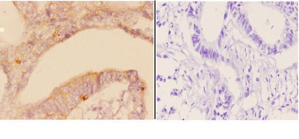
| Sample | Paraffin-embedded human rectum carcinoma tissue; fixative not specified (datasheet A07382) |
| Fixation | Image fixative and duration unreported (datasheet A07382); verify before use. |
| Sectioning | 4–5 µm sections on charged slides (standard) |
| Deparaffinisation | Xylene, graded ethanol series to water (standard) |
| Antigen retrieval | Heat-induced epitope retrieval in citrate buffer, pH 6.0, 20 min at 95–98 °C (standard rule: cytoplasmic / membrane antigen) |
| Peroxidase block | 3% H2O2, 10 min, room temperature (standard) |
| Blocking | 10% normal serum of the secondary host, 30 min, room temperature (standard) |
| Primary antibody | Rabbit anti-ADAM23, 1:50-1:200 (datasheet A07382) |
| Primary incubation | Overnight at 4 °C (standard) |
| Detection | HRP-polymer secondary, DAB chromogen 5–10 min (standard) |
| Counterstain | Hematoxylin, blue, dehydrate and mount (standard) |
| Expected result | ADAM23-positive staining in respiratory epithelial cells of nasopharynx (HPA tissue IHC: High). HPA tissue profile: General cytoplasmic expression and plasma positivity. No signal in the no-primary control. |
ADAM23 is a membrane-associated protein with an extracellular region and a short cytoplasmic tail; UniProt also lists a secreted location (UniProt O75077 topology and location). In tissue IHC, expect cytoplasmic expression and plasma-membrane positivity, particularly in nasopharyngeal respiratory epithelium and testicular Leydig cells (HPA tissue IHC: Approved; High in both cell types).
| Strong cellular staining in nasopharyngeal respiratory epithelium or testicular Leydig cells (HPA: High in both). | This fits the reported positive-cell pattern; assess cytoplasmic and plasma-membrane signal against the surrounding tissue (HPA: tissue IHC profile). |
| A mainly nuclear IHC signal with little cytoplasmic or membrane staining (HPA: tissue IHC profile). | Treat this as discordant with the tissue IHC profile and investigate artefact or antibody specificity; nucleoplasmic staining is separately approved in ICC-IF (HPA: subcellular ICC-IF). |
| Strong staining in cells outside the reported positive populations (HPA: tissue IHC cell-level results). | Check the cell identification and controls before calling it ADAM23; cross-reactivity or endogenous chromogen activity can mimic a positive result (general IHC practice). |
| Uniform colour across cells and extracellular space, with little cell-specific contrast (general IHC practice). | This favours diffuse background over an interpretable cellular pattern; compare the no-primary control and review blocking, washing and detection (general IHC practice). |
| No signal in a correctly identified nasopharyngeal respiratory epithelium or testicular Leydig-cell control (HPA: High in both). | A negative result is inconclusive until tissue integrity, retrieval, antibody dilution and detection are checked (general IHC practice); HPA levels do not guarantee every section will stain. |
| Cell type and tissue | HPA reports High staining in nasopharyngeal respiratory epithelial cells and Leydig cells, Medium in caudate neurons, and Low in several listed cell types (HPA: tissue IHC); compare like cells rather than whole-section colour. |
| Membrane topology and secretion | The mature chain includes an extracellular region at residues 287–792 and a transmembrane segment at 793–813; a secreted location is also listed (UniProt O75077). HPA cautions that secreted variants can complicate RNA–protein agreement (HPA: reliability note). |
| Processing and isoforms | UniProt lists a signal peptide, propeptide, mature chain and Alpha, Beta and Gamma isoforms (UniProt O75077). Without an antibody epitope in the supplied evidence, these features cannot predict which forms a given IHC stain detects. |
| Antibody validation | CAB033276 is listed as IHC Approved, while HPA012130 has ICC Approved status and no IHC status in the supplied antibody record (HPA: antibody validation). Approval supports the stated application; it does not establish identical staining between antibodies. |
| IF/ICC Q&A: should nuclear staining be expected? | HPA approves nucleoplasm and plasma membrane as ICC-IF locations and lists Rh30 and U2OS images (HPA: subcellular ICC-IF). Assess that IF finding in its own application; the tissue IHC profile reports general cytoplasmic expression and plasma positivity (HPA: tissue IHC). |
| Retrieval and detection | Retrieval, blocking and chromogen controls are routine paraffin-IHC variables (general IHC practice). The supplied UniProt and HPA records do not establish ADAM23-specific fixation sensitivity or an optimal retrieval condition. |
| Situation | Likely cause | Next action |
|---|---|---|
| No staining in a known-positive cell population (HPA: High in nasopharyngeal respiratory epithelium and Leydig cells). | An unsuccessful retrieval, unsuitable dilution or failed detection step can suppress IHC signal (general IHC practice); the supplied sources give no ADAM23-specific retrieval setting. | Verify tissue and cell identity, then check the run control, retrieval record, antibody dilution and detection reagents (general IHC practice). |
| Only nuclear staining appears in tissue IHC (HPA: tissue IHC profile). | The result is discordant with HPA's cytoplasmic and plasma-positive tissue profile; HPA's approved nucleoplasmic location comes from ICC-IF (HPA: tissue IHC; subcellular ICC-IF). | Review controls and compare staining with the expected cell populations; do not transfer the ICC-IF nuclear call to paraffin IHC without application-specific evidence (HPA: antibody validation and location records). |
| Colour appears broadly in unexpected cell types (HPA: tissue IHC cell-level results). | Cross-reactivity or endogenous detection activity can produce misleading chromogenic staining (general IHC practice); the appearance alone cannot identify which cause applies. | Inspect a no-primary control, check endogenous-activity blocking appropriate to the detection chemistry, and reassess cellular morphology (general IHC practice). |
| The whole section has diffuse background (general IHC practice). | Inadequate blocking or washing, or excessive detection signal, can reduce cellular contrast (general IHC practice). | Compare with the no-primary control and review blocking, washes, dilution and chromogen development (general IHC practice). |
| Caudate neurons stain less strongly than a High-staining control (HPA: Medium in caudate neurons; High in the specified nasopharyngeal and testicular cells). | The difference may reflect HPA's reported cell-level intensities; unequal staining alone does not demonstrate assay failure (HPA: tissue IHC). | Score the identified neuronal cells against an appropriate control and avoid requiring High intensity in a population reported as Medium (HPA: tissue IHC; general IHC practice). |
| A Low-staining population looks unstained (HPA: Low in the listed glial, epithelial, glandular, stromal or cholangiocyte populations). | Low reported staining offers a weak benchmark; section conditions and scoring thresholds also affect whether faint colour is called positive (HPA: tissue IHC; general IHC practice). | Use a High-staining cell population to check assay performance, and record the Low population as below the scoring threshold if appropriate (HPA: tissue IHC; general IHC practice). |
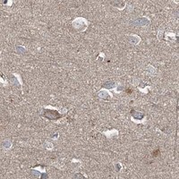


Comprehensive Human Protein Atlas IHC scoring per tissue (reliability: Approved — At least one protein variant secreted, tissue location of RNA and protein might differ and correlation is complex.). Rows are taken directly from the HPA tissue chart — click any row's HPA link to view the source.
| Tissue | Cell type | Level | Evidence | Source |
|---|---|---|---|---|
| Nasopharynx | Respiratory epithelial cells | High | Protein (IHC) | HPA → |
| Testis | Leydig cells | High | Protein (IHC) | HPA → |
| Adipose tissue | Adipocytes | Medium | Protein (IHC) | HPA → |
| Adrenal gland | Glandular cells | Medium | Protein (IHC) | HPA → |
| Appendix | Glandular cells | Medium | Protein (IHC) | HPA → |
| Tissue | Cell type | Level | Evidence | Source |
|---|---|---|---|---|
| None in HPA: ADAM23 is detected in all 45 scored tissues. Use a no-primary (secondary-only) and an isotype control instead. | ||||
Troubleshoot ADAM23 chromogenic IHC in paraffin sections using the stated retrieval setting, the selected antibody image, and compartment aware controls.
A07382 has real IHC data from paraffin-embedded human rectum carcinoma; the catalog also lists mouse reactivity (A07382 image caption; catalog: reactivity).
A07382 is listed for IHC and shows cytoplasmic staining in paraffin-embedded human rectum carcinoma at 1:50 (catalog: applications; A07382 image caption). Mouse reactivity is listed, but no mouse IHC or IF/ICC image is supplied (catalog: reactivity and image fields).
Which to pick: Choose A07382 for tissue IHC: it is a rabbit polyclonal antibody listed for IHC, with an image from paraffin-embedded human rectum carcinoma (catalog: host and applications; A07382 image caption). The caption does not report the fixative (A07382 image caption). A07382 also lists mouse reactivity, but its image shows human tissue; there is no IF/ICC option in this payload (catalog: reactivity, applications and IF fields; A07382 image caption).