This website uses cookies to ensure you get the best experience on our website.
- Table of Contents
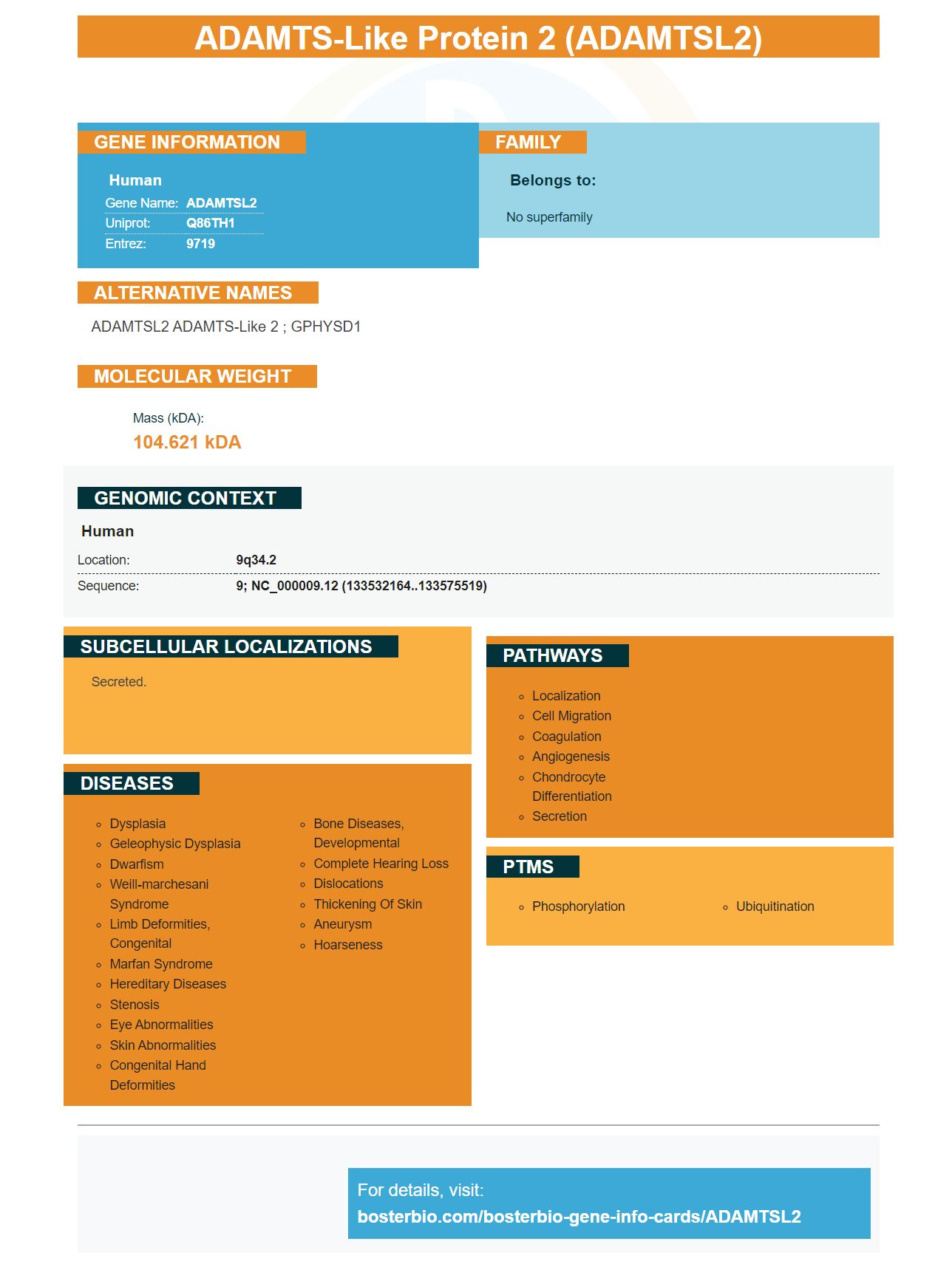
Facts about ADAMTS-like protein 2.
Human | |
---|---|
Gene Name: | ADAMTSL2 |
Uniprot: | Q86TH1 |
Entrez: | 9719 |
Belongs to: |
---|
No superfamily |
ADAMTSL2 ADAMTS-like 2 ; GPHYSD1
Mass (kDA):
104.621 kDA
Human | |
---|---|
Location: | 9q34.2 |
Sequence: | 9; NC_000009.12 (133532164..133575519) |
Secreted.
Boster bio's ADAMTSL2 label is a highly sensitive antibody that can detect the specific protein in your sample. Its primary antibody has been validated for Western blotting, Immunohistochemistry, and ELISA. Boster bio is a well-respected brand that offers high-affinity primary antibody. This product provides optimal efficiency for protein detection and protein transfer.
The ADAMTSL2 gene belongs to the family of secreted glucoproteins. It resembles the ADAMTS protease family but lacks a protease domain. Although its function is not clear, it is known that it regulates fibrillin microfibrils which are key components of the extracellular mat (ECM). Additionally, the gene may control the bioavailability of TGFb and regulate ECM deposition. It may play a significant role in cardiacfibrosis.
Although ADAMTSL2 provides high accuracy in identifying patients who are at risk for fibrosis and is highly sensitive in many clinical settings it is still very sensitive. It has been used for the identification of patients at high risk of liver biopsy or aggressive disease management. This discovery may help patients with NAFLD to be better stratified and managed. Further investigation is warranted into the ADAMTSL2 gene.
ADAMTSL2 inhibits myofibroblast cell differentiation and the fibrotic response of CFBs. Researchers can target therapeutic interventions to patients with high ADAMTSL2 levels if the expression levels of ADAMTSL2 are determined. A study in animals showed that ADAMTSL2 reduced levels of vinculin, p-sMA, and inhibited activation of focal adhesion kinase, and phosphorylation paxillin.
Three cases published in GPHYSD revealed that the ADAMTSL2 mutation was present. ADAMTSL2 variants were detected in three previously published cases. However, there was no genotype/phenotype correlation. Amplification of the gene was performed at PRIMM in Milan, Italy. The results showed a high correlation between the presence and gene of glycoproteins.
Picokine (tm), a new generation ELISA kits that dramatically improve sensitivity, boasts enhanced sensitivity with Picokine (tm). This reagent can be used with a variety samples and is compatible with immunoassays as well as protein purification processes. The Picoband is an additional tool that enables you to easily remove primary antibodies from Western blot membranes without damaging the immobilized antigen, allowing you to reuse the membrane.
Ponceau S solution can be used to rapidly stain proteins on PVDF or nitrocellulose membranes. The dye is sensitive and forms bands with positive-charged amino-acid residues. Ponceau S staining works well on nylon membranes but is very fast and easy. Because of its high sensitivity level and low background, it is an excellent choice to detect protein on polyvinylidene-difluoride membranes. It is also easy for you to bleed proteins from your membrane.
The ECL chemiluminescent detection system by Boster Bio allows you to detect proteins using an antibody as the reporter enzyme. The ECL chemiluminescent substrates include luminol, acridan, and 1,2-dioxetane. Lumenol is the most commonly used chemiluminescent agent. This substance releases photons when it decays to a lower energy state.
The ECL ECL-chemiluminescent detection product can increase the signal upto 1,000-fold. The Bio Rad Clarity Max ECL substrate optimizes target detection sensitivity. This reagent prolongs the signal lifetime to 24 hours, making it possible to reimage samples while maintaining signal intensity.
Adult testis lysate from adult testis was prepared in 50mM Tris (pH 7.4) at 23°C to perform the ECL analysis. The solution also contained 1% NP-40 v/v as well as a protease inhibitor mixture. The membrane was then placed on the X-ray film. The membrane was exposed for 1 min. Light emission is most intense after the substrate has been incubated for five to thirty minutes. CCD chemiluminescent detection systems require a chemiluminescent developer, fixation, and a chemiluminescent detector.
To create the Boster ECL cheiluminescent detector system, two components are mixed together to make a workable solution. To make a working solution, one component (a monoclonal antibody) is diluted in equal amounts with the other component. The reaction produces light emission at 425 nanometers, which is detected with X-ray films. A CCD camera-based imaging system that is cooled can quickly capture data and perform quantitative analysis. This system can also be used to detect proteins with low abundance.
The Clarity Max chemiluminescent Substrates are compatible all horseradish-peroxidase conjugates. They can be used for all types Western Blots. The Clarity Max are extremely sensitive for detecting proteins in western Blots. This is something that is very common in many laboratories. Boster Bio's ECL Chemiluminescent Detection System is compatible with all western Blots. It works with a wide range of horseradish peroxidase conjugates.
The SITVue/DAB Detection System for immunohistochemistry is an Intensification system that significantly increases chromogenic signals. The system includes a horseradish-peroxidase enzyme, which catalyzes deposition of two separate Linkers. The chromogenic detection kit uses diaminobenzidine (DAB) and streptavidin-peroxidase conjugate as reagents.
PMID: 18677313 by Le Goff C., et al. ADAMTSL2 mutations in geleophysic dysplasia demonstrate a role for ADAMTS-like proteins in TGF-beta bioavailability regulation.
PMID: 21415077 by Allali S., et al. Molecular screening of ADAMTSL2 gene in 33 patients reveals the genetic heterogeneity of geleophysic dysplasia.