This website uses cookies to ensure you get the best experience on our website.


- Table of Contents
Plan chromogenic ADCK1 IHC in paraffin sections with a catalog antibody dilution of 1:100–1:300 (datasheet A14988-1). Interpret the observed cytoplasmic tissue staining (HPA tissue IHC) alongside the mitochondrial annotation (UniProt), while accounting for uncertain staining reliability (HPA tissue IHC).

Expected localisation, validated protocols, controls and antibodies — the at-a-glance facts below, then the full design guide.
| Expected localisation | Cytoplasmic staining in several tissues (HPA tissue IHC) | |
| Staining pattern | Cytoplasmic staining in several tissue cell types (HPA tissue IHC) | |
| Antigen retrieval | Tris-EDTA pH 9.0 HIER, heat-mediated (datasheet A14988-1) | |
| Positive control | Appendix+4 more · see all | |
| Negative control | Adipose tissue+4 more · see all |
| Fixation | Keep fixation consistent across sections. (standard IHC practice; not target-specific) | |
| Caveat | Staining awaits external verification (HPA tissue IHC) | |
| Regulation | Expression regulation unreported (UniProt) | |
| Isoform / epitope | 3 isoforms; epitope differences unspecified (UniProt) |
The catalog antibody’s IHC protocol (datasheet A14988-1) is followed by a published ADCK1 IHC protocol for mouse sections and human colorectal tissue (PMC8024291).
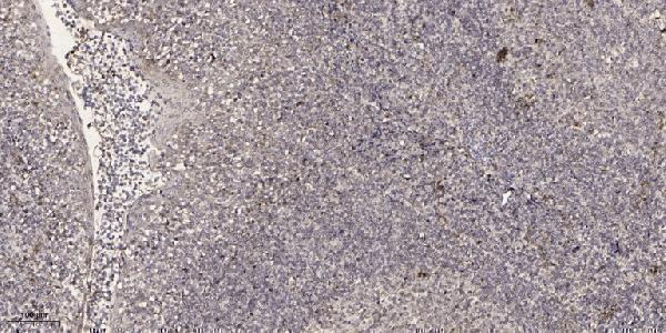
| Sample | Paraffin-embedded human tonsil tissue; fixative not specified (datasheet A14988-1) |
| Fixation | Image fixative and duration unreported (datasheet A14988-1); verify before use. |
| Sectioning | 4–5 µm sections on charged slides (standard) |
| Deparaffinisation | Xylene, graded ethanol series to water (standard) |
| Antigen retrieval | Heat retrieval: Tris-EDTA pH 9.0 (datasheet A14988-1); 20 min, 95–100 °C (standard) |
| Peroxidase block | 3% H2O2, 10 min, room temperature (standard) |
| Blocking | 10% normal serum of the secondary host, 30 min, room temperature (standard) |
| Primary antibody | Rabbit anti-ADCK1, 1:100-1:300 (datasheet A14988-1) |
| Primary incubation | Overnight at 4 °C (standard) |
| Detection | HRP-polymer secondary, DAB chromogen 5–10 min (standard) |
| Counterstain | Hematoxylin, blue, dehydrate and mount (standard) |
| Expected result | ADCK1-positive staining in glandular cells of appendix (HPA tissue IHC: Medium). HPA tissue profile: Cytoplasmic expression in several tissues. No signal in the no-primary control. |
ADCK1 is annotated in mitochondria and has no transmembrane segment (UniProt Q86TW2 topology). In paraffin IHC, expect cytoplasmic staining in selected epithelial and glandular cells, including colon glandular cells and kidney tubule cells, both reported at medium intensity (HPA tissue IHC). Treat this as a provisional pattern: HPA rates its tissue IHC reliability uncertain, pending external verification (HPA tissue IHC).
| Cytoplasmic chromogenic staining is visible in colon glandular cells or kidney tubule cells (HPA tissue IHC). | This fits the reported medium staining in those cells (HPA tissue IHC). Score the specified cells and compartment separately from surrounding tissue. A chromogenic cytoplasmic pattern cannot by itself resolve individual mitochondria, although mitochondrial localisation is annotated (UniProt Q86TW2). |
| Predominantly nuclear staining replaces the expected cytoplasmic pattern in a tissue section (HPA tissue IHC). | Flag a compartment mismatch and review controls and antibody specificity before calling the IHC result positive. HPA reports nucleoplasm in ICC-IF, but labels that location uncertain; UniProt annotates mitochondrion (HPA subcellular; UniProt Q86TW2). The IF observation does not establish a nuclear IHC pattern. |
| Strong staining appears in adipocytes or bone-marrow hematopoietic cells, where staining was not detected (HPA tissue IHC). | Compare a known-positive section processed in the same run before interpreting this as ADCK1. Unexpected staining could reflect cross-reactivity or endogenous detection activity (general IHC practice); HPA's uncertain IHC reliability limits any tissue-specific conclusion (HPA tissue IHC). |
| Color is diffuse across cells and tissue spaces, with no clear cell boundaries or compartment. | Treat the result as background rather than a scorable ADCK1 pattern (general IHC practice). Review primary-antibody concentration, blocking, washes and detection-only control; a genuine positive should be assignable to the reported cell type and cytoplasm (HPA tissue IHC). |
| No staining appears in colon glandular cells or kidney tubule cells, both reported at medium intensity (HPA tissue IHC). | Target-specific fixation effects are not established by the supplied assay evidence. Verify with a matched IHC source before attributing a result to fixation. |
| How should localisation guide scoring? | Use the HPA cytoplasmic tissue pattern for paraffin IHC and record nuclear staining separately (HPA tissue IHC). Mitochondrial localisation is a biological annotation, not proof that individual mitochondria must be visible with chromogenic detection (UniProt Q86TW2; general IHC practice). |
| How strong is the IHC evidence? | HPA calls tissue IHC uncertain and pending external verification; both listed antibodies have uncertain IHC status (HPA tissue IHC; HPA antibodies). Use the reported pattern as a comparison point, and interpret agreement alongside section controls rather than as independent validation. |
| Could isoforms change interpretation? | UniProt lists three ADCK1 isoforms and a protein kinase domain at residues 155–467 (UniProt Q86TW2). The payload does not locate either antibody's epitope or establish isoform-specific IHC staining, so a section cannot be assigned to an isoform from this pattern. |
| Do topology or processing predict staining loss? | UniProt reports no transmembrane segment, signal peptide or propeptide, and no annotated glycosylation sites (UniProt Q86TW2). These annotations do not establish an antigen-retrieval requirement or a fixation effect; choose retrieval conditions from the validated IHC method and controls (general IHC practice). |
| What does the IF/ICC observation mean? | HPA reports nucleoplasm as an uncertain ICC-IF location, while UniProt annotates mitochondrion (HPA subcellular; UniProt Q86TW2). Assess IF/ICC localisation in its own guide; it does not supply a protocol option or override the cytoplasmic tissue IHC observation (HPA tissue IHC). |
| Situation | Likely cause | Next action |
|---|---|---|
| Reported positive cells show no chromogenic signal (HPA tissue IHC). | The staining run may have failed, or retrieval and antibody conditions may be unsuitable (general IHC practice); ADCK1-specific fixation sensitivity is unreported in the supplied sources. | Check a run control and detection reagents, then compare retrieval and primary-antibody conditions with the IHC-validated method. Record the negative result without inferring absent protein. |
| Nuclei dominate the IHC result. | The compartment conflicts with HPA's cytoplasmic tissue profile; HPA's nuclear ICC-IF location is uncertain (HPA tissue IHC; HPA subcellular). | Check counterstain versus chromogen, inspect detection-only and positive controls, and report the compartment mismatch. Do not score nuclear color as confirmed ADCK1 IHC. |
| Adipocytes or bone-marrow hematopoietic cells stain strongly (HPA tissue IHC). | HPA reports staining as not detected in those cells; cross-reactivity or endogenous detection activity is possible (HPA tissue IHC; general IHC practice). | Compare the same run's reported positive cells, inspect a detection-only control, and reassess antibody concentration before assigning specificity. |
| Color is widespread without a cell-specific pattern. | Nonspecific antibody binding, inadequate washes or detection background can produce diffuse color (general IHC practice). | Review blocking, washes, primary-antibody dilution and detection-only control. Score only signal that can be assigned to a cell type and compartment. |
| A weak signal appears in a proposed negative section. | HPA's not-detected calls are observations under its assay conditions, and its tissue IHC reliability is uncertain (HPA tissue IHC). | Describe the observed cells and intensity, compare controls, and avoid treating an HPA not-detected tissue as proof of absolute absence. |
| Two antibodies give different tissue patterns. | Neither listed antibody has enhanced IHC validation; both have uncertain IHC status (HPA antibodies). | Document each antibody's cell types and compartments against the same controls. Treat disagreement as unresolved specificity rather than selecting a pattern solely by signal strength. |


Comprehensive Human Protein Atlas IHC scoring per tissue (reliability: Uncertain — Pending external verification.). Rows are taken directly from the HPA tissue chart — click any row's HPA link to view the source.
| Tissue | Cell type | Level | Evidence | Source |
|---|---|---|---|---|
| Appendix | Glandular cells | Medium | Protein (IHC) | HPA → |
| Breast | Myoepithelial cells | Medium | Protein (IHC) | HPA → |
| Bronchus | Respiratory epithelial cells | Medium | Protein (IHC) | HPA → |
| Colon | Glandular cells | Medium | Protein (IHC) | HPA → |
| Endometrium | Glandular cells | Low | Protein (IHC) | HPA → |
| Tissue | Cell type | Level | Evidence | Source |
|---|---|---|---|---|
| Adipose tissue | Adipocytes | Not detected | Protein (IHC) | HPA → |
| Bone marrow | Hematopoietic cells | Not detected | Protein (IHC) | HPA → |
| Caudate | Glial cells | Not detected | Protein (IHC) | HPA → |
| Cerebellum | Cells in granular layer | Not detected | Protein (IHC) | HPA → |
| Cerebral cortex | Endothelial cells | Not detected | Protein (IHC) | HPA → |
Troubleshoot ADCK1 staining by optimizing paraffin-section conditions and checking cell type and subcellular pattern against independent evidence.
A14988-1 has IHC data from paraffin-embedded human tonsil (IHC image caption). IF is listed for the human- and mouse-reactive antibody, but no IF image is supplied (catalog: applications, reactivity, IF image alts).
A14988-1 will render with its paraffin-section human tonsil IHC figure (IHC image caption). The antibody lists IHC and IF applications and human and mouse reactivity; IF has no supplied figure (catalog: applications, reactivity, IF image alts).
Which to pick: For tissue IHC, choose A14988-1: its caption documents paraffin-embedded human tonsil, antibody at 1:200 overnight at 4 °C, and Tris-EDTA retrieval at pH 9.0 (A14988-1 IHC image caption). For IF, A14988-1 lists a 1:200–1:1000 dilution; ICC validation and an IF figure are unreported (catalog: applications, IF dilution, IF image alts). For cross-species work, A14988-1 lists human and mouse reactivity, though its IHC figure shows only human tonsil; clonality and the tissue fixative are unreported (catalog: reactivity, clone field; A14988-1 IHC image caption).