This website uses cookies to ensure you get the best experience on our website.


- Table of Contents
Plan ADCY9 chromogenic IHC in paraffin sections using the observed cytoplasmic tissue pattern alongside its annotated membrane location (HPA tissue IHC; UniProt). The catalog antibody has an IHC dilution range of 1:50–1:200; compare staining with matched controls and score the relevant cell population (datasheet).

Expected localisation, validated protocols, controls and antibodies — the at-a-glance facts below, then the full design guide.
| Expected localisation | Mostly cytoplasmic in tissue (HPA tissue IHC); membrane protein (UniProt) | |
| Staining pattern | Cytoplasmic staining in most tissues (HPA tissue IHC) | |
| Antigen retrieval | Citrate pH 6.0 HIER, 95–98 °C, 20 min (rule: cytoplasmic / membrane antigen) | |
| Positive control | Gallbladder+4 more · see all | |
| Negative control | Adipose tissue+4 more · see all |
| Fixation | Keep fixation consistent across paraffin sections. (standard IHC practice; not target-specific) | |
| Caveat | Smooth muscle may be unstained despite reported airway expression (HPA tissue IHC; UniProt) | |
| Regulation | Regulation of abundance is unreported (UniProt) | |
| Isoform / epitope | No isoforms annotated; epitope side of membrane is unspecified (UniProt) |
The catalog antibody’s IHC-P protocol is accompanied by published ADCY9 staining protocols for colon cancer tissue arrays (PMC5759510) and lung adenocarcinoma sections (PMC10089850).
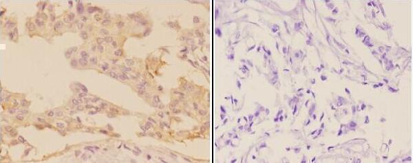
| Sample | Paraffin-embedded human breast carcinoma tissue; fixative not specified (datasheet A06038-1) |
| Fixation | Image fixative and duration unreported (datasheet A06038-1); verify before use. |
| Sectioning | 4–5 µm sections on charged slides (standard) |
| Deparaffinisation | Xylene, graded ethanol series to water (standard) |
| Antigen retrieval | Heat-induced epitope retrieval in citrate buffer, pH 6.0, 20 min at 95–98 °C (standard rule: cytoplasmic / membrane antigen) |
| Peroxidase block | 3% H2O2, 10 min, room temperature (standard) |
| Blocking | 10% normal serum of the secondary host, 30 min, room temperature (standard) |
| Primary antibody | Rabbit anti-ADCY9, 1:50-1:200 (datasheet A06038-1) |
| Primary incubation | Overnight at 4 °C (standard) |
| Detection | HRP-polymer secondary, DAB chromogen 5–10 min (standard) |
| Counterstain | Hematoxylin, blue, dehydrate and mount (standard) |
| Expected result | ADCY9-positive staining in glandular cells of gallbladder (HPA tissue IHC: High). HPA tissue profile: Cytoplasmic expression in most tissues. No signal in the no-primary control. |
ADCY9 is a cell-membrane protein with 12 transmembrane segments (UniProt O60503 topology). In paraffin-section IHC, expect cellular staining in gallbladder glandular cells, with moderate staining in several other listed cell types (HPA tissue IHC: High in gallbladder glandular cells; Medium in adrenal, appendix, breast, colon and duodenal glandular cells, bone-marrow hematopoietic cells and cerebral-cortex glial cells). HPA describes the tissue pattern as cytoplasmic; its IHC reliability is Approved, with medium RNA concordance and external verification pending (HPA tissue IHC).
| Membranous or cytoplasmic chromogenic staining in gallbladder glandular cells, stronger than nearby unstained cells. | This fits a useful positive control: HPA scores these cells High and describes cytoplasmic expression in most tissues (HPA tissue IHC). A membrane component is consistent with ADCY9 topology, but routine IHC may not resolve a sharp membrane rim (UniProt O60503 topology; general IHC practice). |
| Signal confined to nuclei, with no convincing membrane or cytoplasmic staining. | An exclusively nuclear pattern is unsupported by UniProt cell-membrane annotation and HPA's tissue and ICC-IF localisation summaries (UniProt O60503 subcellular location; HPA tissue IHC; HPA subcellular). Treat it as suspect and compare with a positive tissue and detection controls (general IHC practice). |
| Strong staining in adipocytes, prostate glandular cells or heart cardiomyocytes. | HPA reports these specific cells as Not detected, so strong signal calls for a specificity or endogenous-detection check (HPA tissue IHC; general IHC practice). UniProt detection in heart is tissue-level evidence; it does not establish staining in cardiomyocytes (UniProt O60503 tissue specificity; HPA tissue IHC). |
| Uniform haze across cells and surrounding tissue, with little cell-specific contrast. | This is difficult to score as ADCY9 because the HPA reference identifies particular positive and negative cell populations (HPA tissue IHC). Check background in a no-primary control, then review blocking, washes and detection chemistry (general IHC practice). |
| No visible signal in gallbladder glandular cells. | HPA rates these cells High, so an entirely blank positive control calls the run into question (HPA tissue IHC). Check tissue preservation, retrieval, antibody dilution and detection with appropriate controls before interpreting other tissues as ADCY9-negative (general IHC practice). |
| Compartment and epitope access | ADCY9 has 12 membrane spans and large cytoplasmic regions, but the supplied record gives no antibody epitope (UniProt O60503 topology; HPA antibodies). Do not infer which side of the membrane the IHC-validated antibody recognizes or prescribe an ADCY9-specific retrieval condition. |
| Cell-specific tissue comparisons | HPA scores gallbladder glandular cells High, several other listed cell populations Medium, and adipocytes and prostate glandular cells Not detected (HPA tissue IHC). Use the named cells as comparators; a whole-tissue label can conceal differences among its cell types (general IHC practice). |
| Strength of validation | The two listed antibodies are IHC Approved; the tissue profile has medium staining–RNA consistency and awaits external verification (HPA antibodies; HPA tissue IHC). Interpret a matched pattern as supportive rather than definitive proof of antigen identity. |
| IF/ICC: what pattern should be expected? | HPA supports plasma-membrane localisation, marks cytosol uncertain, and lists primary cilium and basal body as uncertain additional locations (HPA subcellular). That ICC-IF evidence can guide compartment interpretation; it does not supply an IF protocol for this IHC section. |
| Retrieval and detection | No ADCY9-specific retrieval or fixation effect is established by these sources (UniProt O60503; HPA tissue IHC). When signal or background is problematic, optimize retrieval and inspect no-primary controls as general paraffin-IHC practice, without attributing an outcome to ADCY9-specific fixation sensitivity. |
| Situation | Likely cause | Next action |
|---|---|---|
| Gallbladder positive control is blank. | The run may have inadequate antigen exposure or detection; HPA expects High staining in gallbladder glandular cells (HPA tissue IHC; general IHC practice). | Verify tissue and control performance, then review the catalog antibody's IHC-P conditions, retrieval and detection steps (general IHC practice). Do not assign an ADCY9-specific fixation effect from these sources. |
| Only a dark nuclear signal is present. | An isolated nuclear compartment conflicts with UniProt cell-membrane annotation and HPA localisation summaries (UniProt O60503 subcellular location; HPA tissue IHC; HPA subcellular). | Inspect a no-primary control and a HPA-listed positive cell population; reassess specificity before scoring nuclei as positive (general IHC practice). |
| Adipocytes or prostate glandular cells stain strongly. | HPA lists those cell populations as Not detected, raising concern about nonspecific binding or endogenous detection activity (HPA tissue IHC; general IHC practice). | Compare a no-primary control, review blocking and detection chemistry, and score the named cells separately from neighboring structures (general IHC practice). |
| Cardiomyocytes stain despite a weak gallbladder control. | HPA lists cardiomyocytes as Not detected; UniProt's detection in heart does not identify the positive heart cell type (HPA tissue IHC; UniProt O60503 tissue specificity). | Resolve the failed positive control first, then compare cardiomyocyte signal with no-primary and adjacent-cell controls (general IHC practice). |
| The whole section has diffuse brown background. | Broad haze obscures the cell-specific pattern described by HPA and can arise from staining or detection background (HPA tissue IHC; general IHC practice). | Check no-primary signal, endogenous activity controls where relevant, blocking, washes and chromogen development before assigning cellular scores (general IHC practice). |
| Cytoplasmic IHC appears broader than membrane staining seen by IF/ICC. | HPA describes cytoplasmic expression in tissue IHC, while its ICC-IF plasma-membrane location is supported and cytosol is uncertain (HPA tissue IHC; HPA subcellular). | Compare cell-specific patterns within each assay and check IHC background controls; do not require identical compartment resolution across chromogenic IHC and ICC-IF (general IHC practice). |
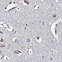


Comprehensive Human Protein Atlas IHC scoring per tissue (reliability: Approved — Medium consistency between antibody staining and RNA expression data. Pending external verification.). Rows are taken directly from the HPA tissue chart — click any row's HPA link to view the source.
| Tissue | Cell type | Level | Evidence | Source |
|---|---|---|---|---|
| Gallbladder | Glandular cells | High | Protein (IHC) | HPA → |
| Adrenal gland | Glandular cells | Medium | Protein (IHC) | HPA → |
| Appendix | Glandular cells | Medium | Protein (IHC) | HPA → |
| Bone marrow | Hematopoietic cells | Medium | Protein (IHC) | HPA → |
| Breast | Glandular cells | Medium | Protein (IHC) | HPA → |
| Tissue | Cell type | Level | Evidence | Source |
|---|---|---|---|---|
| Adipose tissue | Adipocytes | Not detected | Protein (IHC) | HPA → |
| Heart muscle | Cardiomyocytes | Not detected | Protein (IHC) | HPA → |
| Ovary | Ovarian stroma cells | Not detected | Protein (IHC) | HPA → |
| Prostate | Glandular cells | Not detected | Protein (IHC) | HPA → |
| Smooth muscle | Smooth muscle cells | Not detected | Protein (IHC) | HPA → |
Troubleshoot ADCY9 staining in paraffin sections by checking retrieval, compartment, cell type, and controls before comparing chromogenic scores (UniProt O60503; HPA tissue IHC).
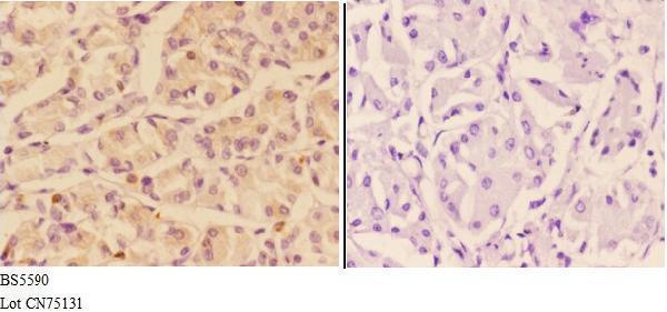
A06038-1 has real IHC images from paraffin sections of human breast and stomach carcinoma (catalog image captions). Human, mouse and rat reactivity is listed (catalog reactivity); IF data are absent (catalog IF image alts).
A06038-1 is listed for IHC in human, mouse and rat (catalog applications/reactivity). Its IHC images show cytoplasmic and membrane staining in paraffin sections of human breast and stomach carcinoma at 1:50 (catalog image captions).
Which to pick: For tissue IHC, choose A06038-1, a rabbit polyclonal antibody with paraffin-section images from human breast and stomach carcinoma (catalog host/image captions). For IF/ICC, no SKU has a listed IF application or IF image (catalog applications/IF image alts). For mouse or rat IHC, A06038-1 has listed reactivity, while its depicted tissue data are human only (catalog reactivity/image captions); the fixative is unreported (catalog image captions).