This website uses cookies to ensure you get the best experience on our website.


- Table of Contents
Plan chromogenic ADH1C IHC using cytoplasmic staining in gastrointestinal glandular cells and strong hepatocyte staining as reference patterns (HPA tissue IHC). The catalog antibody has an IHC dilution range of 1:50–1:200 (datasheet); interpret staining with the HPA warning that antibodies may detect proteins from more than one gene (HPA tissue IHC).

Expected localisation, validated protocols, controls and antibodies — the at-a-glance facts below, then the full design guide.
| Expected localisation | Cytoplasmic staining (HPA tissue IHC) | |
| Staining pattern | Cytoplasmic signal mainly in GI glandular cells (HPA tissue IHC) | |
| Antigen retrieval | Citrate pH 6.0 HIER, 95–98 °C, 20 min (rule: cytoplasmic / membrane antigen) | |
| Positive control | Liver+4 more · see all | |
| Negative control | Bronchus+4 more · see all |
| Fixation | Keep fixation consistent across paraffin sections. (standard IHC practice; not target-specific) | |
| Caveat | Antibody may detect proteins from more than one gene (HPA tissue IHC) | |
| Regulation | Expression regulation not annotated (UniProt) | |
| Isoform / epitope | No isoforms annotated; mature chain spans residues 2–375 (UniProt) |
Compare the catalog antibody’s IHC-P protocol with four published ADH1C IHC protocols (PMC13084684; PMC9855755; PMC8921497; PMC7438573).
| Sample | Paraffin-embedded human esophagus tissue; fixative not specified (datasheet A02650) |
| Fixation | Image fixative and duration unreported (datasheet A02650); verify before use. |
| Sectioning | 4–5 µm sections on charged slides (standard) |
| Deparaffinisation | Xylene, graded ethanol series to water (standard) |
| Antigen retrieval | Heat-induced epitope retrieval in citrate buffer, pH 6.0, 20 min at 95–98 °C (standard rule: cytoplasmic / membrane antigen) |
| Peroxidase block | 3% H2O2, 10 min, room temperature (standard) |
| Blocking | 10% normal serum of the secondary host, 30 min, room temperature (standard) |
| Primary antibody | Rabbit anti-ADH1C, 1:50-1:200 (datasheet A02650) |
| Primary incubation | Overnight at 4 °C (standard) |
| Detection | HRP-polymer secondary, DAB chromogen 5–10 min (standard) |
| Counterstain | Hematoxylin, blue, dehydrate and mount (standard) |
| Expected result | ADH1C-positive staining in hepatocytes of liver (HPA tissue IHC: High). HPA tissue profile: Cytoplasmic expression mainly in glandular cells of gastrointestinal tract. No signal in the no-primary control. |
ADH1C is a cytoplasmic protein without a transmembrane segment (UniProt P00326: subcellular location and topology). In paraffin section IHC, expect cytoplasmic staining, especially in hepatocytes, which show High staining; the HPA profile also describes expression mainly in gastrointestinal glandular cells (HPA: tissue IHC). Interpret the pattern cautiously: HPA rates tissue staining Supported, reports medium agreement with RNA data, and warns that the antibodies target proteins from more than one gene (HPA: tissue IHC reliability).
| Hepatocytes show strong cytoplasmic staining, while surrounding structures remain distinguishable on the counterstained section. | This matches the reported High hepatocyte signal and cytoplasmic location (HPA: liver tissue IHC; UniProt P00326: subcellular location). Assess cell identity and compartment together; a dark area without recognizable stained hepatocytes is less persuasive than a clear cellular pattern (general IHC practice). |
| Signal appears predominantly nuclear, or outlines cell borders without appreciable cytoplasmic staining. | This does not match the expected tissue IHC pattern (HPA: tissue IHC profile; UniProt P00326: subcellular location). HPA ICC-IF lists plasma membrane as uncertain and nucleoplasm as an additional uncertain location; those observations do not establish a membrane or nuclear IHC pattern (HPA: subcellular ICC-IF). Review staining controls before assigning ADH1C (general IHC practice). |
| Prominent staining occurs in cells scored Not detected by HPA, such as bronchial respiratory epithelium or pancreatic exocrine glandular cells. | Such staining conflicts with those specific HPA tissue observations (HPA: bronchus and pancreas tissue IHC). Possible explanations include nonspecific antibody binding or endogenous chromogenic detection activity (general IHC practice). Because HPA cautions that its antibodies target proteins from more than one gene, staining alone cannot identify which related protein produced the signal (HPA: tissue IHC reliability). |
| Color spreads across extracellular areas, section edges, or many unrelated cell populations, obscuring cellular boundaries. | A diffuse deposit cannot be scored confidently as the reported cytoplasmic cell pattern (HPA: tissue IHC profile). Check the detection and blocking controls, wash conditions, and counterstain; excess background can make a genuinely positive compartment difficult to distinguish (general IHC practice). Do not treat overall section darkness as evidence of ADH1C expression (general IHC practice). |
| Hepatocytes show no detectable signal in a section intended as a positive control. | This disagrees with the reported High hepatocyte staining (HPA: liver tissue IHC). First check section integrity, primary antibody application, detection reagents, and the run controls (general IHC practice). An absent signal in one run does not establish that ADH1C is absent from the sampled hepatocytes; technical performance and tissue quality need assessment (general IHC practice). |
| Choice of tissue and cell population | Liver hepatocytes provide a reported High staining reference; appendix and duodenum endocrine cells, and gallbladder glandular cells, are reported Medium (HPA: tissue IHC). Compare the same named cell population when judging agreement; different cells within one organ need not have the listed level (general IHC practice). |
| Specificity of the reported tissue pattern | HPA labels tissue IHC Supported and reports medium consistency with RNA expression; it also cautions that the antibodies target proteins from more than one gene (HPA: tissue IHC reliability). Treat a matching pattern as supportive evidence, while recognizing that this record does not prove ADH1C-specific staining (HPA: tissue IHC reliability). |
| Molecular location and related class I enzymes | UniProt places ADH1C in the cytoplasm and reports no transmembrane segment; it can form dimers with class I alcohol dehydrogenase chains ADH1A or ADH1B (UniProt P00326: location, topology, subunit). Dimerization describes the protein complex and does not by itself validate an antibody signal as ADH1C-specific (general IHC interpretation). |
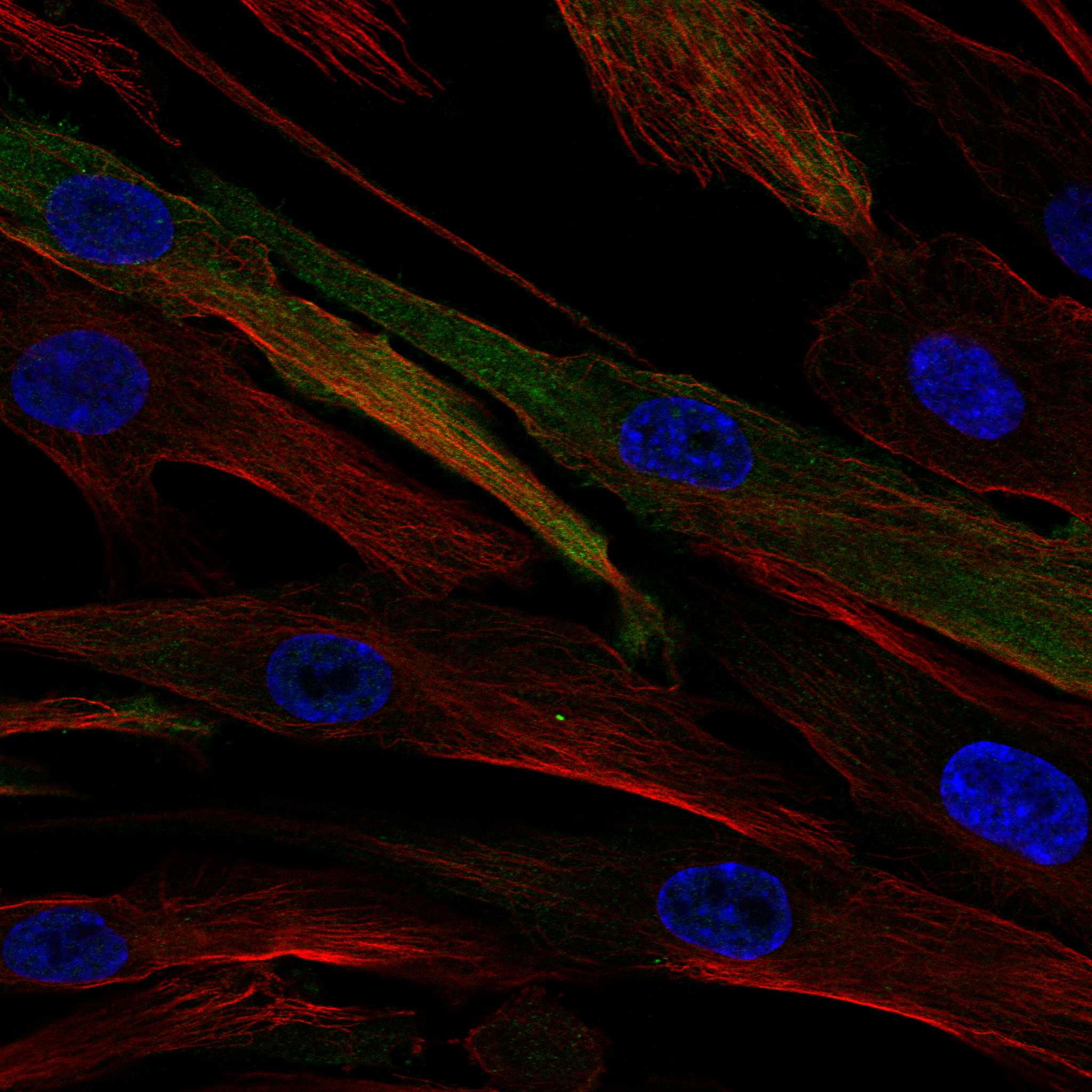
| IF/ICC Q: Should membrane or ciliary staining be expected here? | A: HPA ICC-IF supports cytosol, calls plasma membrane uncertain, and lists primary cilium, transition zone, and basal body as additional uncertain locations; it cautions about antibodies targeting multiple genes (HPA: subcellular ICC-IF). Use those observations only as qualified IF context, not as a paraffin section IHC acceptance criterion (HPA: subcellular ICC-IF; HPA: tissue IHC profile). |
| Situation | Likely cause | Next action |
|---|---|---|
| No hepatocyte staining in the positive reference section | The result conflicts with reported High hepatocyte staining (HPA: liver tissue IHC); a failed staining run or unsuitable section is possible (general IHC practice). | Verify the section, primary antibody step, detection reagents, and run controls; repeat the affected step after identifying a control failure (general IHC practice). Avoid concluding biological absence from this slide alone (general IHC practice). |
| Weak, uneven cytoplasmic signal in liver | Uneven section processing, reagent coverage, or detection can produce patchy staining (general IHC practice); HPA's hepatocyte reference is High (HPA: liver tissue IHC). | Compare intact areas and a concurrently stained control, then review section quality, reagent coverage, and detection performance (general IHC practice). The supplied sources give no ADH1C-specific fixation sensitivity or retrieval setting (UniProt P00326; HPA: tissue IHC). |
| Diffuse brown background obscures cells | Incomplete blocking, inadequate washing, or excess detection signal can reduce contrast in chromogenic IHC (general IHC practice). | Inspect the control without primary antibody, confirm appropriate blocking for the detection system, and optimize washes and detection conditions (general IHC practice). Score only staining that can be assigned to recognizable cells and compartments (general IHC practice). |
| Nuclear or sharply membrane-only staining dominates | This conflicts with cytoplasmic ADH1C and the tissue IHC profile (UniProt P00326: location; HPA: tissue IHC); membrane and nucleoplasm assignments in ICC-IF are uncertain (HPA: subcellular ICC-IF). | Check control sections and detection background, then reassess whether cytoplasmic signal is present (general IHC practice). Do not reinterpret uncertain ICC-IF locations as proof of a nuclear or membrane IHC result (HPA: subcellular ICC-IF). |
| Strong signal appears in a reported negative cell population | For example, HPA reports bronchial respiratory epithelial cells and pancreatic exocrine glandular cells as Not detected (HPA: tissue IHC). Nonspecific binding or endogenous detection activity may contribute (general IHC practice). | Review the control without primary antibody and the relevant blocking steps; compare the exact cell type with HPA's listing (general IHC practice; HPA: tissue IHC). Consider the multiple-gene antibody caution before attributing any residual signal to ADH1C (HPA: tissue IHC reliability). |
| A Medium or Low tissue appears negative while the liver control stains | HPA reports differing levels across cell types, including Medium appendix endocrine cells and Low thyroid glandular cells (HPA: tissue IHC). A weak expected signal may be difficult to separate from background (general IHC practice). | Confirm that the scored cells match the HPA cell population, inspect background and detection performance, and report the observed result with the tissue and cell type (general IHC practice; HPA: tissue IHC). Do not extrapolate the liver intensity to every organ (HPA: tissue IHC). |



Comprehensive Human Protein Atlas IHC scoring per tissue (reliability: Supported — Medium consistency between antibody staining and RNA expression data. Caution, targets protein from more than one gene.). Rows are taken directly from the HPA tissue chart — click any row's HPA link to view the source.
| Tissue | Cell type | Level | Evidence | Source |
|---|---|---|---|---|
| Liver | Hepatocytes | High | Protein (IHC) | HPA → |
| Appendix | Endocrine cells | Medium | Protein (IHC) | HPA → |
| Breast | Adipocytes | Medium | Protein (IHC) | HPA → |
| Duodenum | Endocrine cells | Medium | Protein (IHC) | HPA → |
| Epididymis | Glandular cells | Medium | Protein (IHC) | HPA → |
| Tissue | Cell type | Level | Evidence | Source |
|---|---|---|---|---|
| Bronchus | Respiratory epithelial cells | Not detected | Protein (IHC) | HPA → |
| Cerebral cortex | Glial cells | Not detected | Protein (IHC) | HPA → |
| Cervix | Glandular cells | Not detected | Protein (IHC) | HPA → |
| Endometrium | Cells in endometrial stroma | Not detected | Protein (IHC) | HPA → |
| Nasopharynx | Respiratory epithelial cells | Not detected | Protein (IHC) | HPA → |
Troubleshoot ADH1C staining in paraffin sections by checking cytoplasmic localisation, cell type, antibody specificity and processing controls.
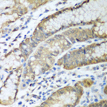
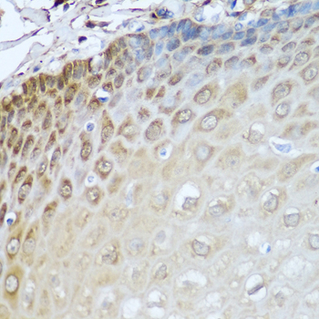
A02650 has real paraffin-section IHC images from human esophagus and stomach (IHC image captions) and listed Human, Mouse and Rat reactivity (catalog: A02650); no IF data are supplied (catalog: A02650).
A02650 will render with IHC images of human esophagus and stomach paraffin sections at 1:100 (IHC image captions). Its listed applications are IHC and WB, and its listed reactivity is Human, Mouse and Rat (catalog: A02650).
Which to pick: Choose A02650 for paraffin-section tissue IHC, supported by its human esophagus and stomach images (IHC image captions); the fixative is unreported (IHC image captions). There is no IF/ICC option here because A02650 has no listed IF application or IF image, and its clonality is unreported (catalog: A02650). For Mouse or Rat IHC, A02650 has listed reactivity, but the supplied tissue images show Human samples only (catalog: A02650; IHC image captions).