This website uses cookies to ensure you get the best experience on our website.
- Table of Contents
1 Citations 1 Q&As
Facts about Adiponectin receptor protein 1.
ADIPOQ-binding activates a signaling cascade that leads to increased AMPK activity, and ultimately to increased fatty acid oxidation, increased glucose uptake and decreased gluconeogenesis. Has high affinity for globular adiponectin and very low affinity for full size adiponectin (By similarity).
Human | |
---|---|
Gene Name: | ADIPOR1 |
Uniprot: | Q96A54 |
Entrez: | 51094 |
Belongs to: |
---|
ADIPOR family |
ACDCR1; adiponectin receptor 1; adiponectin receptor protein 1; AdipoR1; ADR1; CGI45; FLJ42464; PAQR1; PAQR1FLJ25385; Progestin and adipoQ receptor family member I; TESBP1A
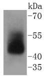
Mass (kDA):
42.616 kDA
Human | |
---|---|
Location: | 1q32.1 |
Sequence: | 1; NC_000001.11 (202940825..202958572, complement) |
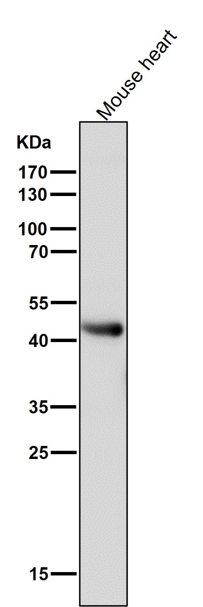
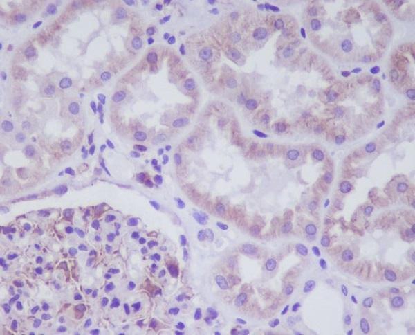
Widely expressed (PubMed:16044242). Highly expressed in heart and skeletal muscle (PubMed:12802337). Expressed at intermediate level in brain, spleen, kidney, liver, placenta, lung and peripheral blood leukocytes (PubMed:12802337). Weakly expressed in colon, thymus and small intestine (PubMed:12802337).
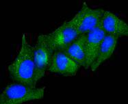
Cell membrane; Multi-pass membrane protein. Localized to the cell membrane and intracellular organelles.
The ADIPOR1 marker is a key part of many biological assays, which are designed to detect biomarkers such as glucose and adipocytes. The marker is a protein produced by adipocytes and is needed for normal body weight. In a biological assay, an antibody that reacts with ADIPOR1 is needed to assess the levels of adiponectin in a sample. Boster Bio develops antibodies to detect the protein in mouse and rabbit samples.
ADIPOQ is a small hormone produced by adipocytes. The hormone has a similar sequence to C1q, but the amino acid sequence of adipoQ is different. In C1q, the amino acid sequence contains conserved cysteines, which form disulfide bonds with the activator molecules. ADIPOQ, however, contains an aspartate residue instead of a conserved cysteine.
Gain-of-function Adipoq models, which overexpress the hormone, have shown profound metabolic effects. Loss-of-function mice, however, show varying metabolic phenotypes. Those carrying loxP-flanked regions of the APN gene have shown increased insulin resistance and hyperlipidemia. Furthermore, loss-of-function mice have a significantly lower survival rate, with nearly 50% dying within five days.
ADIPOQ cDNA generates a protein of approximately 30 kDa. This molecular mass is consistent with its cDNA sequence. Transiently transfected adipoQ cDNA into NIH-3T3 cells, which secreted the protein, and Western blot analysis confirmed that adipoQ is secreted into the medium.
ADIPOQ is a polypeptide of 247 amino acids with globular domain that has been shown to contain significant homology with complement factor C1q, collagen a1(X) and cerebellin subunits. It is a highly regulated hormone that specifically regulates adipocyte differentiation and is specifically expressed in adipose tissue in rodents and humans.
Adiponectin is a natural hormone produced in the body and is abundant in plasma. It accounts for about 0.01% of the total protein concentration in plasma. Interestingly, the concentration of adiponectin is higher than that of the majority of other hormones. In comparison, leptin, cortisol, and tumour necrosis factor alpha are all 1000 times lower in concentration than adiponectin.
Although the precise physiological role of adiponectin has not been established, it is thought to play a role in preventing obesity by reducing inflammation and insulin resistance. While it is still poorly understood, ADIPOQ has numerous benefits for human health. It improves the sensitivity of the immune system, prevents atherosclerosis, and reduces inflammation. In addition, it inhibits the transformation of macrophages into foam cells.
ELISA kits, antibodies, and other immunological reagents are designed to detect biomarkers in cancer, neurosciences, developmental biology, and inflammation. Their products range from picogram to microgram level sensitivity, and they can detect biomarkers in a variety of biological systems, including the gastrointestinal tract. Boster Bio's products can be purchased through tebu-bio.
Primary and secondary antibodies require different species. Generally, goats, donkeys, sheep, rabbits, and mice are the hosts of monoclonal and polyclonal secondary antibodies. Studies usually cite goat or donkey secondary antibodies. Table 4 lists the most common target species for goat anti-mouse and mouse secondary antibodies. Table 4 also lists the host species for rabbit and mouse anti-mouse secondary antibodies.
Primary Abs were prepared by incubating the target tissue overnight at 4degC. Secondary antibodies were prepared by adding goat anti-mouse or goat anti-rabbit IgG to polymers carrying the target antibody. Secondary antibodies were then added to the mixture according to manufacturer's recommendations. After incubating the primary antibodies, the polymers containing the goat anti-mouse or goat anti-rabbit IgG were counterstained with rabbit or mouse HRP conjugated anti-isotype antibodies.
Nano-Secondary reagents for HeLa cells were used to stain the primary antibodies with Alexa Fluor 488. TP1170 was used for nanobody detection. TP1129 and TP1170 can be used together to increase staining intensity. The mice and rabbit polyclonal antibodies were used in parallel to perform immunoblotting.
Rabbit and mouse antibodies are essential tools for numerous basic research techniques and medical diagnostic assays. Polyclonal anti-IgG secondary antibodies are frequently used to detect primary antibodies. To ensure a reliable supply, producers must kill and immunize large numbers of animals, which is expensive and poses a significant animal welfare problem. Additionally, each new batch of serum contains a heterogeneous mix of antibodies. Thus, it is crucial to carefully control the quality of anti-IgG sera every time.
The main drawback of polyclonal secondary antibodies is their high cost. They require intensive animal care, aggressive hyperimmunization strategies, and increased bleedings. Although this method is highly effective in basic research, it has serious animal welfare and ethical issues. The recent Santa Cruz Biotechnology scandal has highlighted these issues. These antibodies can be extremely costly and are often not very useful for clinical trials.
The primary antibodies were not as robust as the secondary ones, but both showed higher variability and could be used multiple times. Some were re-usable, but only twice. The robustness of antibodies was tested in discussion threads, and some investigators have reported success with re-use. Some also recommend using goat anti-mouse secondary antibodies to detect glioma cells.
Secondary antibodies were purchased from MilliporeSigma and can be reused multiple times. However, not all antibodies are reusable, and researchers need to determine whether the cost of purchasing a new batch of antibodies will be worthwhile. Each laboratory will have different requirements and budgets, and the costs of a failed experiment may outweigh any cost savings. For this reason, it is important to ensure that secondary antibodies are re-usable.
PMID: 16044242 by Tang Y.T., et al. PAQR proteins: a novel membrane receptor family defined by an ancient 7-transmembrane pass motif.
PMID: 12802337 by Yamauchi T., et al. Cloning of adiponectin receptors that mediate antidiabetic metabolic effects.
*More publications can be found for each product on its corresponding product page