Click image to see more details
-

-
-
-
-
+4
Product Info Summary
| SKU: | M01869 |
|---|---|
| Size: | 100 μl |
| Reactive Species: | Human, Mouse, Rat |
| Host: | Rabbit |
| Application: | Flow Cytometry, IF, IHC, ICC, WB |
Customers Who Bought This Also Bought

Product info
Product Name
Anti-ADIPOR1 Rabbit Monoclonal Antibody
SKU/Catalog Number
M01869
BM4566 is an alternative SKU for this antibody, used in previous lots.
Size
100 μl
Form
Liquid
Description
Boster Bio Anti-ADIPOR1 Rabbit Monoclonal Antibody catalog # M01869. Tested in WB, IHC, ICC/IF, Flow Cytometry applications. This antibody reacts with Human, Mouse, Rat.
Storage & Handling
Store at -20°C for one year. For short term storage and frequent use, store at 4°C for up to one month. Avoid repeated freeze-thaw cycles.
Cite This Product
Anti-ADIPOR1 Rabbit Monoclonal Antibody (Boster Biological Technology, Pleasanton CA, USA, Catalog # M01869)
Host
Rabbit
Contents
Rabbit IgG in stabilizing components, phosphate buffered saline, pH 7.4, 150mM NaCl, 0.02% sodium azide and 50% glycerol.
*This antibody is supplied in a stabilized formulation.
Compatibility with conjugation reactions depends on the chemistry of the conjugation method used.
For conjugation methods that are not compatible with the stabilizing components present in this formulation, a carrier-free antibody format is required.
Clonality
Monoclonal
Clone Number
FIH-1
Isotype
Rabbit IgG
Immunogen
A synthesized peptide derived from human ADIPOR1
Reactive Species
M01869 is reactive to ADIPOR1 in Human, Mouse, Rat
Observed Molecular Weight
43 kDa
Calculated molecular weight
42.6 kDa
Antibody Validation
Boster validates all antibodies on WB, IHC, ICC, Immunofluorescence, and ELISA with known positive control and negative samples to ensure specificity and high affinity, including thorough antibody incubations.
Application & Images
Applications
M01869 is guaranteed for Flow Cytometry, IF, IHC, ICC, WB Boster Guarantee
Recommend Dilution
WB 1:500-2000
IHC 1:50-200
ICC/IF 1:50-200
FC 1:20
Tested application
Use TE buffer pH 9.0 for antigen retrieval; (*) citrate buffer pH 6.0 is an alternative.
Validation Images & Assay Conditions
Click image to see more details
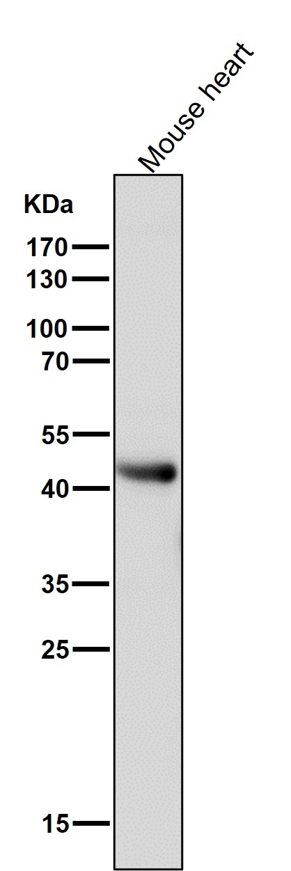
All lanes use the Antibody at 1:1K dilution for 1 hour at room temperature.

Click image to see more details
Immunohistochemical analysis of paraffin-embedded human kidney, using ADIPOR1 Antibody.

Click image to see more details
Mechanisms for local medulla injection of ADPN promoting bone healing. (A) Serum Osteoprotegerin (OPG) levels at weeks 2, 4, and 6. (B) WB analysis of alkaline phosphatase (ALP), bone morphogenic protein 2 (BMP-2), osteocalcin (OCN), and adiponectin receptor 1 (AdipoR1) expressions with corresponding quantification, (C) Immunofluorescent staining of ALP, OCN, BMP-2, and AdipoR1 in G2 (ADPN 1 mg/kg) and G3 (ADPN 2 mg/kg), and the corresponding quantification, n = 5. The white arrow indicates the periosteum, and the blue arrow indicates the lacuna, scale bar = 100 μm.
Index in PubMed under a CC BY license. PMID: 34790669

Click image to see more details
The immunofluorescent staining of ALP, OCN, BMP-2, and AdipoR1 on BMSCs with ADPN 10 μg/ml and AdipoR1 siRNA + ADPN 10 μg/ml, and the corresponding quantification, scale bar = 100 μm.
Index in PubMed under a CC BY license. PMID: 34790669

Click image to see more details
Expression of AdipoR1 and AdipoR2 in BV2 cells and microglia in mice brain. a RT-PCR analysis of AdipoR1 and AdipoR2 in BV2 cells. b Western blot analysis of AdipoR1 and AdipoR2 expression in BV2 cells. Expression of AdipoR1 and AdipoR2 from cerebral cortex homogenates was used as a positive control. α-Tubulin was used as a loading control. c , d Co-immunocytochemistry staining of microglia (Iba1) and AdipoR1 or AdipoR2 in BV2 cells and microglia in the cortex of WT mice. Scale bar 50 μm
Index in PubMed under a CC BY license. PMID: 31128596

Click image to see more details
APN suppressed AβO-induced proinflammatory cytokine release in BV2 cells via AdipoR1. a , b Representative Western blot analysis of AdipoR1 and AdipoR2. BV2 cells were transfected with control siRNA, AdipoR1 siRNA, or AdipoR2 siRNA in a concentration-dependent manner (25, 50, 100 nM). c , d ELISA assays of TNFα and IL-1β were conducted after knockdown of AdipoR1. e , f ELISA assays of TNFα and IL-1β were conducted after knockdown of AdipoR2. Data were presented as the mean ± SEM for at least three independent experiments, and each performed in duplicates ( n = 3). Two-way ANOVA with Tukey’s multiple comparison test revealed a difference between groups . *p < 0.05, **p < 0.01, ***p < 0.001; ns, statistically not significant
Index in PubMed under a CC BY license. PMID: 31128596

Click image to see more details
All lanes use the Antibody at 1:1K dilution for 1 hour at room temperature.

Click image to see more details
Western blot analysis of ADIPOR1 expression in Human heart lysate.
Specific Publications For Anti-ADIPOR1 Rabbit Monoclonal Antibody (M01869)
Loading publications
Recommended Resources
Here are featured tools and databases that you might find useful.
- Boster's Pathways Library
- Protein Databases
- Bioscience Research Protocol Resources
- Data Processing & Analysis Software
- Photo Editing Software
- Scientific Literature Resources
- Research Paper Management Tools
- Molecular Biology Software
- Primer Design Tools
- Bioinformatics Tools
- Phylogenetic Tree Analysis
Customer Reviews
Have you used Anti-ADIPOR1 Rabbit Monoclonal Antibody?
Share your experimental results or join a short interview to earn up to $1,000 in product credits or other rewards.
0 Reviews For Anti-ADIPOR1 Rabbit Monoclonal Antibody
Customer Q&As
Have a question?
Find answers in Q&As, reviews.
Can't find your answer?
Submit your question
1 Customer Q&As for Anti-ADIPOR1 Rabbit Monoclonal Antibody
Question
What is the immunogen sequence of M01869 and Homology percentage with mouse?
Verified customer
Asked: 2019-09-25
Answer
The immunogen sequence for the Anti-ADIPOR1 Rabbit Monoclonal Antibody (M01869) is CGAPASNREADTVE. Homology with mouse is 84.6%.
Boster Scientific Support
Answered: 2019-09-26
