This website uses cookies to ensure you get the best experience on our website.


- Table of Contents
Plan chromogenic IHC on paraffin sections around the extracellular staining reported for ADM (HPA tissue IHC). Adrenal glandular cells and kidney glomerular cells are reported as high staining controls (HPA tissue IHC); the catalog antibody A00594 has a 1:100 IHC starting dilution (datasheet: A00594).

Expected localisation, validated protocols, controls and antibodies — the at-a-glance facts below, then the full design guide.
| Expected localisation | Extracellular staining in most tissues (HPA tissue IHC) | |
| Staining pattern | Extracellular positivity, including placenta and endothelium (HPA tissue IHC) | |
| Antigen retrieval | Citrate pH 6.0 HIER, 95–98 °C, 20 min (rule: cytoplasmic / membrane antigen) | |
| Positive control | Adrenal gland+4 more · see all | |
| Negative control | Caudate+4 more · see all |
| Fixation | Matched tissue-IHC evidence does not establish the fixation claim. Validate the specimen-specific method before use. (selected-SKU IHC image A00594) | |
| Caveat | Secreted ADM may stain away from its source cells (HPA tissue IHC) | |
| Regulation | Staining regulation is not established (UniProt) | |
| Isoform / epitope | No isoforms annotated; peptide processing may affect epitope recognition (UniProt) |
The catalog antibody's IHC-P protocol is followed by published ADM protocols for rat spinal cord (PMC5228177), kidney tissue (PMC5802232), and breast tumour sections (PMC2394432).
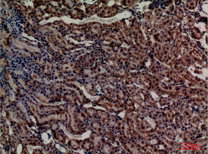
| Sample | Paraffin-embedded rat kidney tissue; fixative not specified (datasheet A00594) |
| Fixation | Image fixative and duration unreported (datasheet A00594); verify before use. |
| Sectioning | 4–5 µm sections on charged slides (standard) |
| Deparaffinisation | Xylene, graded ethanol series to water (standard) |
| Antigen retrieval | Heat-induced epitope retrieval in citrate buffer, pH 6.0, 20 min at 95–98 °C (standard rule: cytoplasmic / membrane antigen) |
| Peroxidase block | 3% H2O2, 10 min, room temperature (standard) |
| Blocking | 10% normal serum of the secondary host, 30 min, room temperature (standard) |
| Primary antibody | Rabbit anti-ADM, 1:100 (datasheet A00594) |
| Primary incubation | Overnight at 4 °C (standard) |
| Detection | HRP-polymer secondary, DAB chromogen 5–10 min (standard) |
| Counterstain | Hematoxylin, blue, dehydrate and mount (standard) |
| Expected result | ADM-positive staining in glandular cells of adrenal gland (HPA tissue IHC: High). HPA tissue profile: Extracellular positivity in most tissues, including placenta and endothelial cells. No signal in the no-primary control. |
ADM is a secreted, non-transmembrane propeptide (UniProt P35318 topology), and HPA reports extracellular positivity in most tissues, including placenta and endothelial cells (HPA tissue IHC). High staining is reported in adrenal glandular cells, bronchial basal cells, and kidney glomerular cells (HPA tissue IHC). HPA rates its tissue IHC evidence Approved, with medium consistency against RNA expression; a secreted protein’s tissue RNA and protein locations may differ (HPA reliability description).
| Extracellular staining with strong signal in adrenal glandular cells, bronchial basal cells, or kidney glomerular cells (HPA tissue IHC). | This fits the reported distribution: HPA describes extracellular positivity in most tissues and rates these cell populations High (HPA tissue IHC). Score the stained compartment and cell population separately; extracellular signal need not identify the cell that produced a secreted protein (UniProt P35318 subcellular location; HPA reliability description). |
| Predominantly nuclear staining, especially without the expected extracellular pattern (HPA tissue IHC; UniProt P35318 subcellular location). | An exclusively nuclear pattern lacks support from the supplied localisation evidence (UniProt P35318; HPA tissue IHC). Treat it as suspect and compare a known-positive section and a no-primary control before interpreting it as ADM (general IHC practice). |
| Strong staining in ovarian stromal cells, caudate glial cells, or smooth muscle cells (HPA tissue IHC). | HPA reports ADM as Not detected in these specific populations (HPA tissue IHC). Check for cross-reactivity or endogenous detection activity with appropriate controls (general IHC practice); the HPA result is cell specific and does not make the entire tissue a universal negative control (HPA tissue IHC). |
| Diffuse colour across tissue spaces and unrelated structures, obscuring cell boundaries (general IHC practice). | Because extracellular ADM staining is reported, extracellular colour alone is insufficient to call background (HPA tissue IHC). Compare its distribution with the expected cells and a no-primary control; widespread colour in that control supports a detection or background problem (general IHC practice). |
| No visible signal in adrenal glandular cells or kidney glomerular cells on an otherwise interpretable section (HPA tissue IHC). | These are High populations in HPA tissue IHC, so an absent result warrants a technical check (HPA tissue IHC; general IHC practice). Confirm that the positive control, antibody application, detection reagents, and counterstain allow a readable result before concluding that ADM is absent from the sample (general IHC practice). |
| Secretion and topology (UniProt P35318). | ADM is secreted and has no transmembrane segment (UniProt P35318 topology). Expect extracellular signal and interpret apparent cellular association in the context of nearby extracellular staining (HPA tissue IHC; UniProt P35318 subcellular location). |
| Precursor processing (UniProt P35318). | The 185-aa precursor has a signal peptide and annotated propeptide regions (UniProt P35318 processing). The supplied evidence does not locate the catalog antibody’s epitope, so staining cannot identify which processed ADM-related product it detects (UniProt P35318 processing; supplied evidence). |
| Tissue and cell context (HPA tissue IHC). | HPA reports High staining in several distinct populations, including appendix and duodenal glandular cells and fallopian-tube ciliated-cell rootlets, but Low staining in colon endothelial cells (HPA tissue IHC). Compare like cells in like tissues when judging intensity (general IHC practice). |
| Evidence strength and RNA comparison (HPA tissue IHC). | The tissue IHC assessment is Approved, with medium consistency against RNA data (HPA reliability description). HPA notes that secretion can separate protein location from tissue RNA location, so an RNA mismatch alone does not establish an IHC artefact (HPA reliability description). |
| Antibody validation scope (HPA antibody records). | HPA068955 and CAB016075 are each IHC Approved; neither has an ICC status in the supplied records (HPA antibody records). Their IHC status supports evaluating tissue staining but does not establish an IF/ICC staining pattern or an IF/ICC protocol (HPA antibody records; HPA subcellular record). |
| Situation | Likely cause | Next action |
|---|---|---|
| No signal in a known-positive population, such as adrenal glandular cells (HPA tissue IHC). | A failed staining run or inadequate detection can give a false negative (general IHC practice). | Run an HPA High tissue as a positive control and check antibody application, detection reagents, and counterstain in the IHC workflow (HPA tissue IHC; general IHC practice). |
| Nuclear staining dominates while extracellular staining is absent (HPA tissue IHC; UniProt P35318). | The compartment disagrees with the reported secreted and extracellular pattern (UniProt P35318; HPA tissue IHC). | Compare with a known-positive section and no-primary control; withhold an ADM-specific call if the discordant pattern persists (HPA tissue IHC; general IHC practice). |
| A cell population reported Not detected stains strongly, such as ovarian stromal cells (HPA tissue IHC). | Cross-reactivity or endogenous detection activity is possible (general IHC practice). | Check a no-primary control and the expected High population in the same run; interpret the unexpected cells separately from other cells in that tissue (HPA tissue IHC; general IHC practice). |
| Colour is widespread and hard to distinguish from reported extracellular positivity (HPA tissue IHC). | Background from the detection workflow can obscure a genuine extracellular pattern (HPA tissue IHC; general IHC practice). | Inspect the no-primary control, then review blocking, washing, and detection conditions as general IHC troubleshooting steps (general IHC practice). |
| Endothelial staining varies between specimens or sites (HPA tissue IHC). | HPA reports extracellular positivity involving endothelial cells, Low colon endothelial staining, and Not detected cerebral-cortex endothelial staining (HPA tissue IHC). | Record the tissue and endothelial population before comparing intensity; avoid applying one site’s HPA level to all endothelium (HPA tissue IHC). |
| IF/ICC question: should fluorescence mark a defined intracellular ADM compartment (HPA subcellular record)? | HPA lists ADM as Secreted but provides no main intracellular location or ICC-IF image cell lines, and the supplied antibodies have no ICC status (HPA subcellular record; HPA antibody records). | Treat a defined intracellular IF/ICC pattern as unverified by these sources; consult the separate IF/ICC guide for assay design (HPA subcellular record; HPA antibody records). |


Comprehensive Human Protein Atlas IHC scoring per tissue (reliability: Approved — Medium consistency between antibody staining and RNA expression data. At least one protein variant secreted, tissue location of RNA and protein might differ and correlation is complex.). Rows are taken directly from the HPA tissue chart — click any row's HPA link to view the source.
| Tissue | Cell type | Level | Evidence | Source |
|---|---|---|---|---|
| Adrenal gland | Glandular cells | High | Protein (IHC) | HPA → |
| Appendix | Glandular cells | High | Protein (IHC) | HPA → |
| Bronchus | Basal cells | High | Protein (IHC) | HPA → |
| Duodenum | Glandular cells | High | Protein (IHC) | HPA → |
| Endometrium | Glandular cells | High | Protein (IHC) | HPA → |
| Tissue | Cell type | Level | Evidence | Source |
|---|---|---|---|---|
| Caudate | Glial cells | Not detected | Protein (IHC) | HPA → |
| Cerebellum | Cells in granular layer | Not detected | Protein (IHC) | HPA → |
| Cerebral cortex | Endothelial cells | Not detected | Protein (IHC) | HPA → |
| Hippocampus | Glial cells | Not detected | Protein (IHC) | HPA → |
| Ovary | Ovarian stroma cells | Not detected | Protein (IHC) | HPA → |
Troubleshoot ADM staining in paraffin sections by checking retrieval, tissue context and extracellular signal before assigning cellular expression (UniProt P35318; HPA tissue IHC).
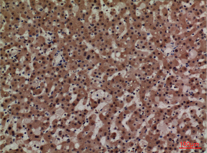
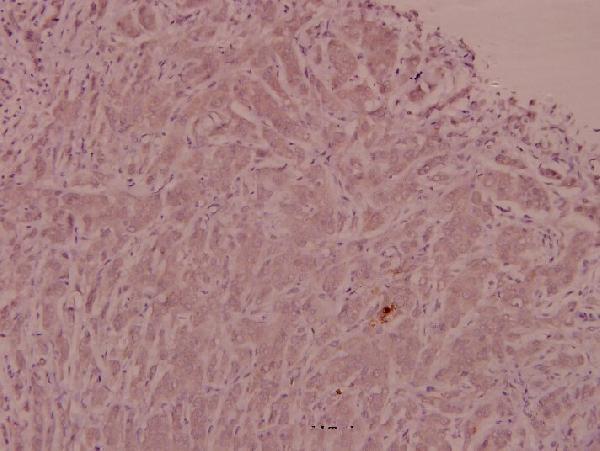
Two anti-ADM antibodies have paraffin-section IHC figures from rat kidney, human liver, and human breast carcinoma (IHC image captions); catalog reactivity spans human, mouse, and rat (catalog: reactivity).
A00594 lists IHC and IF for human and rat, with paraffin-section IHC images from rat kidney and human liver (catalog: applications and reactivity; A00594 IHC image captions). A00594-1 lists IHC for human, mouse, and rat, with a paraffin-section IHC image from human breast carcinoma (catalog: applications and reactivity; A00594-1 IHC image caption).
Which to pick: For human breast carcinoma IHC or listed mouse reactivity, choose A00594-1 at an IHC starting dilution within 1:50–1:200 (A00594-1 IHC image caption; catalog: reactivity and IHC dilution). For IF, choose A00594 at the listed 1:50 dilution; ICC validation is unreported (catalog: applications and IF dilution). Both IHC captions show paraffin sections, and neither reports the fixative (A00594 and A00594-1 IHC image captions).