ADORA2A / Adenosine receptor A2a · IHC design guide

Design Immunohistochemistry for ADORA2A

This guide identifies caudate neuronal projections and a subset of thymic cells as ADORA2A IHC staining references (HPA tissue IHC). It gives a 1:25 starting dilution for the catalog antibody in IHC-P (datasheet M02268).

Evidence assembled Oct 2026 · For research use; verify linked source records and product datasheet before use
Immunohistochemistry protocol sheet for ADORA2A (IHC for ADORA2A): expected localisation Cell membrane expected (UniProt), antibody M02268, validated IHC image, and IHC protocol steps
Printable ADORA2A IHC protocol sheet — expected localisation Cell membrane expected (UniProt), antibody M02268, controls and protocol steps. Open the full ADORA2A IHC guide →

ADORA2A Immunohistochemistry Experimental Design Guide

Expected localisation, validated protocols, controls and antibodies — the at-a-glance facts below, then the full design guide.

Must know before staining
Expected localisation Cell membrane expected (UniProt)
Staining pattern Caudate neuronal projections; a subset of thymic cells (HPA tissue IHC)
Antigen retrieval Citrate pH 6 HIER, heat-mediated (datasheet M02268)
Positive control ⓘ Appendix+4 more · see all
Negative control ⓘ Adipose tissue+4 more · see all
Important caveats
Reasons your staining may differ from the expected pattern.
Fixation Formaldehyde-fixed paraffin sections were used (selected-SKU IHC image M02268)
Caveat Antibody staining and RNA show medium consistency (HPA tissue IHC)
Regulation Expression regulation is not annotated (UniProt)
Isoform / epitope No isoforms annotated; C-terminal region is cytoplasmic (UniProt)
Section 1

Recommended ADORA2A IHC & IF Protocols

The catalog antibody’s IHC-P protocol is paired with one published ADORA2A IHC protocol (PMC9827409).

Recommended immunohistochemistry (IHC-P) protocol parameters
SampleFormaldehyde-fixed, paraffin-embedded Human heart tissue (datasheet M02268)
FixationImage formalin-fixed; duration unreported (datasheet M02268); verify before use.
Sectioning4–5 µm sections on charged slides (standard)
DeparaffinisationXylene, graded ethanol series to water (standard)
Antigen retrievalHeat retrieval: Citrate pH 6 (datasheet M02268); 20 min, 95–100 °C (standard)
Peroxidase block3% H2O2, 10 min, room temperature (standard)
Blocking10% normal serum of the secondary host, 30 min, room temperature (standard)
Primary antibodyRabbit anti-ADORA2A, 1:25 (datasheet M02268)
Primary incubation1 hours at 37°C (datasheet M02268)
DetectionHRP-polymer secondary, DAB chromogen 5–10 min (standard)
CounterstainHematoxylin, blue, dehydrate and mount (standard)
Expected resultADORA2A-positive staining in endocrine cells of appendix (HPA tissue IHC: High). HPA tissue profile: Expression in caudate nucleus and a subset of cells in thymus. No signal in the no-primary control.
💡Decision noteStart with citrate pH 6 heat retrieval (datasheet M02268); the published protocol used microwave EDTA pH 9.0 for 20 min (PMC9827409).
Section 2

What Is the Expected ADORA2A Staining Pattern?

ADORA2A is a seven-transmembrane cell-surface receptor, with reported localization at neuronal processes (UniProt P29274 topology and subcellular location). In paraffin-section IHC, expect strong staining of caudate neuronal projections and scattered gastrointestinal endocrine cells; bone-marrow hematopoietic cells and cerebellar molecular-layer cells show medium staining (HPA tissue IHC). HPA rates the tissue profile Enhanced while describing only medium consistency between staining and RNA expression (HPA tissue IHC).

What am I looking at on my slide?
Strong, selective staining outlines caudate neuronal projections or marks individual endocrine cells in appendix, colon, duodenum, rectum, or small intestine (HPA tissue IHC).This fits the reported high-staining populations (HPA tissue IHC). Judge the labeled structure and cell identity as well as intensity: UniProt places ADORA2A at the cell membrane and notes its localization at neuronal processes (UniProt P29274 subcellular location).
Cerebellar molecular-layer cells show cytoplasmic or membrane staining, or bone-marrow hematopoietic cells show less intense staining than the high-staining examples (HPA tissue IHC).These are reported medium-level patterns, so cytoplasmic signal in the specified cerebellar cells is not automatically an artefact (HPA tissue IHC). Compare like cells across sections; an expectation of equally strong staining in every positive tissue would misread the HPA categories (HPA tissue IHC).
Nuclei dominate the stain, or broad cytoplasmic color obscures cell boundaries and processes.A nuclear-dominant IHC pattern does not match the annotated cell-membrane location (UniProt P29274 subcellular location). Broad cytoplasmic color also needs context: HPA reports cytoplasm/membrane staining in cerebellar molecular-layer cells, while HPA ICC-IF additionally reports vesicles (HPA tissue IHC; HPA subcellular ICC-IF).
Many adipocytes, adrenal glandular cells, or bronchial respiratory epithelial cells stain uniformly despite their reported non-detection (HPA tissue IHC).Treat staining of those specific cell populations as a specificity warning, then check whether color tracks the primary antibody or the detection system (HPA tissue IHC; general IHC practice). An HPA 'Not detected' entry describes its observed cells and assay; it does not prove that every cell in that tissue lacks ADORA2A (HPA tissue IHC).
A caudate section lacks detectable signal in neuronal projections, including when other sections are weak.Caudate neuronal projections are a reported high-staining reference, so their absence makes a negative test section hard to interpret (HPA tissue IHC). Review section quality, the IHC-validated antibody's staining conditions, and the detection controls before calling the sample negative (general IHC practice).
💡Expected ADORA2A appearanceCall the result positive when selective, strong staining follows caudate neuronal projections or scattered gastrointestinal endocrine cells (HPA tissue IHC), in keeping with membrane localization (UniProt P29274); widespread nuclear color or uniform staining of HPA non-detected cell populations is suspect (UniProt P29274 subcellular location; HPA tissue IHC).
How each factor affects the staining
Cell type and tissue choice (HPA tissue IHC)Caudate neuronal projections and specified gastrointestinal endocrine cells provide reported high-staining references; bone marrow and cerebellum provide medium-level examples (HPA tissue IHC). Score the named cells, since the HPA levels do not assign one intensity to every cell in an organ (HPA tissue IHC).
Topology and compartment (UniProt P29274 topology; HPA subcellular ICC-IF)ADORA2A has seven transmembrane segments and a cytoplasmic C-terminal region (UniProt P29274 topology). HPA ICC-IF approves a mainly plasma-membrane location, with additional vesicle and primary-cilium locations; those IF observations should not be imposed as required features of chromogenic tissue IHC (HPA subcellular ICC-IF).
Evidence and antibody validation (HPA tissue IHC; HPA antibodies)The tissue profile is rated Enhanced but has medium staining–RNA consistency (HPA tissue IHC). Listed IHC antibody statuses differ: HPA065566 is Supported, while HPA075997 and CAB001943 are Enhanced (HPA antibodies). Preserve that distinction when comparing images obtained with different antibodies.
Processing and modification (UniProt P29274 processing and glycosylation)UniProt lists one 1–412 chain, no signal peptide or propeptide, and a glycosylation site at residue 154 (UniProt P29274). These annotations alone do not identify the catalog antibody's epitope or establish an ADORA2A-specific retrieval or fixation effect; choose retrieval conditions from the antibody's IHC instructions (general IHC practice).
Why is my staining missing, weak or wrong?
SituationLikely causeNext action
No stain in the caudate positive reference (HPA tissue IHC).Target-specific fixation effects are not established by the supplied assay evidence. Verify with a matched IHC source before attributing a result to fixation.Check the section, antibody concentration and incubation, retrieval conditions specified for that antibody, and detection reagents; repeat with a working caudate reference (general IHC practice; HPA tissue IHC).
Nuclear-dominant color appears across several cell types.This conflicts with ADORA2A's annotated membrane localization, although the image alone cannot identify which reagent caused it (UniProt P29274 subcellular location; general IHC practice).Compare primary-omission and detection controls, inspect counterstain, and assess whether specific membrane or neuronal-process staining remains (general IHC practice; UniProt P29274 subcellular location).
Adipocytes or bronchial respiratory epithelial cells appear strongly positive (HPA tissue IHC).These named cells are reported as Not detected; broad color may reflect off-target binding or endogenous detection activity, pending controls (HPA tissue IHC; general IHC practice).Check cell identity, examine primary-omission controls, and apply the detection system's appropriate endogenous-activity block before interpreting the stain (general IHC practice).
Diffuse color obscures the caudate projections or scattered endocrine cells (HPA tissue IHC).Excess background can mask the selective pattern; its source cannot be assigned from the supplied ADORA2A records alone (general IHC practice; HPA tissue IHC).Inspect blocking, washes, antibody concentration, chromogen development, and a primary-omission control, then reassess the reported high-staining cells (general IHC practice; HPA tissue IHC).
Bone marrow or cerebellum looks weaker than caudate (HPA tissue IHC).That intensity ranking may match the reported medium versus high categories, rather than indicate a failed run (HPA tissue IHC).Score bone-marrow hematopoietic cells or cerebellar molecular-layer cells in their own context; use caudate projections as a high-staining comparator (HPA tissue IHC).
Q: What should an IF/ICC image show?HPA ICC-IF places ADORA2A mainly at the plasma membrane, with additional vesicle and primary-cilium localization (HPA subcellular ICC-IF).A: Evaluate those compartments in the IF/ICC guide; these cell-image locations complement, but do not replace, the tissue and cell-type expectations for IHC (HPA subcellular ICC-IF; HPA tissue IHC).

Sample controls for ADORA2A IHC & IF

🧪Run caudate first and look for staining in neuronal projections (HPA: High in caudate neuronal projections). Use adipose tissue as the negative tissue, focusing on adipocytes (HPA: Not detected in adipocytes); on the caudate slide, nuclei and areas without neuronal projections should show only background staining relative to the stained projections (HPA: High in caudate neuronal projections; standard IHC interpretation).
Positive control tissue: Appendix (Endocrine cells, HPA High)
Negative control tissue: Adipose tissue (HPA Not detected)
ICC-IF cell lines (HPA subcellular resource): HPA ICC-IF images show ADORA2A in HEK293, ASC52telo, RPTEC/TERT1, hTERT-RPE1 (serum starved), with annotated localisation: Plasma membrane (approved) (HPA subcellular).
Technical controls: Include a no-primary (secondary-only) control and an irrelevant IgG control matched to the primary antibody’s host species and isotype, or normal host IgG for a polyclonal primary (standard IHC practice). An ADORA2A knockout specimen provides a biological negative; for chromogenic detection in caudate, block endogenous peroxidase and check any biotin-based detection against a no-primary control (standard IHC practice).
⚠️Feasibility: A target-specific fixation window or fixation effect is unreported, and the selected M02268 tissue-IHC caption reports formaldehyde-fixed paraffin sections (selected-SKU IHC image M02268). The caption uses heat-mediated citrate retrieval at pH 6, but does not establish that ADORA2A staining depends on that retrieval method (selected M02268 caption). The supplied evidence does not establish whether frozen sections or IF are easier; in caudate, endogenous pigment or peroxidase activity can complicate chromogenic interpretation (HPA: High in caudate neuronal projections; standard IHC practice).

HPA tissue IHC evidence for ADORA2A

Comprehensive Human Protein Atlas IHC scoring per tissue (reliability: Enhanced — Medium consistency between antibody staining and RNA expression data.). Rows are taken directly from the HPA tissue chart — click any row's HPA link to view the source.

Positive expression · recommended positive controls

TissueCell typeLevelEvidenceSource
Appendix Endocrine cells High Protein (IHC) HPA →
Caudate Neuronal projections High Protein (IHC) HPA →
Colon Endocrine cells High Protein (IHC) HPA →
Duodenum Endocrine cells High Protein (IHC) HPA →
Rectum Endocrine cells High Protein (IHC) HPA →

Undetected expression · recommended negative controls

TissueCell typeLevelEvidenceSource
Adipose tissue Adipocytes Not detected Protein (IHC) HPA →
Adrenal gland Glandular cells Not detected Protein (IHC) HPA →
Breast Adipocytes Not detected Protein (IHC) HPA →
Bronchus Respiratory epithelial cells Not detected Protein (IHC) HPA →
Cerebral cortex Endothelial cells Not detected Protein (IHC) HPA →
Section 3

Advanced ADORA2A IHC Tips

Troubleshoot ADORA2A staining in paraffin sections by checking retrieval, compartmental pattern, controls and the cell types used for scoring.

What retrieval should I try first if ADORA2A staining is weak?
Use heat-mediated citrate buffer at pH 6 for paraffin-section antigen retrieval (datasheet M02268). The selected tissue image used this retrieval before 1:25 primary incubation for 1 hour at 37°C (caption M02268). If staining remains weak, compare heat exposure times on adjacent sections while keeping the buffer, cooling and detection steps constant (standard IHC practice). Excessive heating can damage section edges, so assess morphology alongside the membrane-associated pattern expected for this 7-pass receptor (UniProt P29274 topology). Include a no-primary control to distinguish retrieval-associated background from specific staining (standard IHC practice).
How should I investigate ADORA2A staining differences between paraffin blocks?
Target-specific sensitivity to fixation time or fixative is unknown from the supplied evidence, so weak staining alone cannot identify a fixation effect (evidence scope). Record fixative, fixation duration, tissue thickness and delay before fixation for each block (standard IHC practice). Process matched sections together and begin with citrate pH 6 retrieval and the reported 1:25 primary dilution (datasheet M02268; caption M02268). Compare morphology and compartmental pattern as well as signal intensity, because processing differences can alter apparent staining (standard IHC practice). Include the same positive control section in each run to check reagent performance (standard IHC practice).
Should ADORA2A signal appear only at the cell surface?
Expect membrane-associated staining because ADORA2A is a 7-transmembrane receptor annotated at the cell membrane (UniProt P29274 topology and subcellular location). A purely nuclear chromogenic pattern would therefore need stronger validation before being scored as receptor staining (UniProt P29274 subcellular location). Vesicular and primary-cilium localisation has also been observed by cellular imaging, but those fine structures may be difficult to resolve in chromogenic sections (HPA subcellular). In caudate, evaluate staining in neuronal projections as well as cell bodies, since projection staining is reported as high (HPA: High in caudate neuronal projections). Compare serial sections and no-primary controls when diffuse cytoplasmic colour obscures these patterns (standard IHC practice).
How does epitope position affect interpretation of ADORA2A staining?
Check the antibody's stated immunogen or mapped epitope before interpreting a negative section; the supplied record does not map the catalog antibody's epitope (evidence scope). ADORA2A has 7 membrane-spanning segments, an extracellular region at residues 144–173, and a cytoplasmic tail at 291–412 (UniProt P29274 topology). A glycosylation site is annotated at residue 154, which may matter when evaluating access to an extracellular epitope, although an effect on this antibody is unproven (UniProt P29274 glycosylation). The supplied record lists 0 isoforms, so it offers no isoform-specific staining explanation (UniProt P29274 isoforms). Compare any discrepant staining with an independently validated epitope and appropriate tissue controls (standard IHC practice).
How can IF help assess an ambiguous chromogenic ADORA2A pattern?
Use IF as a separate localisation check, pairing ADORA2A with a marker for the cell population being evaluated rather than assuming that nearby chromogenic signal belongs to it (standard IF practice). For caudate projections or intestinal endocrine cells, choose a marker validated for the relevant population; those are reported ADORA2A-positive sites (HPA: High in caudate neuronal projections; HPA: High in intestinal endocrine cells). Select fluorophores away from prominent tissue autofluorescence and include single-label controls before interpreting overlap (standard IF practice). If the antibody recognises the cytoplasmic tail at 291–412, permeabilisation is needed; an accessible extracellular epitope may permit surface staining without it (UniProt P29274 topology; standard IF practice). The catalog antibody's epitope is unspecified here (evidence scope).
What should I check when brown staining appears throughout the section?
First compare the stained slide with a no-primary control and inspect whether colour follows tissue edges, damaged areas or the expected cellular pattern (standard IHC practice). The selected image used 3% BSA blocking for 0.5 hour at room temperature, providing a documented starting condition (caption M02268). In a peroxidase-based chromogenic workflow, check the peroxidase block and shorten DAB development if the control also darkens; these are general workflow steps (standard IHC practice). Titrate primary antibody around the reported 1:25 condition while keeping retrieval fixed (caption M02268; standard IHC practice). Prefer interpretable membrane-associated staining over uniform brown colour when judging ADORA2A (UniProt P29274 subcellular location).
How should I score ADORA2A staining across paraffin sections? ⚠ ANSWER MARKED FOR VERIFICATION
Define the anatomical compartment and eligible cell population before scoring, then apply the same definition to every section (standard IHC practice). For discrete cells, report percentage positive and an H-score calculated as 1× weak plus 2× moderate plus 3× strong positive-cell percentages (standard IHC practice). For projection-rich caudate, measure stained area or signal density per mm² of the same defined region rather than forcing a cell count (HPA: High in caudate neuronal projections; standard IHC practice). Normalise counts to eligible cells and area-based measures to analysed tissue area, excluding folds and necrosis (standard IHC practice). Keep retrieval, detection and image thresholds consistent across the comparison (standard IHC practice).
When is a positive ADORA2A IHC result convincing?
A convincing result has a plausible cellular distribution and membrane-associated pattern, supported by controls processed alongside the test section (UniProt P29274 subcellular location; standard IHC practice). High staining in caudate neuronal projections or intestinal endocrine cells provides useful context, while a positive result in another site still needs its own validation (HPA: High in caudate neuronal projections; HPA: High in intestinal endocrine cells). Treat nuclear-only signal, tissue-edge colour, necrotic areas and staining reproduced in a no-primary control as potential artefacts (UniProt P29274 subcellular location; standard IHC practice). Check endogenous peroxidase when using enzyme-based detection (standard IHC practice). HPA reports medium agreement between antibody staining and RNA expression, so interpret discordance cautiously (HPA: Enhanced reliability description).
Boster reagents

Best ADORA2A / Adenosine receptor A2a IHC Antibodies

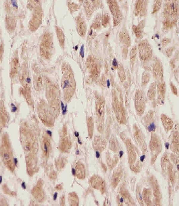
M02268 has real ADORA2A IHC staining data from human heart paraffin sections (M02268 image caption). No IF/ICC data are listed (catalog: M02268 applications and image alts).

Real IHC data M02268 staining ADORA2A in Human heart tissue sections by Immunohistochemistry (IHC-P -paraformaldehyde-fixed, paraffin-embedded sections). Tissue was fixed with formaldehyde and blocked with 3% BSA for 0. 5 hour at room temperature; antigen retrieval was by heat mediation with a citrate buffer (pH6). Samples were incubated with primary antibody (1/25) for 1 hours at 37°C. A undiluted biotinylated goat polyvalent antibody was used as the secondary antibody.
Anti-ADORA2A Antibody (C-term)
Cat # M02268

M02268 is listed for human IHC-P (catalog: M02268 applications and reactivity). Its image documents staining in human heart paraffin sections (M02268 image caption).

Which to pick: Choose M02268 for human tissue IHC-P: its caption reports paraformaldehyde-fixed paraffin sections, formaldehyde fixation, citrate pH 6 retrieval, and a 1:25 primary dilution (M02268 image caption). No IF/ICC-validated or cross-species option is listed: M02268 is a human-reactive polyclonal antibody with no IF/ICC application listed (catalog: M02268 reactivity, dilution_raw, and applications).

Each figure is that product's own IHC / IF validation image from its datasheet.

References

  1. UniProt Consortium. UniProt entry P29274 (AA2AR_HUMAN, Adenosine receptor A2a).
  2. Human Protein Atlas. ADORA2A tissue IHC expression (reliability: Enhanced).
  3. Human Protein Atlas. ADORA2A subcellular location (ICC-IF): Mainly localized to the plasma membrane. In addition localized to vesicles and primary cilium..
  4. Human Protein Atlas. ADORA2A antibody validation summary (3 antibodies).
  5. A lncRNA-immune checkpoint-related gene signature predicts metastasis-free survival in prostate adenocarcinoma. Translational andrology and urology 2022 — PMC9827409.
  6. Adenosine A₂A receptors in striatal glutamatergic terminals and GABAergic neurons oppositely modulate psychostimulant action and DARPP-32 phosphorylation. PloS one 2013 — PMC3842921.
  7. Blockade of endothelial adenosine receptor 2 A suppresses atherosclerosis in vivo through inhibiting CREB-ALK5-mediated endothelial to mesenchymal transition. Pharmacological research 2024 — PMC12674621.
  8. Targeting adenosine 2A receptor signaling suppresses vascular calcification by restraining smooth muscle osteogenic differentiation. Pharmacological research 2025 — PMC13331241.
  9. PubMed PMID:1331670 — UniProt-cited evidence.
  10. PubMed PMID:8670304 — UniProt-cited evidence.
  11. PubMed PMID:15461802 — UniProt-cited evidence.