This website uses cookies to ensure you get the best experience on our website.


- Table of Contents
Plan AFAP1L2 chromogenic IHC around the cytoplasmic connective tissue staining reported in spleen and thyroid (HPA tissue IHC). Compare thyroid glandular cells, reported as high, with adipocytes, reported as not detected, while accounting for low agreement between staining and RNA data (HPA tissue IHC).

Expected localisation, validated protocols, controls and antibodies — the at-a-glance facts below, then the full design guide.
| Expected localisation | Cytoplasm in spleen and thyroid connective tissue (HPA tissue IHC) | |
| Staining pattern | Cytoplasmic staining in spleen and thyroid connective tissue (HPA tissue IHC) | |
| Antigen retrieval | Citrate pH 6.0 HIER, 95–98 °C, 20 min (rule: cytoplasmic / membrane antigen) | |
| Positive control | Thyroid gland+4 more · see all | |
| Negative control | Adipose tissue+4 more · see all |
| Fixation | Keep fixation consistent across sections. (standard IHC practice; not target-specific) | |
| Caveat | Presumed off-target staining; low agreement with RNA data (HPA tissue IHC) | |
| Regulation | Expression regulation is not established (UniProt) | |
| Isoform / epitope | 4 isoforms; effects on the epitope are unknown (UniProt) |
Compare the catalog antibody’s IHC-P protocol with the published cholangiocarcinoma tissue microarray protocol (PMC8592414).
| Sample | Tissue sections; selected-image fixative not specified (standard IHC workflow) |
| Fixation | Image fixative and duration unreported (datasheet A03350); verify before use. |
| Sectioning | 4–5 µm sections on charged slides (standard) |
| Deparaffinisation | Xylene, graded ethanol series to water (standard) |
| Antigen retrieval | Heat-induced epitope retrieval in citrate buffer, pH 6.0, 20 min at 95–98 °C (standard rule: cytoplasmic / membrane antigen) |
| Peroxidase block | 3% H2O2, 10 min, room temperature (standard) |
| Blocking | 10% normal serum of the secondary host, 30 min, room temperature (standard) |
| Primary antibody | Rabbit anti-AFAP1L2, 2.5 μg/mL (datasheet A03350) |
| Primary incubation | Overnight at 4 °C (standard) |
| Detection | HRP-polymer secondary, DAB chromogen 5–10 min (standard) |
| Counterstain | Hematoxylin, blue, dehydrate and mount (standard) |
| Expected result | AFAP1L2-positive staining in glandular cells of thyroid gland (HPA tissue IHC: High). HPA tissue profile: Cytoplasmic expression in connective tissue of spleen and thyroid gland. No signal in the no-primary control. |
AFAP1L2 is cytoplasmic and has no transmembrane segment (UniProt Q8N4X5: localization and topology). In IHC, expect cytoplasmic staining in thyroid glandular cells and selected other cells; HPA also describes connective tissue staining in spleen and thyroid (HPA: tissue IHC). Treat the pattern as provisional: HPA rates its IHC evidence Approved but reports low staining–RNA consistency and presumed off-target binding (HPA: reliability description).
| Thyroid glandular cells show clear cytoplasmic chromogen, with surrounding tissue architecture visible. | This is the strongest listed cell-level reference: High glandular-cell staining (HPA: thyroid IHC). Score the cytoplasm of identifiable cells, not the whole tissue indiscriminately (general IHC practice). |
| Cytoplasmic signal appears in cerebellar granular-layer cells, kidney glomerular cells, or parathyroid glandular cells. | These are plausible additional positives, each listed at Medium staining (HPA: tissue IHC). Their lower reported level makes them less decisive than thyroid glandular cells when checking a weak run. |
| The dominant signal is nuclear, or it follows a crisp membrane outline without convincing cytoplasm. | A nuclear-dominant pattern conflicts with cytoplasmic localization (UniProt Q8N4X5; HPA: tissue IHC). HPA reports plasma-membrane localization as uncertain in ICC-IF, so membrane signal alone cannot establish a correct IHC result (HPA: subcellular). |
| Strong color fills cells listed as unstained, or a uniform haze obscures many cell types. | Adipocytes and bone-marrow hematopoietic cells are listed as not detected (HPA: tissue IHC). Unexpected cell staining warrants an off-target or endogenous-enzyme check; a broad haze also warrants a background-control check (HPA: reliability description; general IHC practice). |
| No interpretable signal appears in thyroid glandular cells. | This conflicts with the listed High thyroid staining (HPA: thyroid IHC). First check whether the positive-control section and detection run worked; a failed control cannot distinguish technical failure from a negative test specimen (general IHC practice). |
| Tissue and cell choice | Thyroid glandular cells provide a listed High reference, while adipocytes are listed as not detected (HPA: tissue IHC). Compare named cell types within preserved tissue architecture (general IHC practice). |
| Antibody evidence | The listed rabbit polyclonal antibody HPA037843 is IHC Approved, and HPA reports low staining–RNA consistency plus presumed off-target binding (HPA: antibody record and reliability description). Resolve unexpected patterns with controls rather than intensity alone (general IHC practice). |
| Isoforms and epitope | UniProt lists 4 isoforms and two PH domains (UniProt Q8N4X5). The supplied record does not locate the catalog antibody's epitope, so isoform coverage and domain-dependent staining cannot be predicted from it. |
| Protein topology and processing | AFAP1L2 has no transmembrane segment or signal peptide and is annotated as one chain spanning residues 1–818 (UniProt Q8N4X5). These annotations support a cytoplasmic expectation but do not establish a shedding pattern or a fixation effect. |
| Retrieval and chromogenic detection | No AFAP1L2-specific retrieval condition is supplied. Check retrieval, blocking, detection reagents and counterstain against the run controls as general paraffin-section IHC practice; do not infer target-specific fixation sensitivity from HPA staining levels. |
| IF/ICC Q: What localization should be checked? | A: Look primarily for cytosol; plasma-membrane localization is uncertain (HPA: subcellular ICC-IF). HPA lists images from A-431 and A-549 and rates HPA037843 ICC as Supported (HPA: subcellular and antibody record). |
| Situation | Likely cause | Next action |
|---|---|---|
| Thyroid glandular cells and the run's positive control are both blank. | A shared staining or detection failure is possible (general IHC practice); HPA lists thyroid glandular cells as High (HPA: thyroid IHC). | Confirm section integrity and the run controls, then review retrieval, primary-antibody application and chromogenic detection records (general IHC practice). |
| Thyroid is weak, but several lower-level tissues look equally dark. | A flat intensity pattern conflicts with HPA's High thyroid and Medium cerebellar granular-layer or glomerular references (HPA: tissue IHC). | Compare the named cells at the same staining exposure and score their cytoplasmic signal; examine unexpected cells with a control section (general IHC practice). |
| Nuclei dominate the signal. | Nuclear dominance conflicts with the annotated cytoplasmic location (UniProt Q8N4X5; HPA: tissue IHC). | Check whether counterstain or precipitate is being mistaken for specific chromogen, and compare with the no-primary control (general IHC practice). |
| Adipocytes or bone-marrow hematopoietic cells stain strongly. | Both are listed as not detected; HPA also notes presumed off-target binding (HPA: tissue IHC and reliability description). Endogenous detection activity is another general IHC possibility. | Review cell identity and use no-primary and appropriate detection controls to investigate background or endogenous activity before calling AFAP1L2 positive (general IHC practice). |
| Diffuse brown haze hides cell borders. | Non-specific reagent binding or inadequate background control may obscure localization (general IHC practice); diffuse haze cannot establish HPA's cell-specific pattern (HPA: tissue IHC). | Inspect no-primary and detection controls, then review blocking, washing, chromogen development and counterstain; score only resolved cellular staining (general IHC practice). |
| A sharp membrane rim is the only reproducible signal. | HPA's plasma-membrane ICC-IF annotation is uncertain, whereas its main location is supported cytosol (HPA: subcellular); UniProt lists no transmembrane segment (UniProt Q8N4X5). | Seek accompanying cytoplasmic staining in the listed positive cells and compare control sections before interpreting a membrane-only IHC pattern (HPA: tissue IHC; general IHC practice). |


Comprehensive Human Protein Atlas IHC scoring per tissue (reliability: Approved — Low consistency between antibody staining and RNA expression data. Presumed off target binding observed and disregarded. Antibody staining in cells/structures not annotated, view images.). Rows are taken directly from the HPA tissue chart — click any row's HPA link to view the source.
| Tissue | Cell type | Level | Evidence | Source |
|---|---|---|---|---|
| Thyroid gland | Glandular cells | High | Protein (IHC) | HPA → |
| Cerebellum | Cells in granular layer | Medium | Protein (IHC) | HPA → |
| Epididymis | Glandular cells | Medium | Protein (IHC) | HPA → |
| Fallopian tube | Glandular cells | Medium | Protein (IHC) | HPA → |
| Kidney | Cells in glomeruli | Medium | Protein (IHC) | HPA → |
| Tissue | Cell type | Level | Evidence | Source |
|---|---|---|---|---|
| Adipose tissue | Adipocytes | Not detected | Protein (IHC) | HPA → |
| Bone marrow | Hematopoietic cells | Not detected | Protein (IHC) | HPA → |
| Bronchus | Respiratory epithelial cells | Not detected | Protein (IHC) | HPA → |
| Caudate | Glial cells | Not detected | Protein (IHC) | HPA → |
| Cervix | Glandular cells | Not detected | Protein (IHC) | HPA → |
Troubleshoot AFAP1L2 staining in paraffin sections by checking retrieval, compartment, cell identity and controls before comparing chromogenic signal across specimens.
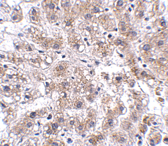
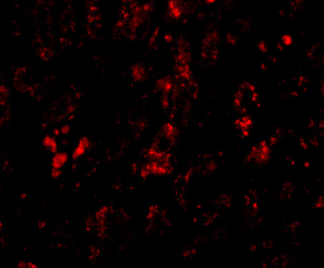
A03350 has IHC and IF images from human liver tissue (catalog image captions); its listed reactivity covers human, mouse, and rat (catalog: reactivity).
A03350 will render with an IHC image of human liver tissue at 2.5 μg/mL (A03350 IHC image caption). The catalog also lists IHC-P and IF applications and shows IF in human liver tissue at 20 μg/mL (catalog: applications; A03350 IF image caption).
Which to pick: Choose A03350 for paraffin-section IHC: IHC-P is listed and its own human liver IHC image shows staining at 2.5 μg/mL; the fixative is unreported (catalog: applications; A03350 IHC image caption). A03350 is also the IF option at 20 μg/mL in human liver tissue, while ICC requires separate validation (catalog: applications; A03350 IF image caption). For mouse or rat samples, A03350 lists reactivity with both species, but its supplied IHC and IF images show human tissue; it is a rabbit antibody with no clone reported (catalog: reactivity, host and clone; A03350 image captions).