AGBL2 / Cytosolic carboxypeptidase 2 · IHC design guide

Design Immunohistochemistry for AGBL2

Plan AGBL2 staining in paraffin sections around the observed cytoplasmic tissue profile (HPA tissue IHC) and the 1:100–1:300 antibody dilution range (datasheet). Interpret staining cautiously because antibody staining and RNA expression show very low consistency (HPA tissue IHC).

Evidence assembled Oct 2026 · For research use; verify linked source records and product datasheet before use
Immunohistochemistry protocol sheet for AGBL2 (IHC for AGBL2): expected localisation Cytoplasmic in tissue (HPA tissue IHC); centrosomal and basal-body localisation annotated (UniProt), antibody A11275-1, validated IHC image, and IHC protocol steps
Printable AGBL2 IHC protocol sheet — expected localisation Cytoplasmic in tissue (HPA tissue IHC); centrosomal and basal-body localisation annotated (UniProt), antibody A11275-1, controls and protocol steps. Open the full AGBL2 IHC guide →

AGBL2 Immunohistochemistry Experimental Design Guide

Expected localisation, validated protocols, controls and antibodies — the at-a-glance facts below, then the full design guide.

Must know before staining
Expected localisation Cytoplasmic in tissue (HPA tissue IHC); centrosomal and basal-body localisation annotated (UniProt)
Staining pattern General cytoplasmic staining (HPA tissue IHC)
Antigen retrieval Citrate pH 6.0 HIER, 95–98 °C, 20 min (rule: cytoplasmic / membrane antigen)
Positive control ⓘ Adrenal gland+4 more · see all
Negative control ⓘ None in HPA (detected in all 44 tissues); use no-primary + isotype controls
Important caveats
Reasons your staining may differ from the expected pattern.
Fixation Keep fixation consistent across sections (standard IHC practice; not target-specific)
Caveat Staining and RNA show very low consistency (HPA tissue IHC)
Regulation No expression regulation annotated (UniProt)
Isoform / epitope 3 isoforms; epitope coverage is unspecified (UniProt)
Section 1

Recommended AGBL2 IHC & IF Protocols

The catalog antibody’s IHC-P protocol is accompanied by published AGBL2 staining protocols for ovarian and renal tissues (PMC6781648; PMC7283229; PMC11417249).

Recommended immunohistochemistry (IHC-P) protocol parameters
SampleParaffin-embedded human heart tissue; fixative not specified (datasheet A11275-1)
FixationImage fixative and duration unreported (datasheet A11275-1); verify before use.
Sectioning4–5 µm sections on charged slides (standard)
DeparaffinisationXylene, graded ethanol series to water (standard)
Antigen retrievalHeat-induced epitope retrieval in citrate buffer, pH 6.0, 20 min at 95–98 °C (standard rule: cytoplasmic / membrane antigen)
Peroxidase block3% H2O2, 10 min, room temperature (standard)
Blocking10% normal serum of the secondary host, 30 min, room temperature (standard)
Primary antibodyRabbit anti-AGBL2, 1:100-1:300 (datasheet A11275-1)
Primary incubationOvernight at 4 °C (standard)
DetectionHRP-polymer secondary, DAB chromogen 5–10 min (standard)
CounterstainHematoxylin, blue, dehydrate and mount (standard)
Expected resultAGBL2-positive staining in glandular cells of adrenal gland (HPA tissue IHC: High). HPA tissue profile: General cytoplasmic expression. No signal in the no-primary control.
💡Decision noteStart with citrate pH 6.0 retrieval at 95–98 °C for 20 min (page antigen-retrieval setting); adjust against the published conditions for your tissue.
Section 2

What Is the Expected AGBL2 Staining Pattern?

AGBL2 is a cytosolic protein without a transmembrane segment; UniProt also places it at centrioles and ciliary basal bodies (UniProt Q5U5Z8). In paraffin sections, expect cytoplasmic staining in selected cells reported as high by HPA, including glandular cells, hematopoietic cells and myoepithelial cells (HPA: tissue IHC). Treat this as a provisional pattern: HPA rates its tissue staining reliability Uncertain because antibody staining and RNA expression have very low consistency (HPA: reliability Uncertain).

What am I looking at on my slide?
Cytoplasmic chromogen in glandular cells of adrenal gland, appendix or cervix, or in bone marrow hematopoietic cells (HPA: High).This matches reported cell types and the general cytoplasmic profile (HPA: tissue IHC). Score the intended cell population separately from surrounding cells; the reported High level is provisional because tissue reliability is Uncertain (HPA: reliability Uncertain).
Chromogen appears exclusively nuclear, membranous or extracellular, with no convincing cytoplasmic component.Review it as a possible compartment mismatch: AGBL2 is cytosolic and has no transmembrane segment (UniProt Q5U5Z8). Nucleoli are reported in ICC-IF (HPA: subcellular), so nuclear signal alone does not establish an IHC artefact.
The strongest staining lies outside the reported cell population in a selected tissue.Check cell identity and staining specificity before calling it AGBL2; cross-reactivity or endogenous detection activity are possible explanations (general IHC practice). HPA reports cell-specific High staining, but its antibody HPA007718 has Uncertain IHC validation (HPA: antibodies).
Diffuse chromogen coats the section, including spaces between cells or tissue-free regions.Treat this as background until controls clarify it (general IHC practice). AGBL2 is intracellular and lacks a transmembrane segment (UniProt Q5U5Z8); widespread surface deposit cannot by itself establish its cellular localisation.
No discernible signal appears in a selected HPA High cell population.First check whether positive-control tissue and detection worked (general IHC practice). Absence may reflect assay performance or variable expression; HPA's High designation is an observation, not a guaranteed positive control (HPA: tissue IHC; reliability Uncertain).
💡Expected AGBL2 appearanceCall a provisional positive when chromogen is predominantly cytoplasmic in a reported High cell population, such as appendix glandular cells (HPA: tissue IHC; UniProt Q5U5Z8); uniform extracellular or unrelated-cell staining warrants specificity review (general IHC practice).
How each factor affects the staining
Tissue and cell selectionHPA reports High staining in several distinct populations, including breast myoepithelial and caudate glial cells, while adipocytes are Low (HPA: tissue IHC). Compare identified cells within each section; these levels do not validate an antibody.
IF/ICC: where should signal localise?HPA reports nucleoli and cytosol as approved locations, and basal body and flagellar centriole as supported locations (HPA: subcellular ICC-IF). UniProt also places AGBL2 at centrioles and ciliary basal bodies (UniProt Q5U5Z8).
Antibody evidenceThe listed HPA antibody, HPA007718, is rated Uncertain for IHC and ICC (HPA: antibodies). Tissue IHC has very low staining–RNA consistency and a splice or transcript discrepancy caution (HPA: reliability Uncertain); interpret isolated positives conservatively.
Isoforms and epitope coverageUniProt lists three AGBL2 isoforms (UniProt Q5U5Z8). The supplied sources do not identify the antibody epitope or establish which isoforms it recognizes; differences between specimens cannot be assigned to isoforms from staining alone.
Processing and topologyUniProt lists a 1–902 cytosolic chain, no signal peptide or propeptide, and no transmembrane segment (UniProt Q5U5Z8). Interpret surface-only staining cautiously; the supplied sources give no AGBL2-specific fixation-sensitivity result.
Why is my staining missing, weak or wrong?
SituationLikely causeNext action
No signal in an HPA High population.Detection failure, antibody dilution or retrieval conditions may contribute (general IHC practice); HPA's tissue calls have Uncertain reliability (HPA: tissue IHC).Check a run control, reagent order and detection chemistry; optimize retrieval and dilution as general IHC variables. Recheck cell identity before treating an HPA High population as a definitive positive (general IHC practice; HPA: reliability Uncertain).
Strong diffuse staining obscures cellular boundaries.Nonspecific binding, excessive chromogen development or insufficient blocking may elevate background (general IHC practice).Inspect the no-primary control, shorten development or adjust blocking and primary dilution (general IHC practice). Score only distinguishable cellular signal against that background; HPA describes general cytoplasmic expression (HPA: tissue IHC).
Signal is confined to the nucleus or nucleoli.HPA reports nucleoli in ICC-IF, while its tissue IHC profile is generally cytoplasmic (HPA: subcellular; tissue IHC).Document the compartment and compare controls and independent staining evidence before accepting nuclear-only IHC as target-specific (general IHC practice). Do not discard nucleolar IF solely because UniProt also reports cytosolic localisation (HPA: subcellular; UniProt Q5U5Z8).
An unexpected cell type stains more strongly than the reported population.Misidentified cells, cross-reactivity or endogenous detection activity are possibilities (general IHC practice); HPA IHC validation is Uncertain (HPA: antibodies).Verify morphology, compare the no-primary control and, where available, independent antibody evidence (general IHC practice). Record the discrepancy instead of assigning it to AGBL2 solely from intensity (HPA: reliability Uncertain).
Brown chromogen persists in the no-primary control.Endogenous enzyme activity or detection-reagent background can create target-independent staining (general chromogenic IHC practice).Review the detection chemistry and apply the matching endogenous-activity block; compare the control at the same development time (general chromogenic IHC practice). Do not score control-matched deposits as AGBL2.
Adipocytes stain as strongly as the selected HPA High cells.HPA lists adipocytes as Low, but its tissue profile has Uncertain reliability (HPA: tissue IHC); background or genuine variation remains possible.Compare cell morphology and background controls, then repeat or corroborate the pattern with independent evidence (general IHC practice). Report the observed distribution rather than forcing a positive or negative call from HPA levels.

Sample controls for AGBL2 IHC & IF

🧪Run adrenal gland first and score its glandular cells for AGBL2 staining (HPA: High in adrenal gland glandular cells). HPA detects AGBL2 in all 44 scored tissues, so use no-primary and isotype controls for the negative reference; any cells designated as internal negatives on the positive slide should show only background staining, and their target-negative status must be verified rather than assumed (HPA: no negative tissue rows).
Positive control tissue: Adrenal gland (Glandular cells, HPA High)
Negative control tissue: None in HPA: AGBL2 is detected in all 44 scored tissues. Use a no-primary (secondary-only) and an isotype control instead.
ICC-IF cell lines (HPA subcellular resource): HPA ICC-IF images show AGBL2 in ASC52telo, RPTEC/TERT1, hTERT-RPE1 (serum starved), Sperm, with annotated localisation: Nucleoli (approved), Basal body (supported), Cytosol (approved), Flagellar centriole (supported) (HPA subcellular).
Technical controls: Include a no-primary (secondary-only) control and an isotype control matched to the primary antibody’s host species and clonality (standard IHC practice). Add a knockout specimen or a peptide-block control as a biological specificity check; the selected A11275-1 tissue-IHC caption describes peptide blocking in human heart (caption: peptide-blocked image). Quench endogenous peroxidase for chromogenic detection and assess background on the adrenal gland control slide (standard chromogenic IHC practice).
⚠️Feasibility: A target-specific fixation window and the effect of fixation are unreported, and the selected A11275-1 paraffin-section caption does not state a fixative (caption: fixative unreported). AGBL2-specific antigen-retrieval requirements are unreported; the supplied evidence does not establish whether frozen sections or IF would be easier than paraffin IHC (supplied target/application evidence). No adrenal gland-specific artefact is established by the supplied evidence, so compare staining with the negative controls before scoring (HPA: High in adrenal gland glandular cells; standard IHC practice).

HPA tissue IHC evidence for AGBL2

Comprehensive Human Protein Atlas IHC scoring per tissue (reliability: Uncertain — Very low consistency between antibody staining and RNA expression data. Pending external verification. Caution, Splice and/or transcript discrepancy exists.). Rows are taken directly from the HPA tissue chart — click any row's HPA link to view the source.

Positive expression · recommended positive controls

TissueCell typeLevelEvidenceSource
Adrenal gland Glandular cells High Protein (IHC) HPA →
Appendix Glandular cells High Protein (IHC) HPA →
Bone marrow Hematopoietic cells High Protein (IHC) HPA →
Breast Myoepithelial cells High Protein (IHC) HPA →
Caudate Glial cells High Protein (IHC) HPA →

Undetected expression · recommended negative controls

TissueCell typeLevelEvidenceSource
None in HPA: AGBL2 is detected in all 44 scored tissues. Use a no-primary (secondary-only) and an isotype control instead.
Section 3

Advanced AGBL2 IHC Tips

Troubleshoot AGBL2 staining in paraffin sections by checking retrieval, compartment, controls and scoring before interpreting chromogenic signal.

How should I adjust retrieval when AGBL2 staining is weak in paraffin sections?
Start with citrate buffer at pH 6.0 for heat induced epitope retrieval at 95–98 °C for 20 min (page retrieval setting). Let sections cool in buffer, then compare a consistently processed positive control with the test section before changing conditions (standard IHC practice). The selected product image shows staining in paraffin embedded human heart, but its caption does not report retrieval conditions (A11275-1 caption). If signal remains weak, compare a carefully titrated alkaline retrieval condition on adjacent sections while keeping antibody and detection conditions fixed (standard IHC practice). Record tissue preservation alongside staining intensity, since excessive heating can make cellular localisation harder to judge (standard IHC practice).
Could fixation explain variable AGBL2 staining between paraffin blocks?
Target specific fixation sensitivity is unknown from the supplied evidence, and the selected paraffin section caption does not state a fixative (A11275-1 caption). Record each block’s fixative and fixation duration, then compare sections processed together using the same retrieval, antibody incubation and chromogenic detection conditions (standard IHC practice). Poor morphology, uneven staining or inconsistent nuclear counterstain can indicate a processing problem, but those findings alone do not identify an AGBL2 specific fixation effect (standard IHC practice). If blocks differ, optimize retrieval on adjacent sections and judge signal within well preserved cells rather than comparing raw colour intensity across batches (standard IHC practice).
Which cellular staining pattern is plausible for AGBL2 in tissue sections?
A cytoplasmic pattern is plausible because AGBL2 is annotated in the cytosol, centrosome, centriole and cilium basal body (UniProt Q5U5Z8 localisation). HPA describes general cytoplasmic tissue staining, while its subcellular data also place AGBL2 in nucleoli, cytosol, basal bodies and flagellar centrioles (HPA tissue IHC; HPA subcellular). In chromogenic sections, score diffuse cytoplasmic staining separately from small perinuclear or apical puncta, and inspect morphology before assigning those puncta to a structure (standard IHC practice; UniProt Q5U5Z8 localisation). Do not treat a purely membranous outline as established localisation: the protein has no transmembrane segment (UniProt Q5U5Z8 topology).
How could isoforms or epitope accessibility affect the AGBL2 IHC pattern?
AGBL2 has 3 annotated isoforms, so an antibody’s epitope must be checked against the sequence shared by the forms under study (UniProt Q5U5Z8 isoforms; standard IHC practice). The peptidase M14 domain spans residues 396–666, but the supplied product caption does not identify the antibody’s epitope (UniProt Q5U5Z8 domains; A11275-1 caption). A peptide blocked image supports competition by that peptide in paraffin embedded human heart, without establishing recognition of every isoform or staining specificity in other tissues (A11275-1 caption). Compare documented immunogen information with isoform sequences and use an independent antibody or orthogonal assay when discordant cellular patterns matter (standard IHC practice).
How can I investigate AGBL2 localisation by IF alongside the chromogenic IHC findings?
For this secondary IF application, multiplex AGBL2 with a marker for the expected cell type so punctate signal can be assigned to the right cells (standard IF practice). Choose fluorophores after checking tissue autofluorescence in unstained and single stained controls, reserving a cleaner spectral channel for weak AGBL2 signal (standard IF practice). Because AGBL2 is cytosolic and associated with intracellular centrosomes and basal bodies, permeabilise fixed cells or sections sufficiently to expose an intracellular epitope, while checking that morphology remains intact (UniProt Q5U5Z8 localisation; standard IF practice). HPA reports cytosol, basal body and flagellar centriole localisation in subcellular data, but its tissue IHC reliability is uncertain, so compare compartments without assuming matched assay performance (HPA subcellular; HPA tissue IHC).
What controls help separate AGBL2 signal from chromogenic background?
Run a no primary control and inspect pigment, folds and section edges before attributing brown colour to AGBL2 (standard IHC practice). Quench endogenous peroxidase before an HRP and DAB workflow, and use an appropriate protein block to reduce nonspecific reagent binding (standard IHC practice). The selected paraffin embedded human heart image includes a section blocked with synthesized peptide, which is useful competition evidence for that product image (A11275-1 caption). Compare the blocked and unblocked patterns within intact cells, but use an independent antibody or orthogonal evidence if specificity remains important, since peptide competition alone cannot resolve every source of background (standard IHC practice).
How should I score AGBL2 staining across tissue sections? ⚠ ANSWER MARKED FOR VERIFICATION
Define the cell population and cytoplasmic scoring compartment before reading slides, since HPA describes general cytoplasmic tissue staining (HPA tissue IHC; standard IHC practice). For comparable sections, record the percentage of positive cells and intensity categories, then calculate an H-score from 0–300 if an intensity weighted summary is needed (standard IHC practice). Normalize counts to the number of eligible intact cells, or report positive cell density per mm² of evaluable tissue when cellularity varies (standard IHC practice). Keep acquisition, counterstain and scoring thresholds consistent, and report punctate centrosomal or basal body patterns separately from diffuse cytoplasmic signal when they can be resolved (standard IHC practice; UniProt Q5U5Z8 localisation).
When should an apparent AGBL2 positive result be treated cautiously?
Look for signal within intact cytoplasm or plausible centrosomal and basal body regions, rather than accepting an isolated cell edge or tissue edge deposit (UniProt Q5U5Z8 localisation; standard IHC practice). Exclude necrotic areas, folds and endogenous peroxidase activity with morphology review and a no primary control (standard IHC practice). Check that the stained cells match the population being scored; HPA lists high staining in several cell populations but rates its tissue IHC reliability uncertain because staining and RNA data have very low consistency (HPA tissue IHC). A plausible compartment and appropriate controls strengthen interpretation, while discordance warrants an independent antibody or orthogonal measurement before claiming AGBL2 expression (standard IHC practice).
Boster reagents

Best AGBL2 / Cytosolic carboxypeptidase 2 IHC Antibodies

Both anti-AGBL2 antibodies have human-heart paraffin-section IHC images (catalog image captions), list human and mouse reactivity, and list IF as an application (catalog applications and reactivity).

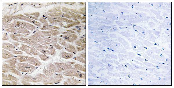
Real IHC data Immunohistochemistry analysis of paraffin-embedded human heart tissue, using CBCP2 Antibody. The picture on the right is blocked with the synthesized peptide.
Anti-CCP2 AGBL2 Antibody
Cat # A11275-1
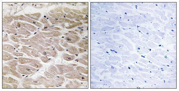
Real IHC data Immunohistochemistry analysis of paraffin-embedded human heart tissue, using CBCP2 Antibody. The picture on the right is blocked with the synthesized peptide.
Anti-CBCP2 AGBL2 Antibody
Cat # A30633

A11275-1 lists IHC and IF for human and mouse (catalog: A11275-1); its own image shows human-heart paraffin-section IHC with a peptide-blocking comparison (A11275-1 image caption). A30633 lists IHC, IF, and ICC for human and mouse (catalog: A30633); its own image shows human-heart paraffin-section IHC with a peptide-blocking comparison (A30633 image caption).

Which to pick: For tissue IHC, either SKU has its own human-heart paraffin-section image (A11275-1 and A30633 image captions); the fixative is unreported in both captions. For IF, both SKUs list the application (catalog applications); choose A30633 if ICC is required, since it alone lists ICC and is described as polyclonal (A30633 applications and catalog description). For human and mouse samples, either SKU lists reactivity with both species (catalog reactivity); the supplied IHC images show human tissue only (A11275-1 and A30633 image captions).

Each figure is that product's own IHC / IF validation image from its datasheet.

References

  1. UniProt Consortium. UniProt entry Q5U5Z8 (CBPC2_HUMAN, Cytosolic carboxypeptidase 2).
  2. Human Protein Atlas. AGBL2 tissue IHC expression (reliability: Uncertain).
  3. Human Protein Atlas. AGBL2 subcellular location (ICC-IF): Mainly localized to the nucleoli, cytosol, basal body and flagellar centriole. In addition localized to the mid piece, principal piece and end piece..
  4. Human Protein Atlas. AGBL2 antibody validation summary (1 antibodies).
  5. High expression of AGBL2 is a novel prognostic factor of adverse outcome in patients with ovarian carcinoma. Oncology letters 2019 — PMC6781648.
  6. Clinical implications of AGBL2 expression and its inhibitor latexin in breast cancer. World journal of surgical oncology 2014 — PMC4069086.
  7. Expression of RARRES1 and AGBL2 and progression of conventional renal cell carcinoma. British journal of cancer 2020 — PMC7283229.
  8. AGBL2 promotes renal cell carcinoma cells proliferation and migration via α-tubulin detyrosination. Heliyon 2024 — PMC11417249.
  9. PubMed PMID:14702039 — UniProt-cited evidence.
  10. PubMed PMID:16554811 — UniProt-cited evidence.
  11. PubMed PMID:15489334 — UniProt-cited evidence.