This website uses cookies to ensure you get the best experience on our website.


- Table of Contents
Plan chromogenic IHC on paraffin sections using cytoplasmic staining in hepatocytes and skeletal myocytes as reference patterns (HPA tissue IHC). Compare control tissues and interpret nuclear staining cautiously because the tissue IHC assessment considers it off target (HPA tissue IHC).

Expected localisation, validated protocols, controls and antibodies — the at-a-glance facts below, then the full design guide.
| Expected localisation | Cytoplasmic staining in several tissues (HPA tissue IHC) | |
| Staining pattern | Cytoplasmic staining in hepatocytes and skeletal myocytes (HPA tissue IHC) | |
| Antigen retrieval | EDTA pH 8.0 HIER, heat-mediated (datasheet A02555) | |
| Positive control | Cervix+4 more · see all | |
| Negative control | Adrenal gland+4 more · see all |
| Fixation | Keep fixation consistent across sections (standard IHC practice; not target-specific) | |
| Caveat | Disregard observed nuclear tissue staining as off target (HPA tissue IHC) | |
| Regulation | Glycogenolysis can redistribute AGL to nuclei (UniProt) | |
| Isoform / epitope | Three listed isoforms (1, 5, 6); epitope coverage unknown (UniProt) |
The catalog antibody’s paraffin IHC protocol is paired with one published AGL staining protocol using frozen human muscle sections (PMC2753483).
| Sample | Paraffin-embedded human placenta tissue; fixative not specified (datasheet A02555) |
| Fixation | Image fixative and duration unreported (datasheet A02555); verify before use. |
| Sectioning | 4–5 µm sections on charged slides (standard) |
| Deparaffinisation | Xylene, graded ethanol series to water (standard) |
| Antigen retrieval | Heat retrieval: EDTA pH 8.0 (datasheet A02555); 20 min, 95–100 °C (standard) |
| Peroxidase block | 3% H2O2, 10 min, room temperature (standard) |
| Blocking | 10% goat serum (datasheet A02555) |
| Primary antibody | Rabbit anti-AGL, 2-5 μg/ml (datasheet A02555) |
| Primary incubation | Overnight at 4 °C (datasheet A02555) |
| Detection | HRP-conjugated secondary, DAB chromogen (datasheet A02555) |
| Counterstain | Hematoxylin, blue, dehydrate and mount (standard) |
| Expected result | AGL-positive staining in squamous epithelial cells of cervix (HPA tissue IHC: Medium). HPA tissue profile: Cytoplasmic expression in several tissues, including skeletal muscle and liver. Additional nuclear expression in squamous epithelia and brain. No signal in the no-primary control. |
AGL is a soluble, non-membrane enzyme found mainly in the cytoplasm; UniProt also reports nuclear localisation under glycogenolytic conditions (UniProt P35573: localisation; topology). In paraffin-section IHC, expect cytoplasmic staining in hepatocytes and skeletal myocytes, each reported at medium intensity (HPA: liver and skeletal muscle). HPA rates its tissue IHC profile Approved but notes low agreement with RNA data and disregards the nuclear tissue staining as off-target (HPA: tissue IHC reliability).
| Cytoplasmic colour in hepatocytes or skeletal myocytes, with identifiable cell borders and little surrounding colour. | This fits the reported medium staining in both cell types (HPA: liver hepatocytes; skeletal muscle myocytes) and AGL’s principal location (UniProt P35573: cytoplasm). Compare cells within the same section before calling a weaker region negative (general IHC interpretation). |
| Strong nuclear colour dominates a tissue section, especially in squamous epithelium, with little cytoplasmic colour. | Treat it as suspect: HPA observed nuclear tissue staining but disregarded it as off-target (HPA: tissue IHC reliability). UniProt reports conditional nuclear localisation, which alone cannot authenticate this IHC pattern (UniProt P35573: localisation). |
| Colour appears in cells reported as unstained, such as bone-marrow hematopoietic cells, or follows blood, edges, or tissue debris. | HPA reports no detection in bone-marrow hematopoietic cells (HPA: bone marrow). A mismatched cellular pattern raises cross-reactivity or detection-background concerns; edge and debris staining also warrant a reagent-only control (general IHC practice). |
| Broad, hazy colour covers cells and extracellular areas without resolving cytoplasm. | This cannot establish AGL localisation (general IHC interpretation). Check whether the no-primary control also colours the section, then review blocking, wash stringency, antibody concentration, and chromogen development as general IHC variables (general IHC practice). |
| No cytoplasmic colour appears in adequately preserved liver hepatocytes or skeletal myocytes. | These are useful positive comparators because HPA reports medium staining in each (HPA: liver; skeletal muscle). A blank result warrants checking the antibody’s documented IHC-P conditions and detection controls; HPA supplies no AGL-specific fixation-sensitivity finding here (HPA: tissue IHC; general IHC practice). |
| Cell type and compartment | Use hepatocytes and skeletal myocytes as medium-staining references (HPA: tissue IHC); AGL has no transmembrane segment and is principally cytoplasmic (UniProt P35573: topology; localisation). |
| Tissue-IHC confidence | HPA labels its tissue IHC Approved while flagging low staining–RNA consistency and a splice/transcript discrepancy (HPA: reliability). Interpret borderline positives with that limitation; Approved does not resolve a disputed nuclear pattern (HPA: tissue IHC reliability). |
| Isoform context | UniProt reports differing isoform expression across liver and muscle (UniProt P35573: tissue specificity). The supplied record gives no antibody epitope or isoform coverage, so a difference in staining cannot be assigned to a particular isoform. |
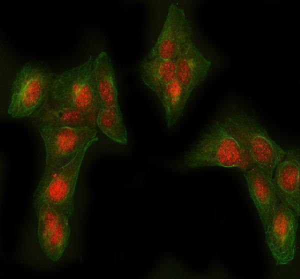
| IF/ICC Q&A: should nuclear signal match tissue IHC? | No direct equivalence is established: ICC-IF lists supported nucleoplasm, nuclear bodies, and cytosol (HPA: subcellular ICC-IF), whereas HPA disregards nuclear tissue-IHC staining (HPA: tissue IHC reliability). Assess each application against its own validation. |
| Chromogenic detection | Endogenous enzyme activity can mimic signal when enzyme-based detection is used; a no-primary control and the matching detection block help identify it (general IHC practice). This is a workflow consideration, not an AGL-specific tissue claim. |
| Situation | Likely cause | Next action |
|---|---|---|
| Liver or skeletal muscle is blank. | A failed staining or detection step is possible because HPA reports medium signal in hepatocytes and myocytes (HPA: tissue IHC); a blank slide alone does not identify which step failed. | Confirm tissue morphology, the antibody’s documented IHC-P conditions, and a working detection control; review retrieval and dilution against the antibody instructions (general IHC practice). |
| Nuclei stain more strongly than cytoplasm in tissue IHC. | HPA observed nuclear tissue staining and explicitly disregarded it as off-target (HPA: tissue IHC reliability); UniProt’s conditional nuclear localisation does not settle this slide’s specificity (UniProt P35573: localisation). | Score cytoplasm separately, inspect the no-primary control, and compare with the expected positive cell types (HPA: liver; skeletal muscle; general IHC practice). |
| A reportedly negative cell population stains. | For example, hematopoietic cells in bone marrow were not detected by HPA (HPA: bone marrow); cross-reactivity or endogenous chromogenic activity may explain a discordant result (general IHC interpretation). | Check the no-primary and detection controls, then reassess whether colour is cellular and confined to the expected compartment (general IHC practice; UniProt P35573: cytoplasm). |
| The entire section has diffuse brown colour. | Residual detection background, concentrated antibody, or overdevelopment can obscure cell boundaries (general IHC practice); diffuse colour is not evidence of the reported cytoplasmic pattern (HPA: tissue IHC). | Compare a no-primary section; review blocking, wash steps, antibody dilution, and development time using the selected detection system’s instructions (general IHC practice). |
| Muscle and liver differ in intensity. | HPA reports medium staining in skeletal myocytes and hepatocytes but warns of low staining–RNA consistency (HPA: tissue IHC reliability). UniProt describes tissue-dependent isoform expression without establishing this antibody’s isoform recognition (UniProt P35573: tissue specificity). | Compare like cells under matched staining conditions and report the observed intensity; avoid assigning the difference to an isoform without epitope evidence (general IHC interpretation). |
| An ICC-IF image shows nuclei, while the paraffin IHC section is cytoplasmic. | HPA supports nucleoplasmic ICC-IF localisation but disregards nuclear staining in its tissue-IHC profile (HPA: subcellular ICC-IF; tissue IHC reliability). | Use the application-specific reference when scoring: assess this IHC section against the cytoplasmic tissue pattern, and evaluate ICC-IF separately under its own guide (HPA: tissue IHC; subcellular ICC-IF). |
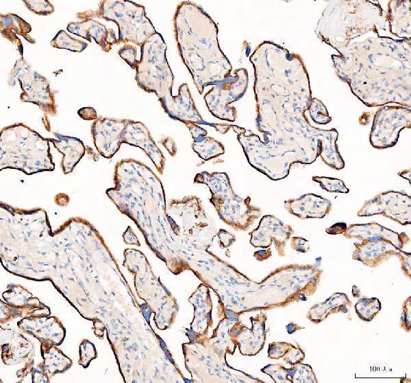


Comprehensive Human Protein Atlas IHC scoring per tissue (reliability: Approved — Low consistency between antibody staining and RNA expression data. Caution, Splice and/or transcript discrepancy exists. Nuclear staining considered off target binding and disregarded.). Rows are taken directly from the HPA tissue chart — click any row's HPA link to view the source.
| Tissue | Cell type | Level | Evidence | Source |
|---|---|---|---|---|
| Cervix | Squamous epithelial cells | Medium | Protein (IHC) | HPA → |
| Esophagus | Squamous epithelial cells | Medium | Protein (IHC) | HPA → |
| Liver | Hepatocytes | Medium | Protein (IHC) | HPA → |
| Nasopharynx | Respiratory epithelial cells | Medium | Protein (IHC) | HPA → |
| Placenta | Trophoblastic cells | Medium | Protein (IHC) | HPA → |
| Tissue | Cell type | Level | Evidence | Source |
|---|---|---|---|---|
| Adrenal gland | Glandular cells | Not detected | Protein (IHC) | HPA → |
| Appendix | Glandular cells | Not detected | Protein (IHC) | HPA → |
| Bone marrow | Hematopoietic cells | Not detected | Protein (IHC) | HPA → |
| Breast | Adipocytes | Not detected | Protein (IHC) | HPA → |
| Caudate | Glial cells | Not detected | Protein (IHC) | HPA → |
Troubleshoot AGL staining in paraffin sections by checking retrieval, compartment, cell type, and controls before interpreting chromogenic signal.
A02555 has IHC data from a human placenta paraffin section and IF data from U2OS cells (A02555 image captions). Both antibodies list human, mouse and rat reactivity (catalog: A02555, M02555).
A02555 lists IHC and IF and has images from a human placenta paraffin section and U2OS cells, respectively (catalog: A02555; A02555 image captions). M02555 lists IHC and ICC/IF for human, mouse and rat, but has no IHC or IF image caption in this payload (catalog: M02555).
Which to pick: For tissue IHC, choose A02555: its own caption documents a human placenta paraffin section, EDTA retrieval at pH 8.0 and 2 μg/ml primary antibody; the fixative is unreported (A02555 IHC caption). For IF/ICC, A02555 has a U2OS IF image at 5 μg/ml (A02555 IF caption); M02555 is a rabbit monoclonal that lists ICC/IF but has no image caption here (catalog: M02555). Both list human, mouse and rat reactivity for cross-species planning, while the supplied IHC and IF images document only the A02555 samples named above (catalog: A02555, M02555; A02555 image captions).