This website uses cookies to ensure you get the best experience on our website.


- Table of Contents
Plan paraffin-section AHCY IHC around nuclear and cytoplasmic staining (HPA tissue IHC). Use kidney tubule cells as a high-staining reference and adipocytes as an undetected reference (HPA tissue IHC), and titrate the catalog antibody within 2–5 μg/ml (datasheet A04236-1).

Expected localisation, validated protocols, controls and antibodies — the at-a-glance facts below, then the full design guide.
| Expected localisation | Nuclear and cytoplasmic staining (HPA tissue IHC) | |
| Staining pattern | Widespread nuclear and cytoplasmic cellular staining (HPA tissue IHC) | |
| Antigen retrieval | EDTA pH 8.0 HIER, heat-mediated (datasheet A04236-1) | |
| Positive control | Epididymis+4 more · see all | |
| Negative control | Adipose tissue |
| Fixation | Keep fixation consistent across sections. (standard IHC practice; not target-specific) | |
| Caveat | Staining and RNA show medium consistency (HPA tissue IHC) | |
| Regulation | Specific regulators unreported (UniProt) | |
| Isoform / epitope | 2 isoforms; epitope coverage is unknown (UniProt) |
The catalog antibody’s IHC-P protocol (datasheet A04236-1) is accompanied by one published AHCY staining protocol for paraffin sections (PMC9345734).
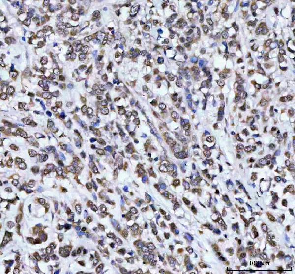
| Sample | Paraffin-embedded human breast cancer tissue; fixative not specified (datasheet A04236-1) |
| Fixation | Image fixative and duration unreported (datasheet A04236-1); verify before use. |
| Sectioning | 4–5 µm sections on charged slides (standard) |
| Deparaffinisation | Xylene, graded ethanol series to water (standard) |
| Antigen retrieval | Heat retrieval: EDTA pH 8.0 (datasheet A04236-1); 20 min, 95–100 °C (standard) |
| Peroxidase block | 3% H2O2, 10 min, room temperature (standard) |
| Blocking | 10% goat serum (datasheet A04236-1) |
| Primary antibody | Rabbit anti-AHCY, 2-5 μg/ml (datasheet A04236-1) |
| Primary incubation | Overnight at 4 °C (datasheet A04236-1) |
| Detection | HRP-conjugated secondary, DAB chromogen (datasheet A04236-1) |
| Counterstain | Hematoxylin, blue, dehydrate and mount (standard) |
| Expected result | AHCY-positive staining in glandular cells of epididymis (HPA tissue IHC: High). HPA tissue profile: Ubiquitous nuclear and cytoplasmic expression. No signal in the no-primary control. |
AHCY should show nuclear and cytoplasmic staining across many tissue cell types, with particularly strong staining in selected glandular, tubular, seminiferous and urothelial cells (HPA tissue IHC). HPA rates the tissue pattern Approved, with medium consistency between staining and RNA data (HPA tissue IHC). AHCY has no transmembrane segment, so a membrane-restricted pattern needs scrutiny (UniProt P23526 topology).
| Nuclear and cytoplasmic staining in kidney tubule cells, with stronger signal than nearby weakly stained cells. | This fits the reported high tubule-cell signal and broad nuclear–cytoplasmic distribution (HPA tissue IHC). Judge intensity within the same section; ubiquitous expression does not mean every cell stains equally (HPA tissue IHC). |
| Signal confined to cell membranes or extracellular spaces, without convincing intracellular staining. | That distribution conflicts with HPA tissue IHC and the absence of a transmembrane segment (UniProt P23526 topology). Review morphology and controls before calling it AHCY; UniProt also lists nucleus and endoplasmic reticulum as locations (UniProt P23526). |
| Strong adipocyte staining, or dominant signal in a cell population expected to stain weakly. | Adipocytes were not detected in the HPA tissue panel, while glial, alveolar and muscle cells had low signal (HPA tissue IHC). Unexpected staining may reflect cross-reactivity or endogenous detection activity; compare with appropriate controls (general IHC practice). |
| Uniform colour over cells and empty spaces, obscuring cell boundaries. | Diffuse background prevents a reliable compartment call. Check the no-primary control, blocking and detection chemistry before scoring the section (general IHC practice); HPA's Approved rating does not validate this individual slide (HPA tissue IHC). |
| No detectable signal in an otherwise interpretable kidney tubule or thyroid gland section. | Both are reported high-staining cell populations (HPA tissue IHC). First assess tissue preservation, retrieval, antibody dilution and the detection system using suitable controls (general IHC practice); a negative slide alone does not establish absent AHCY. |
| Tissue and cell selection | High signal is reported in epididymal and thyroid glandular cells, kidney tubule cells, seminiferous-duct cells and urothelium (HPA tissue IHC). Adipocytes were not detected, making them an unsuitable sole positive control (HPA tissue IHC). |
| Compartment used for scoring | HPA describes tissue staining as nuclear and cytoplasmic, while its supported ICC-IF main location is cytosol (HPA tissue IHC; HPA subcellular ICC-IF). UniProt also lists nucleus, endoplasmic reticulum and melanosome; melanosome identification came from fraction mass spectrometry, not a stated routine IHC pattern (UniProt P23526). |
| IHC antibody evidence | The tissue profile is Approved, with medium staining–RNA consistency; HPA041225 and HPA044675 each have Approved IHC status (HPA tissue IHC; HPA antibodies). These ratings support a reference pattern but do not establish specificity for an untested staining run (general IHC practice). |
| IF/ICC: what pattern is supported? | Cytosolic signal is the supported main ICC-IF location, with images listed for CACO-2, Hep-G2 and KOLF2.1J (HPA subcellular ICC-IF). HPA044675 has Supported ICC status; those IF observations should not be treated as an IHC-P protocol (HPA antibodies). |
| Target-specific preparation limits | The supplied UniProt and HPA records do not report an AHCY-specific fixation effect or antigen-retrieval condition. Select and document IHC-P preparation using validated assay instructions and controls (general IHC practice), without inferring retrieval sensitivity from tissue intensity (HPA tissue IHC). |
| Situation | Likely cause | Next action |
|---|---|---|
| Known high-staining tissue is blank. | An assay-step failure is possible; kidney tubules and thyroid glandular cells are reported high (HPA tissue IHC). | Check section integrity, retrieval, primary-antibody preparation and chromogenic detection against run controls (general IHC practice). |
| Membrane-only colour dominates. | AHCY has no transmembrane segment, and HPA reports nuclear–cytoplasmic tissue staining (UniProt P23526 topology; HPA tissue IHC). | Inspect cellular boundaries and compare a no-primary control; withhold an AHCY call until intracellular staining is credible (general IHC practice). |
| Adipocytes stain strongly. | HPA reports AHCY as not detected in adipocytes; cross-reactivity or endogenous detection activity is possible (HPA tissue IHC; general IHC practice). | Compare no-primary and detection controls, then assess the staining pattern in a reported high-staining cell population (general IHC practice; HPA tissue IHC). |
| Colour coats the whole section. | Nonspecific background or endogenous detection activity may obscure the cellular pattern (general IHC practice). | Review blocking, reagent concentration, washing and no-primary controls before assigning nuclear or cytoplasmic scores (general IHC practice). |
| Only weak signal appears in lung alveolar cells or muscle cells. | HPA reports low signal in alveolar cells, cardiomyocytes and skeletal myocytes (HPA tissue IHC). | Compare with a reported high-staining tissue in the same run before changing assay settings (HPA tissue IHC; general IHC practice). |
| Tissue IHC looks nuclear, but ICC-IF looks mainly cytosolic. | These are the reported observations for the respective applications (HPA tissue IHC; HPA subcellular ICC-IF). | Score each preparation against its own supported reference pattern and controls; investigate only if staining conflicts with that pattern (general IHC practice). |


Comprehensive Human Protein Atlas IHC scoring per tissue (reliability: Approved — Medium consistency between antibody staining and RNA expression data.). Rows are taken directly from the HPA tissue chart — click any row's HPA link to view the source.
| Tissue | Cell type | Level | Evidence | Source |
|---|---|---|---|---|
| Epididymis | Glandular cells | High | Protein (IHC) | HPA → |
| Kidney | Cells in tubules | High | Protein (IHC) | HPA → |
| Testis | Cells in seminiferous ducts | High | Protein (IHC) | HPA → |
| Thyroid gland | Glandular cells | High | Protein (IHC) | HPA → |
| Urinary bladder | Urothelial cells | High | Protein (IHC) | HPA → |
| Tissue | Cell type | Level | Evidence | Source |
|---|---|---|---|---|
| Adipose tissue | Adipocytes | Not detected | Protein (IHC) | HPA → |
Troubleshoot AHCY staining in paraffin section chromogenic IHC using the selected antibody’s tissue image and independent expression evidence.
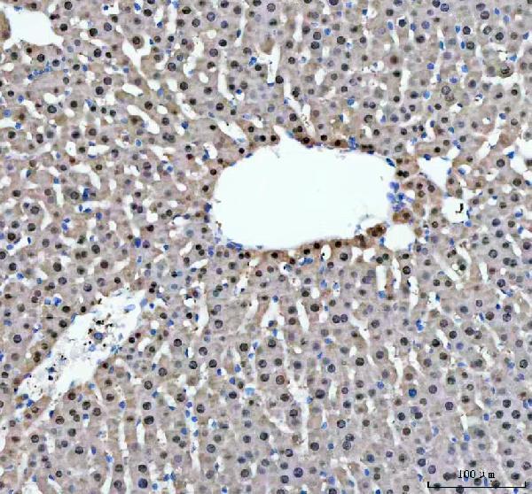
The catalog antibody has IHC data from paraffin sections of human breast cancer and rat liver (IHC image captions); human, mouse and rat reactivity is listed (catalog).
A04236-1 is listed for IHC and has images from paraffin sections of human breast cancer and rat liver (catalog applications; IHC image captions). Its listed reactivity is human, mouse and rat, while its pictured IHC examples cover human and rat tissue (catalog reactivity; IHC image captions).
Which to pick: Choose A04236-1 for paraffin-section IHC: its own captions show staining at 2 μg/ml after EDTA pH 8.0 retrieval; the fixative is unreported (IHC image captions). For IF/ICC, this SKU has no listed IF/ICC application or IF image, so the payload provides no IF/ICC validation (catalog applications; IF image alts). For cross-species work, A04236-1 lists human, mouse and rat reactivity, but its pictured IHC evidence covers human and rat only (catalog reactivity; IHC image captions).