This website uses cookies to ensure you get the best experience on our website.


- Table of Contents
Plan AHCYL1 IHC in paraffin sections around the cytoplasmic pattern reported across most tissue types (HPA tissue IHC). Use duodenal glandular cells with medium staining as a reference (HPA tissue IHC), and start with the catalog antibody’s 2–5 μg/ml IHC range (datasheet A08908-2).

Expected localisation, validated protocols, controls and antibodies — the at-a-glance facts below, then the full design guide.
| Expected localisation | Cytoplasmic staining in most tissue types (HPA tissue IHC) | |
| Staining pattern | Medium cytoplasmic signal in duodenal glandular cells (HPA tissue IHC) | |
| Antigen retrieval | EDTA pH 8.0 HIER, heat-mediated (datasheet A08908-2) | |
| Positive control | Duodenum+4 more · see all | |
| Negative control | Adipose tissue+4 more · see all |
| Fixation | Matched tissue-IHC evidence does not establish the fixation claim. Validate the specimen-specific method before use. (selected-SKU IHC image A08908-2) | |
| Caveat | Staining shows low consistency with RNA expression (HPA tissue IHC) | |
| Regulation | Apoptotic stress reduces MAM localization (UniProt) | |
| Isoform / epitope | Two isoforms; epitope coverage is unspecified (UniProt) |
Compare the catalog antibody’s IHC-P protocol with published AHCYL1 staining conditions for chicken oviduct and human lung tissue (PMC9107370; PMC3492294; PMC12068309).
| Sample | Paraffin-embedded human bladder adenosquamous carcinoma tissue; fixative not specified (datasheet A08908-2) |
| Fixation | Image fixative and duration unreported (datasheet A08908-2); verify before use. |
| Sectioning | 4–5 µm sections on charged slides (standard) |
| Deparaffinisation | Xylene, graded ethanol series to water (standard) |
| Antigen retrieval | Heat retrieval: EDTA pH 8.0 (datasheet A08908-2); 20 min, 95–100 °C (standard) |
| Peroxidase block | 3% H2O2, 10 min, room temperature (standard) |
| Blocking | 10% goat serum (datasheet A08908-2) |
| Primary antibody | Rabbit anti-AHCYL1, 2-5 μg/ml (datasheet A08908-2) |
| Primary incubation | Overnight at 4 °C (datasheet A08908-2) |
| Detection | Streptavidin-biotin complex (SABC), DAB chromogen (datasheet A08908-2) |
| Counterstain | Hematoxylin, blue, dehydrate and mount (standard) |
| Expected result | AHCYL1-positive staining in glandular cells of duodenum (HPA tissue IHC: Medium). HPA tissue profile: Cytoplasmic expression in most tissue types. No signal in the no-primary control. |
AHCYL1 should appear predominantly cytoplasmic in most tissue types (HPA tissue IHC), with medium staining in glandular cells of the duodenum, endometrium, fallopian tube and thyroid gland, and exocrine glandular cells of the pancreas (HPA tissue IHC). Cytosolic and endoplasmic reticulum localisation, with possible apical membrane association, is consistent with a protein lacking a transmembrane segment (UniProt O43865). HPA rates its tissue IHC evidence Approved but notes low agreement with RNA expression and pending external verification (HPA tissue IHC).
| Medium cytoplasmic staining in duodenal glandular cells or pancreatic exocrine glandular cells (HPA tissue IHC). | This matches the reported cell type, compartment and intensity (HPA tissue IHC). Judge individual cells rather than the whole section: adjacent unstained structures do not negate a positive result. The HPA tissue pattern is Approved but pending external verification (HPA tissue IHC). |
| A strong nuclear-only deposit, with no corresponding cytoplasmic staining. | Treat this as a localisation mismatch: HPA reports cytoplasmic tissue staining, while UniProt lists cytosol, endoplasmic reticulum, microsomes and apical membrane, not a nuclear location (HPA tissue IHC; UniProt O43865). Review morphology and detection controls before scoring it as AHCYL1. |
| Signal in cells listed as undetected, such as adipocytes or cardiomyocytes (HPA tissue IHC). | Check whether staining belongs to those cells rather than nearby structures. If it does, cross-reactivity or endogenous detection activity is possible; the HPA result is cell-specific and does not make every cell in adipose tissue or heart muscle negative (HPA tissue IHC). |
| Diffuse colour across cells, stroma and empty areas, obscuring cellular boundaries. | This is background rather than an interpretable cytoplasmic pattern. Review no-primary controls, blocking, washes and chromogen development as general IHC checks. HPA reports cytoplasmic expression in most tissue types, so a broad tissue distribution alone does not establish background (HPA tissue IHC). |
| No cytoplasmic signal in glandular cells of an expected positive section. | Duodenal, endometrial, fallopian tube and thyroid glandular cells, and pancreatic exocrine glandular cells, have reported medium staining (HPA tissue IHC). A blank result warrants checks of tissue identity, morphology, antibody and detection controls before calling the sample negative; HPA does not establish fixation sensitivity for AHCYL1. |
| Cell-level tissue distribution | HPA reports cytoplasmic expression in most tissue types and low RNA tissue specificity, yet its examples distinguish medium-staining glandular cells from specific cells scored not detected (HPA tissue IHC). Select and score controls by the named cell population; do not treat an entire organ as uniformly positive or negative. |
| Membrane-associated localisation | AHCYL1 has no transmembrane segment, but UniProt lists endoplasmic reticulum, microsomes and apical cell membrane; phosphorylation can promote membrane association through ITPR1 (UniProt O43865). An apical component can therefore coexist with cytoplasmic staining; assess it against the cellular pattern before dismissing it. |
| Protein forms and modifications | UniProt lists 2 isoforms, a mature chain spanning residues 2–530, and several modified residues, including phosphoserines (UniProt O43865). The payload gives no antibody epitope or isoform-specific staining data, so these annotations cannot predict differential IHC intensity or establish an antigen retrieval requirement. |
| Strength of tissue evidence | HPA labels the tissue stain Approved but reports low consistency between antibody staining and RNA expression, with external verification pending; HPA042589 has IHC Approved status (HPA tissue IHC; HPA antibodies). Use the listed cells as practical reference patterns while keeping a discordant stain provisional until controls support it. |
| Situation | Likely cause | Next action |
|---|---|---|
| Expected positive glandular cells show no signal (HPA tissue IHC). | The section may lack identifiable target cells, or the staining run may have failed; the payload gives no target-specific fixation or retrieval failure mechanism. | Confirm cell identity on the counterstain and inspect a reported medium-staining control cell population (HPA tissue IHC). Check the catalog antibody, detection reagents and routine IHC run controls before changing conditions. |
| Nuclear-only staining dominates the section. | The distribution conflicts with the reported cytoplasmic tissue pattern and listed nonnuclear locations (HPA tissue IHC; UniProt O43865); its precise cause is undetermined. | Inspect no-primary and detection controls, compare cytoplasmic staining in a reported positive cell population, and avoid scoring isolated nuclear colour as confirmed AHCYL1 (HPA tissue IHC). |
| Colour appears in adipocytes or cardiomyocytes, which HPA lists as not detected (HPA tissue IHC). | Cell identification may be mistaken; cross-reactivity or endogenous detection activity is also possible. HPA's entries apply to the named cell types (HPA tissue IHC). | Recheck morphology, compare a reported positive cell population, and use no-primary and detection controls to investigate staining that persists outside the expected cells. |
| Diffuse colour prevents cell-level scoring. | Nonspecific background, endogenous detection activity or excessive chromogen development are general IHC possibilities; the supplied sources do not assign an AHCYL1-specific cause. | Review blank and no-primary controls, blocking, washes and development time. Score only resolvable cellular staining; HPA's broad cytoplasmic tissue profile alone is not evidence of background (HPA tissue IHC). |
| An apical edge stains alongside cytoplasm. | Apical membrane localisation and phosphorylation-dependent membrane association are reported (UniProt O43865), so this pattern is not automatically artefactual. | Assess whether the edge belongs to intact cells and whether a cytoplasmic component is present. Compare morphology and controls before accepting a membrane-only deposit as specific. |
| IF/ICC Q: Where should AHCYL1 fluorescence appear? | HPA approves cytosol as the main ICC-IF location and lists images from A-431, U-251MG and U2OS cells (HPA subcellular; HPA antibodies). | Expect cytosolic fluorescence (HPA subcellular). Treat that observation as the IF/ICC localisation reference; the tissue IHC intensity ratings do not define an IF/ICC intensity threshold (HPA tissue IHC; HPA subcellular). |
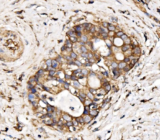
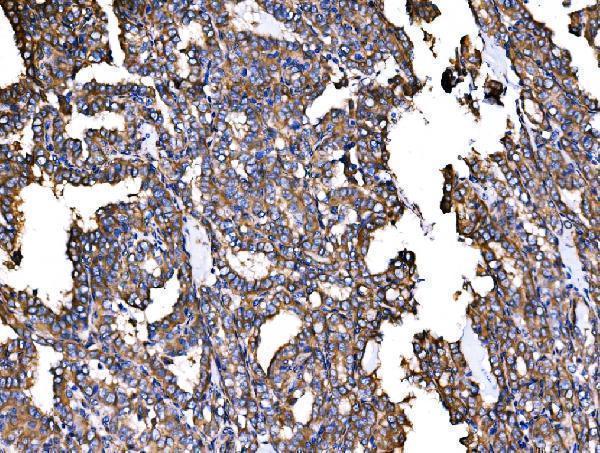
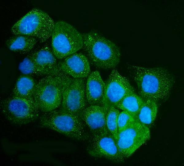


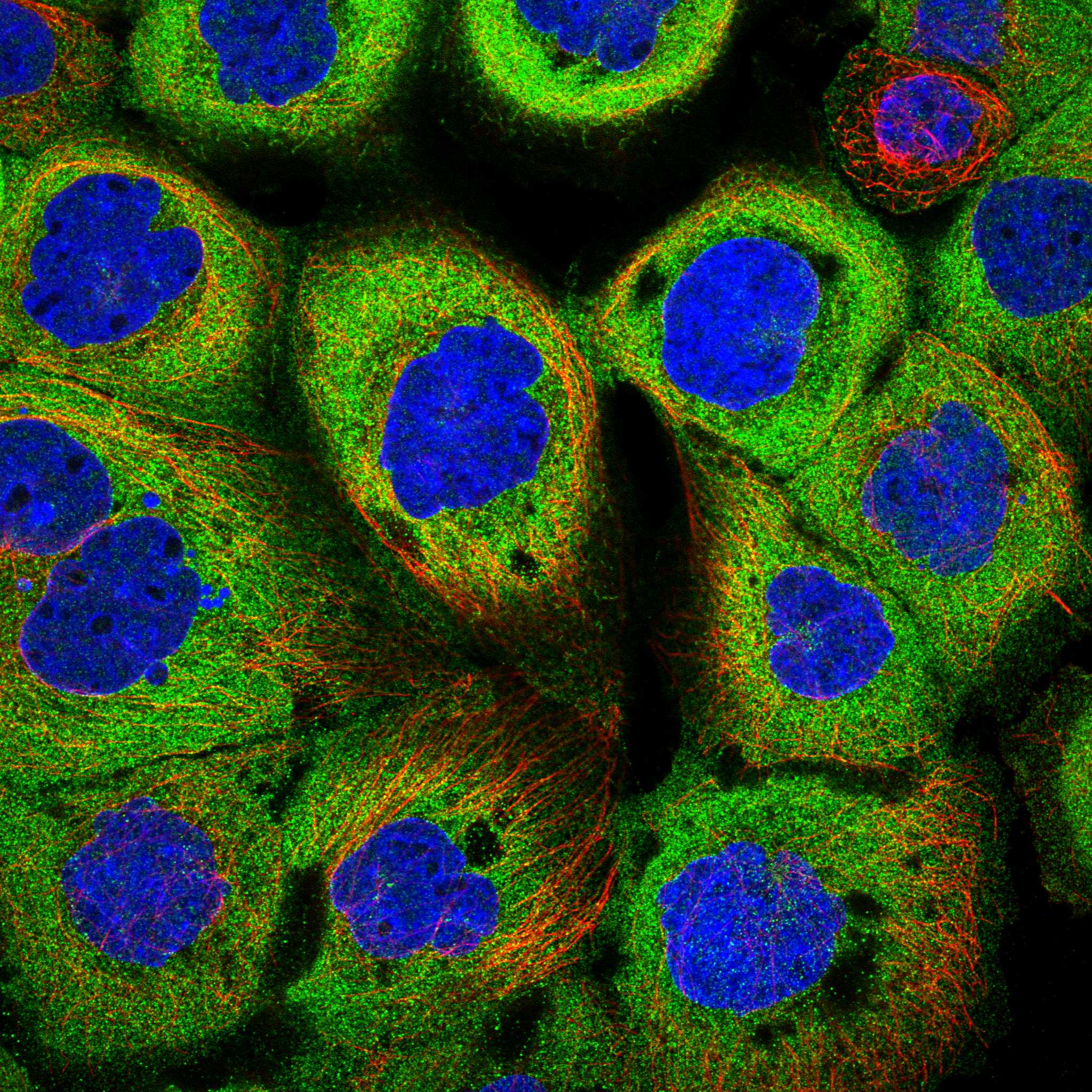
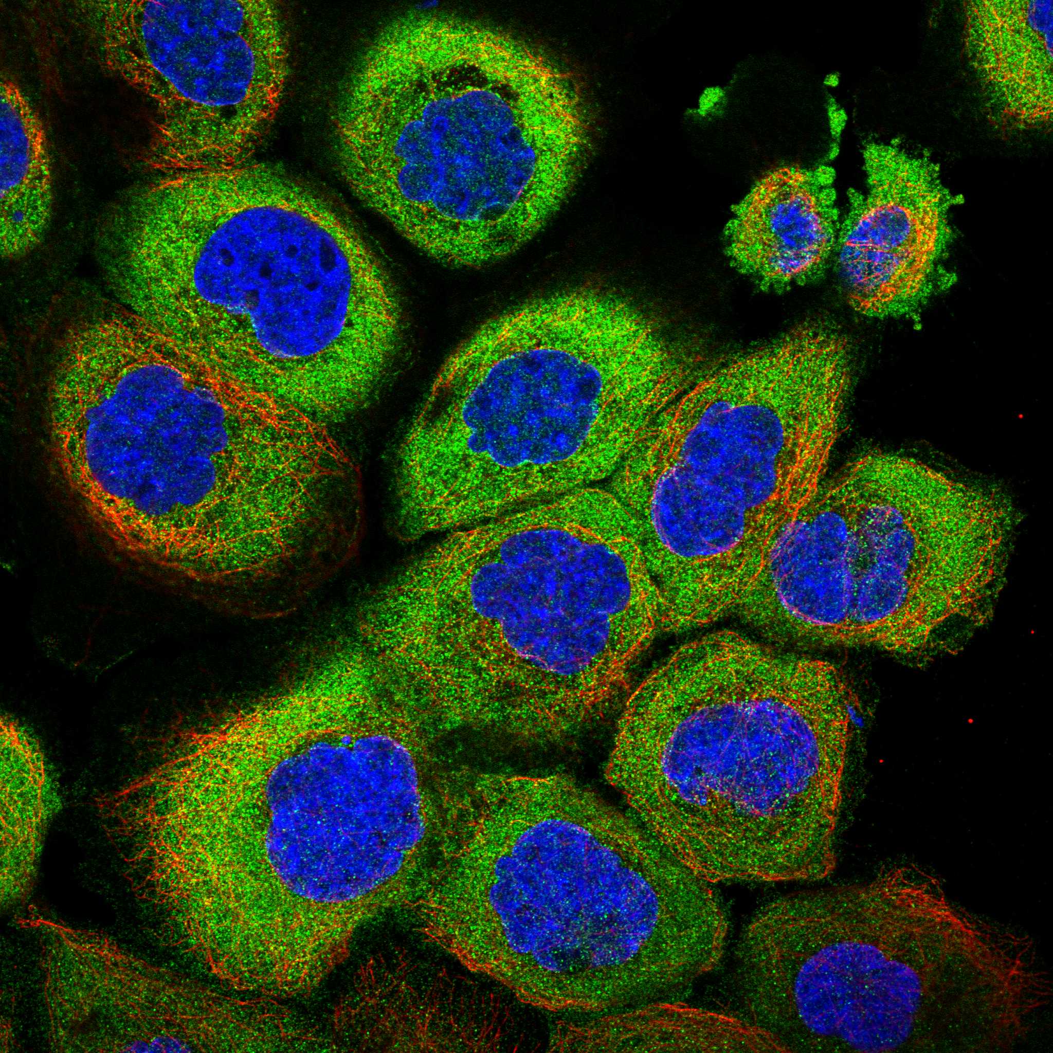
Comprehensive Human Protein Atlas IHC scoring per tissue (reliability: Approved — Low consistency between antibody staining and RNA expression data. Pending external verification.). Rows are taken directly from the HPA tissue chart — click any row's HPA link to view the source.
| Tissue | Cell type | Level | Evidence | Source |
|---|---|---|---|---|
| Duodenum | Glandular cells | Medium | Protein (IHC) | HPA → |
| Endometrium | Glandular cells | Medium | Protein (IHC) | HPA → |
| Fallopian tube | Glandular cells | Medium | Protein (IHC) | HPA → |
| Pancreas | Exocrine glandular cells | Medium | Protein (IHC) | HPA → |
| Thyroid gland | Glandular cells | Medium | Protein (IHC) | HPA → |
| Tissue | Cell type | Level | Evidence | Source |
|---|---|---|---|---|
| Adipose tissue | Adipocytes | Not detected | Protein (IHC) | HPA → |
| Appendix | Glandular cells | Not detected | Protein (IHC) | HPA → |
| Bone marrow | Hematopoietic cells | Not detected | Protein (IHC) | HPA → |
| Cerebellum | Cells in granular layer | Not detected | Protein (IHC) | HPA → |
| Esophagus | Squamous epithelial cells | Not detected | Protein (IHC) | HPA → |
Troubleshoot AHCYL1 staining in paraffin sections by checking retrieval, cell and compartment patterns, controls, and scoring before interpreting chromogenic signal.
A08908-2 has IHC images from human paraffin sections and an IF image from A431 cells (catalog image captions). Listed reactivity covers human, mouse and rat (catalog reactivity).
A08908-2 will render with IHC evidence from human paraffin sections of bladder adenosquamous carcinoma, Hashimoto thyroiditis, thyroid papillary carcinoma and laryngeal squamous cell carcinoma (A08908-2 IHC image captions). Its IF/ICC evidence comes from A431 cells (A08908-2 IF image caption), and the catalog lists IHC, ICC and IF applications (catalog applications).
Which to pick: Choose A08908-2 for chromogenic IHC on paraffin sections: its caption documents EDTA retrieval at pH 8.0, a 2 μg/ml primary concentration and SABC-DAB detection; the fixative is unreported (A08908-2 IHC image caption). For IF/ICC, A08908-2 has an A431 cell image using 5 μg/ml primary antibody (A08908-2 IF image caption). For cross-species planning, A08908-2 lists human, mouse and rat reactivity (catalog reactivity), while the shown IHC sections are human (A08908-2 IHC image captions); its host is rabbit and clonality is unreported (catalog host/clone).