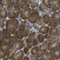
AIMP1 / Aminoacyl tRNA synthase complex-interacting multifunctional protein 1 · IHC design guide

Design Immunohistochemistry for AIMP1

Plan chromogenic AIMP1 IHC in paraffin sections using the observed cytoplasmic tissue pattern and high staining in neuronal cells and pancreas (HPA tissue IHC). Interpret staining with AIMP1 secretion and processing in mind (UniProt).

Evidence assembled Oct 2026 · For research use; verify linked source records and product datasheet before use
Immunohistochemistry protocol sheet for AIMP1 (IHC for AIMP1): expected localisation Cytoplasmic staining in tissue (HPA tissue IHC), antibody A03109, validated IHC image, and IHC protocol steps
Printable AIMP1 IHC protocol sheet — expected localisation Cytoplasmic staining in tissue (HPA tissue IHC), antibody A03109, controls and protocol steps. Open the full AIMP1 IHC guide →

AIMP1 Immunohistochemistry Experimental Design Guide

Expected localisation, validated protocols, controls and antibodies — the at-a-glance facts below, then the full design guide.

Must know before staining
Expected localisation Cytoplasmic staining in tissue (HPA tissue IHC)
Staining pattern Widespread cytoplasmic staining; high in neurons and pancreas (HPA tissue IHC)
Antigen retrieval Tris-EDTA pH 9.0 HIER, 95–98 °C, 20 min (rule: nuclear antigen)
Positive control ⓘ Caudate+4 more · see all
Negative control ⓘ None in HPA (detected in all 45 tissues); use no-primary + isotype controls
Important caveats
Reasons your staining may differ from the expected pattern.
Fixation Keep formalin fixation consistent across paraffin sections (standard IHC practice; not target-specific)
Caveat Secreted AIMP1 can weaken RNA–staining concordance (HPA tissue IHC)
Regulation Hypoxia induces secretion (UniProt)
Isoform / epitope 2 isoforms; EMAP2 (147–312) lacks the N-terminal region (UniProt)
Section 1

Recommended AIMP1 IHC & IF Protocols

The catalog antibody’s IHC-P protocol is followed by a published AIMP1 staining workflow for multiple myeloma bone marrow biopsies (PMC9648396).

Recommended immunohistochemistry (IHC-P) protocol parameters
SampleTissue sections; selected-image fixative not specified (standard IHC workflow)
FixationImage fixative and duration unreported (datasheet A03109); verify before use.
Sectioning4–5 µm sections on charged slides (standard)
DeparaffinisationXylene, graded ethanol series to water (standard)
Antigen retrievalHeat-induced epitope retrieval in Tris-EDTA buffer, pH 9.0, 20 min at 95–98 °C (standard rule: nuclear antigen)
Peroxidase block3% H2O2, 10 min, room temperature (standard)
Blocking10% normal serum of the secondary host, 30 min, room temperature (standard)
Primary antibodyRabbit anti-AIMP1, 1:100–1:500 starting range (standard)
Primary incubationOvernight at 4 °C (standard)
DetectionHRP-polymer secondary, DAB chromogen 5–10 min (standard)
CounterstainHematoxylin, blue, dehydrate and mount (standard)
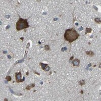
Expected resultAIMP1-positive staining in neuronal cells of caudate (HPA tissue IHC: High). HPA tissue profile: Ubiquitous cytoplasmic expression. High expression in neuronal cells and pancreas. No signal in the no-primary control.
💡Decision noteStart with Tris-EDTA pH 9.0 retrieval at 95–98 °C for 20 min (page retrieval rule: nuclear antigen); the published workflow does not specify retrieval (PMC9648396).
Section 2

What Is the Expected AIMP1 Staining Pattern?

AIMP1 should appear predominantly cytoplasmic in tissue IHC, with strong staining expected in neuronal cells and pancreatic tissue (HPA tissue IHC: ubiquitous cytoplasmic expression; high in neuronal cells and pancreas). Cytosolic enrichment is supported by ICC-IF (HPA subcellular ICC-IF: cytosol). The tissue pattern has Supported reliability with medium RNA–protein consistency (HPA tissue IHC: Supported); AIMP1 has no transmembrane segment (UniProt Q12904 topology).

What am I looking at on my slide?
Clear cytoplasmic staining in neuronal cells of caudate, cerebral cortex, or hippocampus.This matches the reported high neuronal signal and broad cytoplasmic pattern (HPA tissue IHC: High in neuronal cells; ubiquitous cytoplasmic expression). Compare cells within the same section before scoring intensity; a stained section alone does not establish antibody specificity (general IHC practice).
Strong signal confined chiefly to nuclei or crisp cell outlines, with little cytoplasmic signal.Check this compartment mismatch against a known cytoplasmic positive: HPA reports cytoplasmic tissue staining and cytosolic ICC-IF localization (HPA tissue IHC; HPA subcellular ICC-IF). Nuclear signal is not automatically false, because UniProt also lists the nucleus; AIMP1 has no transmembrane segment, so surface-dominant staining needs scrutiny (UniProt Q12904 localization and topology).
Strong staining appears mainly in cells reported as low, such as cardiomyocytes or adipocytes.Treat the contrast as a specificity question, not proof of cross-reactivity: those cells have low reported staining, while AIMP1 is broadly expressed (HPA tissue IHC: Low in cardiomyocytes and adipocytes; ubiquitous cytoplasmic expression). Review morphology and negative detection controls for nonspecific binding or endogenous chromogen activity (general IHC practice).
Diffuse color covers stroma, luminal material, and cells without clear boundaries.Scoring becomes unreliable when background obscures intracellular signal (general IHC practice). Secreted AIMP1 may complicate interpretation of extracellular material, but extracellular color alone cannot identify AIMP1 (UniProt Q12904: secreted; HPA tissue IHC: secreted variants can shift RNA–protein agreement). Examine detection controls before assigning it as specific.
No staining is visible in a neuronal region expected to be strongly positive.A missing signal conflicts with the reported high neuronal staining, though HPA rates tissue reliability Supported with only medium RNA–protein consistency (HPA tissue IHC). Check section quality, retrieval, antibody application, and detection controls as general IHC workflow checks; this result alone cannot distinguish specimen variation from assay failure (general IHC practice).
💡Expected AIMP1 appearanceCall a result consistent when neuronal cells show clear, predominantly cytoplasmic signal with strong intensity (HPA tissue IHC: High in neuronal cells; ubiquitous cytoplasmic expression); isolated surface outlines or widespread color without cell boundaries warrant control review (UniProt Q12904 topology; general IHC practice).
How each factor affects the staining
Tissue and cell contextHPA reports high signal in neuronal cells and lung alveolar type II cells, and high expression in pancreatic tissue (HPA tissue IHC). It reports low signal in cardiomyocytes, myocytes, and adipocytes; these are low-expression comparisons, not established antigen-negative controls (HPA tissue IHC).
Intracellular location and secretionHPA describes ubiquitous cytoplasmic tissue staining and cytosolic ICC-IF localization (HPA tissue IHC; HPA subcellular ICC-IF). UniProt also lists nucleus, ER, Golgi, and secretion; these annotations allow context-dependent signal but do not make every stain in those compartments specific (UniProt Q12904 localization).
Processing and isoformsUniProt lists 2 isoforms and a processed endothelial monocyte-activating polypeptide 2 chain spanning residues 147–312 (UniProt Q12904 processing and isoforms). Interpret differences between intracellular and extracellular staining cautiously because the supplied record does not map the IHC antibody epitope to either product.
IHC evidence strengthThe listed antibodies have Supported IHC status, while the overall tissue profile has Supported reliability and medium consistency with RNA (HPA antibodies: HPA018476 and CAB017618; HPA tissue IHC). Those labels support a reference pattern, not certainty for every specimen, cell, or staining compartment.
IF/ICC Q: what location should appear?A: Predominantly cytosolic signal is the reported ICC-IF result (HPA subcellular ICC-IF: Cytosol, enhanced). The listed antibody validation differs by antibody: ICC Enhanced for HPA018476 and ICC Supported for CAB017618 (HPA antibodies). Use the separate IF/ICC guide for that application's design.
Why is my staining missing, weak or wrong?
SituationLikely causeNext action
A neuronal positive is blank.The result conflicts with high neuronal staining, but its cause is undetermined (HPA tissue IHC: High in neuronal cells).Verify tissue identity and assay controls, then review retrieval and detection steps (general IHC practice).
The whole section has diffuse chromogen.Background can arise from nonspecific binding or endogenous detection activity (general IHC practice).Inspect negative detection controls, blocking, washes, and development time before scoring cells (general IHC practice).
Only nuclear signal is prominent.This differs from HPA's predominant cytoplasmic pattern, although UniProt lists nuclear localization (HPA tissue IHC; UniProt Q12904 localization).Compare a known cytoplasmic positive and controls; record the compartment rather than calling it confirmed AIMP1 (general IHC practice).
Crisp membrane outlines dominate.AIMP1 lacks a transmembrane segment; surface-dominant staining is therefore unexpected from its topology alone (UniProt Q12904 topology).Check background and antibody specificity controls, then compare intracellular staining in a high-expression region (general IHC practice; HPA tissue IHC).
A low-expression tissue stains as strongly as a neuronal positive.The comparison differs from HPA's reported levels; it does not by itself identify cross-reactivity (HPA tissue IHC).Compare cell types within matched sections and inspect negative controls for nonspecific or endogenous signal (general IHC practice).
Extracellular material stains while nearby cells are weak.Secretion makes a cell-to-protein-location mismatch plausible, but extracellular staining is not independently validated by that annotation (UniProt Q12904: secreted; HPA tissue IHC: secreted variant caveat).Score intracellular and extracellular observations separately, and use controls before attributing extracellular color to AIMP1 (general IHC practice).

Sample controls for AIMP1 IHC & IF

🧪Run caudate first and confirm staining in neuronal cells (HPA: High in caudate neuronal cells). HPA detects AIMP1 in all 45 scored tissues, so use no-primary and isotype controls for the negative; cells without visible specific chromogen on the positive slide can serve as a background reference, but no cell type is established as AIMP1-negative (HPA: no negative tissue rows; detected in all 45 scored tissues).
Positive control tissue: Caudate (Neuronal cells, HPA High)
Negative control tissue: None in HPA: AIMP1 is detected in all 45 scored tissues. Use a no-primary (secondary-only) and an isotype control instead.
ICC-IF cell lines (HPA subcellular resource): HPA ICC-IF images show AIMP1 in A-431, U-251MG, U2OS, U2OS, siRNA 1 (10x), U2OS, siRNA 1 (40x), U2OS, scrambled (10x), U2OS, scrambled (40x), with annotated localisation: Cytosol (enhanced) (HPA subcellular).
Technical controls: Include a no-primary, secondary-only control, a host- and clonality-matched nonimmune IgG control, and an AIMP1 knockout specimen where available as a biological negative (standard IHC practice). For brain IHC, quench endogenous peroxidase and assess pigment background; for brain IF, check tissue autofluorescence (standard IHC/IF practice).
⚠️Feasibility: No target-specific fixation window or antigen-retrieval dependence is reported in the supplied evidence; the 5 µg/mL A03109 mouse-brain IHC caption leaves the fixative unreported (A03109 caption: 5 µg/mL; fixative not stated). The evidence does not establish whether frozen sections or IF are easier than paraffin IHC, although ICC-IF images show cytosolic AIMP1 in listed cell lines (HPA subcellular: cytosol, enhanced). Brain pigment and autofluorescence can complicate interpretation, so compare staining with the matched controls (standard IHC/IF practice).

HPA tissue IHC evidence for AIMP1

Comprehensive Human Protein Atlas IHC scoring per tissue (reliability: Supported — Medium consistency between antibody staining and RNA expression data. At least one protein variant secreted, tissue location of RNA and protein might differ and correlation is complex.). Rows are taken directly from the HPA tissue chart — click any row's HPA link to view the source.

Positive expression · recommended positive controls

TissueCell typeLevelEvidenceSource
Caudate Neuronal cells High Protein (IHC) HPA →
Cerebellum GLUC cells - cytoplasm/membrane High Protein (IHC) HPA →
Cerebral cortex Neuronal cells High Protein (IHC) HPA →
Hippocampus Neuronal cells High Protein (IHC) HPA →
Lung Alveolar cells type II High Protein (IHC) HPA →

Undetected expression · recommended negative controls

TissueCell typeLevelEvidenceSource
None in HPA: AIMP1 is detected in all 45 scored tissues. Use a no-primary (secondary-only) and an isotype control instead.
Section 3

Advanced AIMP1 IHC Tips

Troubleshoot AIMP1 staining in paraffin sections by checking retrieval, compartment, cell identity, and controls before comparing chromogenic signal across specimens.

Which retrieval condition should I start with for AIMP1 in paraffin sections?
Start with Tris-EDTA at pH 9.0, heated to 95–98 °C for 20 min (page retrieval setting). Allow sections to cool in the retrieval buffer, then compare test and control sections processed together so heating differences do not masquerade as biological differences (standard IHC practice). The catalog antibody has a mouse brain tissue IHC image at 5 µg/mL, but its caption does not identify a retrieval method or fixative (catalog antibody A03109 caption). If staining is weak, test an alternative retrieval buffer on adjacent sections while keeping detection conditions fixed (standard IHC practice). Assess whether added signal retains the expected predominantly cytoplasmic pattern (HPA: ubiquitous cytoplasmic expression).
How should I investigate weak AIMP1 staining when fixation history differs between blocks?
AIMP1-specific sensitivity to fixation is unknown from the supplied evidence; the mouse brain IHC caption does not state a fixative (catalog antibody A03109 caption). Record each block’s documented fixative and fixation duration, then stain comparable sections in the same run with the page’s Tris-EDTA pH 9.0, 95–98 °C, 20 min retrieval condition (page retrieval setting; standard IHC practice). Include a control section with a known staining pattern to distinguish a block problem from a run problem (standard IHC practice). Do not infer fixation tolerance from AIMP1’s cellular locations or modified residues (UniProt Q12904). Treat differences between blocks as provisional until section quality and staining controls are comparable (standard IHC practice).
Which cellular staining pattern is credible for AIMP1 in tissue IHC?
Expect predominantly cytoplasmic staining across many cell types, with strong neuronal staining reported in several brain regions (HPA: ubiquitous cytoplasmic expression; HPA: High in caudate, cerebral cortex, and hippocampal neuronal cells). Cytosolic localisation is also supported by cell imaging, while UniProt lists nuclear, cytosolic, secreted, endoplasmic reticulum, and Golgi locations (HPA: Cytosol enhanced; UniProt Q12904 subcellular location). Compare the chromogen with cell morphology and a matched counterstain before assigning a compartment (standard IHC practice). Investigate exclusively nuclear or diffuse extracellular staining with controls rather than calling either pattern AIMP1 positive on location alone (HPA: Cytosol enhanced; UniProt Q12904 subcellular location). The secreted variant can complicate agreement between tissue protein staining and RNA location (HPA: Supported reliability description).
Can this antibody distinguish intact AIMP1 from its processed product in IHC?
Do not assign an IHC signal to a specific AIMP1 form without an epitope map and validation for the antibody used (standard IHC practice). AIMP1 has 2 isoforms, and UniProt annotates both a 2–312 chain and a 147–312 endothelial monocyte-activating polypeptide 2 chain (UniProt Q12904 isoforms and processing). An epitope within residues 147–312 could be present in both annotated chains, whereas an epitope before residue 147 would not be present in the shorter chain (UniProt Q12904 processing). Request the antibody’s immunogen or epitope information and assess validated controls before interpreting compartment differences (standard IHC practice). Retrieval can alter epitope accessibility, so compare sections under the same pH 9.0 condition (page retrieval setting; standard IHC practice).
How can IF help check an ambiguous AIMP1 IHC pattern?
Use IF as a separate confirmation experiment and multiplex AIMP1 with a validated marker for the cell population being assessed, such as the neuronal population highlighted in brain tissue (HPA: High in neuronal cells; standard IF practice). Choose fluorophores after checking the specimen’s autofluorescence in each channel, and include single-label controls to evaluate spectral overlap (standard IF practice). Permeabilise cells when testing access to a cytosolic epitope, while matching the treatment to the antibody’s mapped epitope and any extracellular staining question (HPA: Cytosol enhanced; UniProt Q12904 subcellular location; standard IF practice). HPA cell imaging reports cytosolic localisation, but that observation does not establish performance of catalog antibody A03109 in IF (HPA: Cytosol enhanced; catalog antibody A03109 caption). Compare IF and IHC by cell identity and compartment, rather than by signal intensity (standard microscopy practice).
What should I change when AIMP1 DAB staining is diffuse or uneven?
First compare stained sections with a no-primary control and inspect whether diffuse colour tracks section edges, folds, or damaged areas (standard IHC practice). In a chromogenic workflow, check the peroxidase block, washing, and DAB development time before changing antibody concentration (standard IHC practice). The catalog mouse brain image used 5 µg/mL; treat that as a documented image condition, not proof that it is optimal for every block (catalog antibody A03109 caption). If background remains high, titrate the primary antibody and detection reagent on adjacent sections while holding retrieval at Tris-EDTA pH 9.0, 95–98 °C, 20 min (page retrieval setting; standard IHC practice). Preserve a readable cytoplasmic pattern during optimisation (HPA: ubiquitous cytoplasmic expression).
How should I quantify AIMP1 IHC across samples with different cell composition? ⚠ ANSWER MARKED FOR VERIFICATION
Define the cell population and tissue region before scoring, because AIMP1 staining varies by cell type and is prominent in neuronal cells and pancreas (HPA: tissue IHC profile). For cellular staining, record the percentage of positive cells and intensity categories to calculate an H-score; report the scoring thresholds and use the same thresholds throughout (standard IHC quantification practice). Normalise positive counts to the number of eligible cells, or report positive-cell density per mm² of analysed tissue when cell counting is impractical (standard IHC quantification practice). Exclude folds, edges, and necrotic regions by a prespecified rule (standard IHC practice). Keep section thickness, retrieval, imaging, and DAB development consistent across the comparison (standard IHC practice).
How do I distinguish genuine AIMP1 signal from misleading tissue staining?
A credible result aligns with cell morphology and the broadly cytoplasmic pattern reported for AIMP1, especially in neuronal cells in brain tissue (HPA: ubiquitous cytoplasmic expression; HPA: High in neuronal cells). Check unexpected exclusively nuclear, necrotic, or edge-restricted colour against a no-primary control and neighbouring intact tissue before assigning positivity (HPA: Cytosol enhanced; standard IHC practice). Endogenous peroxidase activity can produce chromogenic signal, so evaluate the peroxidase block and detection controls when staining appears independent of the primary antibody (standard IHC practice). AIMP1 can be secreted, and HPA rates tissue IHC reliability as Supported with medium RNA–protein consistency; extracellular signal or RNA disagreement alone cannot settle its identity (UniProt Q12904 subcellular location; HPA: Supported reliability description). Document the control results alongside any biological interpretation (standard IHC practice).
Boster reagents

Best AIMP1 / Aminoacyl tRNA synthase complex-interacting multifunctional protein 1 IHC Antibodies

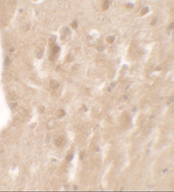
A03109 has a mouse brain IHC image (catalog image caption) and listed human, mouse and rat reactivity (catalog reactivity); no IF data are supplied (catalog IF image alts).

Real IHC data Immunohistochemistry of AIMP1 in mouse brain tissue with AIMP1 antibody at 5 μg/mL.
Anti-AIMP1 Antibody
Cat # A03109

A03109 is listed for IHC-P and shown in mouse brain IHC at 5 μg/mL (catalog applications; image caption). Human, mouse and rat reactivity is listed, but the supplied IHC image documents mouse tissue only (catalog reactivity; image caption).

Which to pick: Choose A03109 for paraffin-section tissue IHC because IHC-P is listed and its own image shows mouse brain staining; the fixative is unreported (catalog applications; image caption). No SKU is documented for IF/ICC (catalog applications; IF image alts). For cross-species work, A03109 lists human, mouse and rat reactivity, while the stated IHC validation is limited to mouse samples (catalog reactivity; dilution_raw).

Each figure is that product's own IHC / IF validation image from its datasheet.

References

  1. UniProt Consortium. UniProt entry Q12904 (AIMP1_HUMAN, Aminoacyl tRNA synthase complex-interacting multifunctional protein 1).
  2. Human Protein Atlas. AIMP1 tissue IHC expression (reliability: Supported).
  3. Human Protein Atlas. AIMP1 subcellular location (ICC-IF): Localized to the cytosol..
  4. Human Protein Atlas. AIMP1 antibody validation summary (2 antibodies).
  5. Response to anti-angiogenic therapy is affected by AIMP protein family activity in glioblastoma and lower-grade gliomas. bioRxiv : the preprint server for biology 2025 — PMC11952521.
  6. AIMP1 promotes multiple myeloma malignancy through interacting with ANP32A to mediate histone H3 acetylation. Cancer communications (London, England) 2022 — PMC9648396.
  7. AIMP1 downregulation restores chondrogenic characteristics of dedifferentiated/degenerated chondrocytes by enhancing TGF-β signal. Cell death & disease 2016 — PMC5399188.
  8. Altered Smooth Muscle Cell Histone Acetylome by the SPHK2/S1P Axis Promotes Pulmonary Hypertension. Circulation research 2023 — PMC10543610.
  9. PubMed PMID:7929199 — UniProt-cited evidence.
  10. PubMed PMID:14702039 — UniProt-cited evidence.
  11. PubMed PMID:15489334 — UniProt-cited evidence.