This website uses cookies to ensure you get the best experience on our website.


- Table of Contents
Plan AK5 IHC in paraffin sections using neuronal cytoplasmic staining as the expected pattern (HPA tissue IHC). Compare staining across consistently fixed sections and assess unexpected cell staining in light of HPA's reported off-target binding (HPA tissue IHC).

Expected localisation, validated protocols, controls and antibodies — the at-a-glance facts below, then the full design guide.
| Expected localisation | Neuronal cytoplasm (HPA tissue IHC) | |
| Staining pattern | Cytoplasmic staining in neuronal cells; texture unreported (HPA tissue IHC) | |
| Antigen retrieval | Citrate pH 6.0 HIER, 95–98 °C, 20 min (rule: cytoplasmic / membrane antigen) | |
| Positive control | Cerebral cortex+4 more · see all | |
| Negative control | Adipose tissue+4 more · see all |
| Fixation | Keep fixation consistent across sections. (standard IHC practice; not target-specific) | |
| Caveat | Presumed off-target staining was disregarded (HPA tissue IHC) | |
| Regulation | Brain-enriched RNA expression (HPA tissue RNA) | |
| Isoform / epitope | 3 isoforms; epitope coverage unknown (UniProt) |
Compare the catalog antibody’s IHC-P protocol (datasheet) with the published AK5 colorectal tumour tissue microarray method (PMC8209216).
| Sample | Paraffin-embedded human brain tissue; fixative not specified (datasheet A11378) |
| Fixation | Image fixative and duration unreported (datasheet A11378); verify before use. |
| Sectioning | 4–5 µm sections on charged slides (standard) |
| Deparaffinisation | Xylene, graded ethanol series to water (standard) |
| Antigen retrieval | Heat-induced epitope retrieval in citrate buffer, pH 6.0, 20 min at 95–98 °C (standard rule: cytoplasmic / membrane antigen) |
| Peroxidase block | 3% H2O2, 10 min, room temperature (standard) |
| Blocking | 10% normal serum of the secondary host, 30 min, room temperature (standard) |
| Primary antibody | Rabbit anti-AK5, 1:100-1:300 (datasheet A11378) |
| Primary incubation | Overnight at 4 °C (standard) |
| Detection | HRP-polymer secondary, DAB chromogen 5–10 min (standard) |
| Counterstain | Hematoxylin, blue, dehydrate and mount (standard) |
| Expected result | AK5-positive staining in neuronal cells of cerebral cortex (HPA tissue IHC: High). HPA tissue profile: Cytoplasmic expression in neuronal cells. No signal in the no-primary control. |
AK5 is a cytoplasmic protein with no transmembrane segment (UniProt Q9Y6K8). In paraffin-section IHC, expect cytoplasmic staining chiefly in neuronal cells, particularly in cerebral cortex and hippocampus (HPA: cytoplasmic neuronal expression; High in both regions). HPA rates the tissue IHC profile Enhanced, while noting medium agreement with RNA data and presumed off-target binding that was disregarded (HPA: reliability description).
| Cytoplasmic chromogen in cortical or hippocampal neurons; signal is stronger than the local background. | This matches the reported compartment and cell type (HPA: cytoplasmic neuronal expression). Both regions have High neuronal staining (HPA: cerebral cortex and hippocampus). Judge the pattern by cell identity and localisation as well as intensity; a dark deposit alone does not establish a specific AK5 signal. |
| Predominantly nuclear, sharply outlined membrane, or broad extracellular staining. | These patterns conflict with the expected cytoplasmic localisation (UniProt Q9Y6K8: cytoplasm; no transmembrane segment; HPA: cytoplasmic neuronal expression). Treat a dominant signal in another compartment as suspect. Check morphology and compare the slide with appropriate staining controls before assigning that signal to AK5. |
| Strong staining in adipocytes, marrow hematopoietic cells, or bronchial respiratory epithelium. | Those cell types are reported Not detected in HPA tissue IHC (HPA: adipose tissue, bone marrow, bronchus). Investigate antibody cross-reactivity or chromogenic background, including endogenous detection activity. Cell type matters: HPA also reports High staining in duodenal and small-intestinal endocrine cells, so staining outside neurons is not automatically an artefact (HPA: tissue IHC). |
| A diffuse wash of colour covers cells and spaces, obscuring cytoplasmic boundaries. | The expected readout is cellular and cytoplasmic (HPA: cytoplasmic neuronal expression). As general IHC practice, assess the no-primary control, blocking, washing, and detection conditions for nonspecific background. Avoid scoring a uniform haze as AK5 merely because it appears in a tissue with reported positive cells. |
| No discernible neuronal signal in cerebral cortex or hippocampus. | Both are reported High in neuronal cells (HPA: cerebral cortex and hippocampus). First confirm that neurons and the tissue section are intact. Then review the antibody's IHC-P validation and the run controls; general IHC variables such as retrieval, antibody dilution, and detection may need optimisation. The supplied sources do not establish AK5-specific fixation sensitivity. |
| Tissue and cell selection | Cerebral cortex and hippocampus provide reported High neuronal staining (HPA: tissue IHC). HPA also lists High intestinal endocrine staining and several Medium non-neuronal patterns (HPA: tissue IHC). Choose and score a defined cell population; UniProt describes AK5 as brain specific, so the non-brain IHC observations warrant cautious interpretation (UniProt Q9Y6K8: tissue specificity; HPA: reliability description). |
| Compartment and topology | Cytoplasm is the annotated location, and AK5 has no transmembrane segment or signal peptide (UniProt Q9Y6K8: subcellular location, topology, processing). The primary IHC readout is cytoplasmic neuronal staining (HPA: tissue IHC). These annotations support a compartment check; they do not identify an antibody epitope or predict retrieval performance. |
| Antibody evidence | Two listed rabbit polyclonal antibodies have Enhanced IHC status (HPA: HPA019128 and HPA057255). HPA nevertheless reports medium consistency between staining and RNA expression and notes presumed off-target binding that was disregarded (HPA: reliability description). Use the reported cell and compartment pattern when interpreting an unfamiliar signal. |
| IF/ICC: what localisation should I expect? | Mainly cytosolic signal, with an additional centriolar-satellite location, is reported for ICC-IF (HPA: subcellular summary). The main cytosol location is Supported and the additional location Approved (HPA: subcellular validation). This is context for IF interpretation; tissue IHC reports cytoplasmic neuronal expression (HPA: tissue IHC). |
| Isoform coverage | Three isoforms are listed (UniProt Q9Y6K8: isoforms 1, 2, 3). The supplied sources do not map the listed antibodies' epitopes to those isoforms, so isoform-specific staining cannot be inferred. If isoform coverage matters to the experiment, check antibody documentation before treating a negative section as absence of every AK5 isoform. |
| Situation | Likely cause | Next action |
|---|---|---|
| Positive-control neurons are blank. | Cortical and hippocampal neurons are reported High (HPA: tissue IHC); a blank result could reflect a failed staining run or a section issue. | Confirm neuron preservation and run-control performance. As general IHC practice, review retrieval, primary dilution, incubation, and detection against the antibody's IHC-P instructions; the sources provide no AK5-specific fixation-effect claim. |
| Signal appears mainly in nuclei or along plasma membranes. | A dominant nuclear or membrane pattern conflicts with cytoplasmic AK5 localisation (UniProt Q9Y6K8; HPA: tissue IHC). | Recheck cell boundaries and counterstain, then compare positive tissue and no-primary controls. Repeat with an independently validated antibody if the discordant compartment persists; both listed HPA antibodies have Enhanced IHC status (HPA: antibodies). |
| Chromogen covers the section uniformly. | Uniform colour can obscure the reported cytoplasmic neuronal pattern (HPA: tissue IHC); background may arise from detection chemistry or nonspecific binding in a chromogenic IHC run. | Use a no-primary control to localise the background source. As general IHC practice, review endogenous-enzyme blocking where applicable, reagent concentration, blocking, and washes before scoring individual cells. |
| Strong signal occurs in an HPA Not detected cell population. | Staining in adipocytes, marrow hematopoietic cells, or bronchial respiratory epithelium disagrees with their reported levels (HPA: tissue IHC). Cross-reactivity or detection background is possible. | Verify cell identity and compare controls and positive neurons. Consider a second IHC-validated antibody if the signal is reproducible; the HPA tissue profile itself notes presumed off-target binding (HPA: reliability description). |
| Intestinal endocrine cells stain despite the brain-enriched RNA profile. | HPA reports High IHC staining in duodenal and small-intestinal endocrine cells, while its RNA profile is brain enriched (HPA: tissue IHC and RNA specificity). | Record the observed cell type and compartment without assuming specificity from intensity alone. Interpret with HPA's medium staining–RNA consistency and presumed off-target-binding caveat (HPA: reliability description). |
| Only faint neuronal staining is visible. | Cortical and hippocampal neurons are reported High in HPA IHC (HPA: tissue IHC), but visual intensity can vary between staining runs as a general IHC consideration. | Compare the positive tissue, negative control, and cell-level background in the same run. If separation is poor, optimise the catalog antibody's IHC-P conditions and detection while preserving a readable counterstain; do not assign an AK5-specific fixation cause from these sources. |
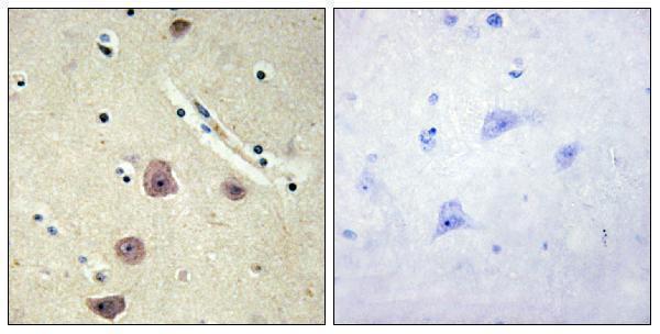
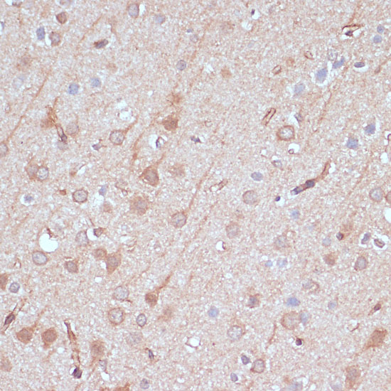
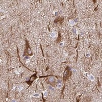
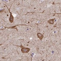


Comprehensive Human Protein Atlas IHC scoring per tissue (reliability: Enhanced — Medium consistency between antibody staining and RNA expression data. Presumed off target binding observed and disregarded.). Rows are taken directly from the HPA tissue chart — click any row's HPA link to view the source.
| Tissue | Cell type | Level | Evidence | Source |
|---|---|---|---|---|
| Cerebral cortex | Neuronal cells | High | Protein (IHC) | HPA → |
| Duodenum | Endocrine cells | High | Protein (IHC) | HPA → |
| Hippocampus | Neuronal cells | High | Protein (IHC) | HPA → |
| Small intestine | Endocrine cells | High | Protein (IHC) | HPA → |
| Breast | Glandular cells | Medium | Protein (IHC) | HPA → |
| Tissue | Cell type | Level | Evidence | Source |
|---|---|---|---|---|
| Adipose tissue | Adipocytes | Not detected | Protein (IHC) | HPA → |
| Bone marrow | Hematopoietic cells | Not detected | Protein (IHC) | HPA → |
| Bronchus | Respiratory epithelial cells | Not detected | Protein (IHC) | HPA → |
| Cervix | Glandular cells | Not detected | Protein (IHC) | HPA → |
| Endometrium | Cells in endometrial stroma | Not detected | Protein (IHC) | HPA → |
Troubleshoot chromogenic AK5 staining in paraffin sections using tissue and subcellular evidence, with IF/ICC guidance for cross-checking localisation.
Two rabbit polyclonal anti-AK5 antibodies list IHC and IF applications for human and mouse samples; one also lists rat reactivity (catalog: applications, reactivity, clonality). Both have IHC images (catalog: IHC image captions).
A11378 lists human and mouse reactivity and IF and IHC applications; its IHC image shows paraffin-embedded human brain with a peptide-blocked comparison (catalog: A11378 applications, reactivity and IHC image caption). A11378-1 lists human, mouse and rat reactivity and IF, ICC and IHC applications; its IHC image shows paraffin-embedded rat brain (catalog: A11378-1 applications, reactivity and IHC image caption).
Which to pick: For human brain paraffin-section IHC, start with A11378 because its own IHC image uses that sample; the caption does not report the fixative (catalog: A11378 IHC image caption). For IF/ICC, choose A11378-1 if ICC is required: it lists both applications, whereas A11378 lists IF; both are rabbit polyclonal antibodies, and neither has an IF image in the supplied catalog (catalog: applications, clonality and IF image captions). For rat work, choose A11378-1 because it lists rat reactivity and shows rat brain paraffin-section IHC; its caption does not report the fixative (catalog: A11378-1 reactivity and IHC image caption).