This website uses cookies to ensure you get the best experience on our website.


- Table of Contents
Plan paraffin-section AKAP12 IHC with catalog antibody A02303 at 1:100–1:300 (datasheet). Use the cytoplasmic tissue profile to choose scoring regions, and review unannotated stained structures when interpreting results (HPA tissue IHC).

Expected localisation, validated protocols, controls and antibodies — the at-a-glance facts below, then the full design guide.
| Expected localisation | Cytoplasmic (HPA tissue IHC); cortical cytoskeleton possible (UniProt) | |
| Staining pattern | Cytoplasmic in seminiferous ducts, endothelium and connective tissue (HPA tissue IHC) | |
| Antigen retrieval | Citrate pH 6.0 HIER, 95–98 °C, 20 min (rule: cytoplasmic / membrane antigen) | |
| Positive control | Kidney+4 more · see all | |
| Negative control | Adrenal gland+4 more · see all |
| Fixation | Keep fixation consistent across sections (standard IHC practice; not target-specific) | |
| Caveat | Unannotated stained cells may complicate scoring (HPA tissue IHC) | |
| Regulation | Expression regulation not specified (UniProt) | |
| Isoform / epitope | 3 isoforms; epitope coverage is unresolved (UniProt) |
The catalog antibody protocol uses citrate retrieval (page antigen-retrieval rule). Two published chromogenic IHC protocols provide additional conditions (PMC11695186; PMC10202715).
| Sample | Paraffin-embedded human brain tissue; fixative not specified (datasheet A02303) |
| Fixation | Image fixative and duration unreported (datasheet A02303); verify before use. |
| Sectioning | 4–5 µm sections on charged slides (standard) |
| Deparaffinisation | Xylene, graded ethanol series to water (standard) |
| Antigen retrieval | Heat-induced epitope retrieval in citrate buffer, pH 6.0, 20 min at 95–98 °C (standard rule: cytoplasmic / membrane antigen) |
| Peroxidase block | 3% H2O2, 10 min, room temperature (standard) |
| Blocking | 10% normal serum of the secondary host, 30 min, room temperature (standard) |
| Primary antibody | Rabbit anti-AKAP12, 1:100-1:300 (datasheet A02303) |
| Primary incubation | Overnight at 4 °C (standard) |
| Detection | HRP-polymer secondary, DAB chromogen 5–10 min (standard) |
| Counterstain | Hematoxylin, blue, dehydrate and mount (standard) |
| Expected result | AKAP12-positive staining in bowman's capsule of kidney (HPA tissue IHC: High). HPA tissue profile: Cytoplasmic expression mainly in seminiferous ducts, endothelial cells and connective tissue. No signal in the no-primary control. |
AKAP12 should show predominantly cytoplasmic staining, sometimes accentuated at the cell edge, in selected structures such as testicular Sertoli cells, cells of the kidney Bowman’s capsule, endothelium and connective tissue (HPA: tissue IHC; UniProt Q02952: cell cortex and cytoskeleton). It has no transmembrane segment (UniProt Q02952: topology). HPA rates the tissue IHC evidence “Enhanced” while noting medium consistency with RNA expression (HPA: reliability description).
| Cytoplasmic staining in Sertoli cells or cells of the kidney Bowman’s capsule, with little staining in adjacent cells. | This matches the reported high staining in those specific cell populations (HPA: Testis, Sertoli cells, High; Kidney, Bowman’s capsule, High). Judge the named cells within each tissue rather than treating an entire section as uniformly positive (HPA: tissue IHC). |
| Cytoplasmic staining with some cell-edge accentuation; no dominant nuclear signal. | This fits the reported cytoplasmic IHC profile and AKAP12’s cortical cytoskeleton and membrane associations (HPA: tissue IHC profile; UniProt Q02952: subcellular location). A predominantly nuclear result warrants review of morphology and staining controls; neither source lists the nucleus as an expected main location (HPA: subcellular location; UniProt Q02952: subcellular location). |
| Strong nuclear staining or an exclusively crisp surface outline. | Neither is the expected IHC pattern: HPA describes mainly cytoplasmic tissue staining, and UniProt reports no transmembrane segment (HPA: tissue IHC profile; UniProt Q02952: topology). Check whether counterstain, precipitate or another staining artifact is being mistaken for specific signal (standard IHC practice). |
| Signal dominates HPA-negative cell populations, such as bone marrow hematopoietic cells or adrenal glandular cells. | Those named populations were reported as not detected (HPA: Bone marrow, hematopoietic cells; Adrenal gland, glandular cells). Consider nonspecific antibody binding or endogenous detection activity, and compare primary-antibody omission and detection controls before assigning the signal to AKAP12 (standard IHC practice). |
| Diffuse color across tissue, or no signal in a known-positive cell population. | A widespread haze cannot establish the reported cell-specific pattern (HPA: tissue IHC profile). An absent signal in Sertoli cells or cells of the Bowman’s capsule conflicts with HPA’s high staining reports, but by itself does not identify the failed step (HPA: Testis; Kidney). Review controls and morphology before interpreting the specimen (standard IHC practice). |
| Cell population and tissue | Expected intensity depends on the named cells: Sertoli cells and cells of the Bowman’s capsule are High; lung endothelial, ovarian follicle and smooth muscle cells are Medium (HPA: tissue IHC). Colon endothelial cells are Not detected, so endothelial identity alone does not predict positivity (HPA: Colon). |
| Intracellular distribution | A cytoplasmic pattern can include cortical or membrane-associated emphasis because AKAP12 is reported at the cell cortex, cytoskeleton and membrane (UniProt Q02952: subcellular location). Its lack of a transmembrane segment does not require an uninterrupted surface outline in paraffin IHC (UniProt Q02952: topology). |
| Antibody evidence and isoforms | HPA lists Enhanced IHC status for HPA006344, HPA056230 and CAB026379 (HPA: antibodies). UniProt lists three isoforms, but these records do not establish which isoforms a given antibody detects; check its documented epitope before drawing isoform-specific conclusions (UniProt Q02952: isoforms; standard IHC practice). |
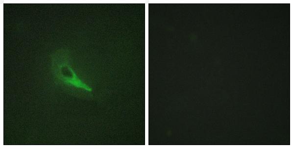
| IF/ICC Q&A: Where should fluorescence appear? | Predominantly in cytosol and at the plasma membrane, both rated enhanced in HPA’s ICC-IF summary (HPA: subcellular). This supports compartment interpretation in IF/ICC; it does not turn the ICC-IF images into a paraffin-IHC protocol or establish matching signal intensity across methods (HPA: subcellular; standard IHC practice). |
| Situation | Likely cause | Next action |
|---|---|---|
| No signal in Sertoli cells or cells of the kidney Bowman’s capsule. | The result differs from HPA’s High reports, but the records cannot identify whether tissue quality, detection or antibody performance is responsible (HPA: Testis; Kidney; standard IHC practice). | Confirm the named cells are present, then inspect a concurrent positive control and each detection step. Review the antibody’s documented IHC conditions; do not infer an AKAP12-specific fixation effect from these sources (standard IHC practice). |
| Only nuclei stain strongly. | A dominant nuclear pattern conflicts with the reported cytoplasmic and cortical locations (HPA: tissue IHC profile; UniProt Q02952: subcellular location). | Check the hematoxylin counterstain and chromogen distribution, and compare a primary-antibody omission control. Treat the result as unconfirmed until expected cytoplasmic staining appears in a known-positive cell population (standard IHC practice; HPA: Testis; Kidney). |
| Most cell types show diffuse color. | This obscures the selective cytoplasmic pattern reported by HPA and may reflect nonspecific binding or background detection (HPA: tissue IHC profile; standard IHC practice). | Compare the primary-antibody omission control and a documented negative cell population; inspect blocking, washes, antibody dilution and detection background under the chosen IHC workflow (standard IHC practice; HPA: Bone marrow, hematopoietic cells, Not detected). |
| A nominally negative region stains. | The HPA negative calls apply to specified cells, such as adrenal glandular cells and bone marrow hematopoietic cells, rather than every structure in those tissues (HPA: tissue IHC negatives). | Identify the stained cell type before calling a discrepancy. If the specifically named negative cells stain, compare omission and detection controls and reassess specificity (HPA: tissue IHC negatives; standard IHC practice). |
| Endothelial staining varies between tissues. | HPA reports Medium staining in lung endothelial cells but Not detected in colon endothelial cells (HPA: Lung; Colon). | Score each tissue and cell population against its own HPA observation. Use morphology and appropriate controls when a result differs; do not use lung endothelium as a universal endothelial intensity benchmark (HPA: Lung; Colon; standard IHC practice). |
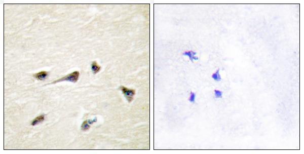
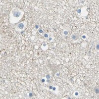


Comprehensive Human Protein Atlas IHC scoring per tissue (reliability: Enhanced — Medium consistency between antibody staining and RNA expression data. Antibody staining in cells/structures not annotated, view images.). Rows are taken directly from the HPA tissue chart — click any row's HPA link to view the source.
| Tissue | Cell type | Level | Evidence | Source |
|---|---|---|---|---|
| Kidney | Bowman's capsule | High | Protein (IHC) | HPA → |
| Testis | Sertoli cells | High | Protein (IHC) | HPA → |
| Cerebellum | Processes in molecular layer | Medium | Protein (IHC) | HPA → |
| Lung | Endothelial cells | Medium | Protein (IHC) | HPA → |
| Ovary | Follicle cells | Medium | Protein (IHC) | HPA → |
| Tissue | Cell type | Level | Evidence | Source |
|---|---|---|---|---|
| Adrenal gland | Glandular cells | Not detected | Protein (IHC) | HPA → |
| Appendix | Glandular cells | Not detected | Protein (IHC) | HPA → |
| Bone marrow | Hematopoietic cells | Not detected | Protein (IHC) | HPA → |
| Breast | Adipocytes | Not detected | Protein (IHC) | HPA → |
| Bronchus | Respiratory epithelial cells | Not detected | Protein (IHC) | HPA → |
Troubleshoot AKAP12 staining in paraffin sections by checking retrieval, compartment, cell identity, and matched controls before interpreting chromogenic signal.
The IHC-validated antibody has paraffin-section human brain data and HeLa IF data (catalog image captions); catalog reactivity covers human, mouse and rat (catalog: A02303).
A02303 will render with an IHC image of paraffin-embedded human brain tissue and peptide blocking (catalog: A02303 IHC caption). Its IF image shows HeLa cells with peptide blocking, and its listed reactivity is human, mouse and rat (catalog: A02303 IF caption; catalog: A02303 reactivity).
Which to pick: For tissue IHC, choose A02303: its own image documents paraffin-embedded human brain tissue; the fixative is unreported (catalog: A02303 IHC caption). For IF, A02303 has a HeLa image; for ICC, A02303-4 lists ICC and IF but has no supplied image, and both antibodies are rabbit polyclonals (catalog: A02303 IF caption; catalog: A02303-4 applications and image alts; catalog: antibody descriptions and host). For work across human, mouse and rat, either SKU lists all three species, though the supplied IHC image documents human tissue only for A02303 (catalog: both reactivity lists; catalog: A02303 IHC caption).