AKAP4 / A-kinase anchor protein 4 · IHC design guide

Design Immunohistochemistry for AKAP4

Plan chromogenic IHC of AKAP4 in paraffin sections using testis spermatids as the positive reference (HPA tissue IHC). The IHC-validated antibody starts at 5 μg/mL (datasheet: 5 μg/mL); compare staining across spermatid stages because the reported stage differs between sources (UniProt; HPA tissue IHC).

Evidence assembled Oct 2026 · For research use; verify linked source records and product datasheet before use
Immunohistochemistry protocol sheet for AKAP4 (IHC for AKAP4): expected localisation Spermatids in testis (HPA tissue IHC); flagellar principal piece expected (UniProt), antibody A07362, validated IHC image, and IHC protocol steps
Printable AKAP4 IHC protocol sheet — expected localisation Spermatids in testis (HPA tissue IHC); flagellar principal piece expected (UniProt), antibody A07362, controls and protocol steps. Open the full AKAP4 IHC guide →

AKAP4 Immunohistochemistry Experimental Design Guide

Expected localisation, validated protocols, controls and antibodies — the at-a-glance facts below, then the full design guide.

Must know before staining
Expected localisation Spermatids in testis (HPA tissue IHC); flagellar principal piece expected (UniProt)
Staining pattern Selective spermatid staining in seminiferous ducts (HPA tissue IHC)
Antigen retrieval Citrate pH 6.0 HIER, 95–98 °C, 20 min (rule: cytoplasmic / membrane antigen)
Positive control ⓘ Testis
Negative control ⓘ Adipose tissue+4 more · see all
Important caveats
Reasons your staining may differ from the expected pattern.
Fixation Keep fixation consistent across sections (standard IHC practice; not target-specific)
Caveat Reported stage differs: round versus late spermatids (UniProt; HPA tissue IHC)
Regulation High in late spermatids (HPA tissue IHC)
Isoform / epitope 2 isoforms; mature chain starts at 189—check epitope mapping (UniProt)
Section 1

Recommended AKAP4 IHC & IF Protocols

The catalog antibody’s IHC-P protocol is accompanied by four published AKAP4 IHC protocols (PMC3667910; PMC5462453; PMC3579772; PMC8974088).

Recommended immunohistochemistry (IHC-P) protocol parameters
SampleTissue sections; selected-image fixative not specified (standard IHC workflow)
FixationImage fixative and duration unreported (datasheet A07362); verify before use.
Sectioning4–5 µm sections on charged slides (standard)
DeparaffinisationXylene, graded ethanol series to water (standard)
Antigen retrievalHeat-induced epitope retrieval in citrate buffer, pH 6.0, 20 min at 95–98 °C (standard rule: cytoplasmic / membrane antigen)
Peroxidase block3% H2O2, 10 min, room temperature (standard)
Blocking10% normal serum of the secondary host, 30 min, room temperature (standard)
Primary antibodyRabbit anti-AKAP4, 5 μg/mL (datasheet A07362)
Primary incubationOvernight at 4 °C (standard)
DetectionHRP-polymer secondary, DAB chromogen 5–10 min (standard)
CounterstainHematoxylin, blue, dehydrate and mount (standard)
Expected resultAKAP4-positive staining in elongated or late spermatids of testis (HPA tissue IHC: High). HPA tissue profile: Selective expression in spermatids of seminiferous ducts. No signal in the no-primary control.
💡Decision noteStart with citrate pH 6.0 retrieval at 95–98 °C for 20 min (page retrieval rule); the testis protocol also reports citrate retrieval (PMC5462453).
Section 2

What Is the Expected AKAP4 Staining Pattern?

AKAP4 is a structural component of the sperm fibrous sheath and localizes to the principal piece of the sperm flagellum; it has no transmembrane segment (UniProt Q5JQC9). In paraffin testis sections, expect selective spermatid staining, with High staining reported in elongated or late spermatids (HPA tissue IHC). HPA rates the tissue pattern Enhanced, citing consistency between staining and RNA expression (HPA tissue IHC).

What am I looking at on my slide?
Selective, strong staining in elongated or late spermatids within seminiferous ducts (HPA tissue IHC).This matches the observed testis IHC pattern (HPA tissue IHC). Score the stained cell population and distribution; chromogenic sections may not resolve the principal piece as clearly as sperm IF images (HPA subcellular ICC-IF; general IHC practice).
Predominantly nuclear staining, or staining confined to a compartment other than the sperm flagellar principal piece.That distribution conflicts with the reported principal-piece location (UniProt Q5JQC9; HPA subcellular ICC-IF). Treat it as suspect and compare with a known-positive section and primary-antibody omission control (general IHC practice).
Strong staining in unrelated cells, such as adipocytes or adrenal glandular cells.HPA reports AKAP4 as not detected in those cells (HPA tissue IHC). Consider antibody cross-reactivity or endogenous chromogenic detection activity; the image alone cannot distinguish them (general IHC practice).
Diffuse staining across tissue structures, obscuring individual spermatids.The pattern is difficult to reconcile with selective spermatid staining (HPA tissue IHC). Excess detection signal or inadequate blocking or washing can raise background (general IHC practice).
No discernible staining in a testis section that contains elongated or late spermatids.This disagrees with the reported High signal in that population (HPA tissue IHC). Check that the relevant cells are present before troubleshooting antibody and detection steps (general IHC practice).
💡Expected AKAP4 appearanceCall a result positive when elongated or late spermatids show selective, High staining (HPA tissue IHC); dominant nuclear, diffuse, or unrelated-cell staining warrants control-based review (UniProt Q5JQC9; HPA tissue IHC; general IHC practice).
How each factor affects the staining
Cell stage in the sectionHPA observes staining in elongated or late spermatids, while UniProt describes expression in round spermatids (HPA tissue IHC; UniProt Q5JQC9). Record the visible stage; these sources do not resolve the difference.
Flagellar compartmentUniProt places AKAP4 in the sperm flagellar principal piece; sperm ICC-IF also reports principal-piece localization (UniProt Q5JQC9; HPA subcellular ICC-IF). Section orientation and resolution can limit compartment scoring (general IHC practice).
Protein processing and epitopeUniProt annotates a propeptide at residues 1–188 and a chain at 189–854 (UniProt Q5JQC9). Antibody epitope positions are not supplied, so staining of precursor versus processed protein cannot be assigned.
Isoform coverageUniProt lists 2 isoforms (UniProt Q5JQC9). Without the antibody's epitope or isoform validation, this record cannot establish whether both contribute to IHC signal.
Antibody validationHPA marks IHC for HPA005949 and HPA020046 as Enhanced (HPA antibodies). That supports confidence in the reported pattern, but does not establish performance for a different catalog antibody or every staining run.
IF/ICC localisation?In sperm, ICC-IF reports localization to the principal piece with Enhanced evidence (HPA subcellular ICC-IF; HPA antibodies). Use that as a localisation comparison; assess paraffin-section IHC against the tissue IHC pattern (HPA tissue IHC).
Why is my staining missing, weak or wrong?
SituationLikely causeNext action
No staining in a presumed positive testis section.The sampled area may lack identifiable elongated or late spermatids, the cells reported as High by HPA (HPA tissue IHC).Inspect morphology and select a section containing those cells; if present, verify antibody, retrieval, detection, and counterstain steps against the assay controls (general IHC practice).
Late spermatids stain weakly while the rest of the section is clear.Signal may be below the assay's detection threshold; HPA's High result does not define a dilution or retrieval condition for this antibody (HPA tissue IHC; general IHC practice).Confirm the IHC-validated antibody's supplied conditions and check a known-positive control in the same run; adjust retrieval or detection using the assay instructions (general IHC practice).
Diffuse chromogenic background covers the seminiferous ducts.Background can arise from excess antibody or detection reagent, insufficient blocking, or incomplete washing (general IHC practice).Compare a primary-antibody omission control; then review blocking, washes, reagent concentration, and development time (general IHC practice).
Unrelated cells show strong staining.Cross-reactivity or endogenous detection activity is possible; HPA reports no AKAP4 detection in several non-testis cell populations (HPA tissue IHC; general IHC practice).Check primary-antibody omission and endogenous-enzyme controls, then compare with the selective testis pattern; do not score isolated ectopic staining as AKAP4 without validation (HPA tissue IHC; general IHC practice).
A nuclear pattern dominates the positive section.It conflicts with principal-piece localization reported for AKAP4 (UniProt Q5JQC9; HPA subcellular ICC-IF). The stain alone does not identify its cause.Recheck cell identification, counterstain, and control slides; assess whether expected spermatid staining is present before interpreting the nuclear signal (HPA tissue IHC; general IHC practice).
Dense signal prevents assessment of which spermatids stain.Overdevelopment can obscure cell boundaries and the selective pattern needed for interpretation (HPA tissue IHC; general IHC practice).Reduce chromogen development or detection intensity and compare with a lightly stained control section; score cell type and distribution only when morphology remains readable (general IHC practice).

Sample controls for AKAP4 IHC & IF

🧪Run testis first and expect strong staining in elongated or late spermatids (HPA: High in elongated or late spermatids). Use adipose tissue as a negative specimen (HPA: Not detected in adipocytes); on the testis slide, compare staining in other cells with the spermatids as an internal background check, without assuming those cells are definitively negative (HPA: positive call is specific to elongated or late spermatids).
Positive control tissue: Testis (Elongated or late spermatids, HPA High)
Negative control tissue: Adipose tissue (HPA Not detected)
ICC-IF cell lines (HPA subcellular resource): HPA ICC-IF images show AKAP4 in Sperm, with annotated localisation: Principal piece (enhanced) (HPA subcellular).
Technical controls: Include a no-primary, secondary-only control; an isotype control matched to the primary antibody’s host species and clonality; and, if available, a knockout specimen or validated peptide-block control (standard IHC control practice). For chromogenic testis IHC, quench endogenous peroxidase and check background on the control slide; for IF, assess tissue autofluorescence before assigning signal (standard IHC/IF practice).
⚠️Feasibility: The supplied evidence reports no AKAP4-specific fixation window or antigen-retrieval dependency, and the A07362 human-testis IHC caption does not state its fixative (selected-SKU caption: fixative unreported). The available IHC caption and sperm ICC-IF images do not establish whether frozen sections or IF are easier than paraffin IHC (selected-SKU caption: human-testis IHC; HPA subcellular: sperm ICC-IF images). In IF, distinguish principal-piece signal from specimen autofluorescence using the no-primary control (HPA subcellular: principal piece; standard IF practice).

HPA tissue IHC evidence for AKAP4

Comprehensive Human Protein Atlas IHC scoring per tissue (reliability: Enhanced — High consistency between antibody staining and RNA expression data.). Rows are taken directly from the HPA tissue chart — click any row's HPA link to view the source.

Positive expression · recommended positive controls

TissueCell typeLevelEvidenceSource
Testis Elongated or late spermatids High Protein (IHC) HPA →

Undetected expression · recommended negative controls

TissueCell typeLevelEvidenceSource
Adipose tissue Adipocytes Not detected Protein (IHC) HPA →
Adrenal gland Glandular cells Not detected Protein (IHC) HPA →
Appendix Glandular cells Not detected Protein (IHC) HPA →
Bone marrow Hematopoietic cells Not detected Protein (IHC) HPA →
Breast Adipocytes Not detected Protein (IHC) HPA →
Section 3

Advanced AKAP4 IHC Tips

Troubleshoot AKAP4 chromogenic IHC in paraffin sections against its spermatid expression and flagellar localisation (HPA tissue IHC; UniProt Q5JQC9).

How should I adjust retrieval when AKAP4 staining is weak in paraffin sections?
Use citrate pH 6.0 heat-induced retrieval at 95–98 °C for 20 min before chromogenic IHC (page retrieval setting). If testis staining is weak, compare adjacent sections processed for shorter and longer retrieval times while keeping detection conditions constant (standard IHC practice). Assess signal in spermatids within seminiferous ducts, where selective staining is expected (HPA tissue IHC). The selected antibody has a human testis IHC image at 5 μg/ml, but its caption does not state the fixative or retrieval method (caption A07362). Record retrieval time and tissue integrity alongside staining intensity so stronger colour is not mistaken for better specificity (standard IHC practice).
Could fixation explain inconsistent AKAP4 staining between testis sections?
AKAP4-specific fixation sensitivity is unknown from the supplied evidence, so compare sections with documented fixation histories before attributing a weak result to fixation. Record fixative, fixation duration, processing date and section thickness, then hold citrate pH 6.0 retrieval at 95–98 °C for 20 min constant (page retrieval setting; standard IHC practice). Include a testis reference section in each run and assess spermatid staining in comparable seminiferous ducts (HPA tissue IHC; standard IHC practice). The selected antibody’s human testis IHC caption reports 5 μg/ml but gives no fixative (caption A07362). Do not use staining differences alone to assign a fixation effect to AKAP4 (standard IHC practice).
Where should convincing AKAP4 staining appear in human testis IHC?
Look for staining associated with spermatids in seminiferous ducts, especially elongated or late spermatids reported as high in tissue IHC (HPA tissue IHC). AKAP4 is a major sperm fibrous sheath component, and its mature localisation is the principal piece of the sperm flagellum (UniProt Q5JQC9; HPA subcellular). Assess the pattern at sufficient magnification to distinguish flagellar structures from diffuse colour over neighbouring cells (standard IHC practice). UniProt describes expression in round spermatids, whereas HPA reports strong tissue staining in elongated or late spermatids; preserve that distinction when recording developmental stage (UniProt Q5JQC9; HPA tissue IHC). Compare adjacent ducts under the same 20 min retrieval condition before calling a compartment shift (page retrieval setting; standard IHC practice).
Can this IHC stain distinguish AKAP4 isoforms or precursor from mature protein?
Do not assign an isoform from chromogenic staining alone: AKAP4 has 2 annotated isoforms, and no epitope map for the selected antibody is supplied (UniProt Q5JQC9; supplied antibody evidence). The annotated precursor has a propeptide at residues 1–188 and a mature chain at 189–854, so epitope position would matter for interpreting processing (UniProt Q5JQC9). Several phosphorylated residues are annotated, including serines 190 and 213, but their effect on this antibody’s IHC staining is unknown (UniProt Q5JQC9; supplied antibody evidence). Request epitope information before claiming precursor, mature-chain or phosphoform selectivity (standard IHC interpretation). Meanwhile, score the cellular and flagellar pattern without assigning a molecular form (HPA tissue IHC; HPA subcellular).
How can I check an AKAP4 IHC pattern with multiplex immunofluorescence?
In a separate IF assay, pair AKAP4 with an independently validated spermatid marker and examine whether signal tracks the expected spermatid population and flagellar principal piece (HPA tissue IHC; HPA subcellular; standard IF practice). Choose a far-red fluorophore when testis autofluorescence compromises shorter wavelengths, and include unstained and single-colour controls for spectral interpretation (standard IF practice). Apply gentle permeabilisation after fixation to access intracellular fibrous-sheath epitopes; AKAP4 has no annotated transmembrane segment (UniProt Q5JQC9; standard IF practice). Check whether permeabilisation changes morphology before interpreting lost signal as absent antigen (standard IF practice). The catalog IHC caption reports 5 μg/ml in human testis, but supplies no IF fixation or dilution conditions (caption A07362).
How do I separate AKAP4 signal from chromogenic background?
Include a no-primary control and a tissue control, then inspect whether colour follows spermatids rather than section edges or broadly stained cells (standard IHC practice; HPA tissue IHC). HPA reports AKAP4 as high in testis elongated or late spermatids and undetected in several other sampled cell populations, including adipose adipocytes (HPA tissue IHC). Use a peroxidase block before DAB development and keep development time matched across sections; these are general chromogenic IHC controls (standard IHC practice). If diffuse staining rises with antibody concentration, titrate around the caption’s 5 μg/ml while maintaining citrate pH 6.0 retrieval (caption A07362; page retrieval setting; standard IHC practice). Interpret clean background only alongside preserved tissue morphology and the expected cellular pattern (standard IHC practice; HPA tissue IHC).
What should I count when quantifying AKAP4 IHC in seminiferous ducts? ⚠ ANSWER MARKED FOR VERIFICATION
Score the percentage of positive spermatids and staining intensity within each evaluated seminiferous duct, recording developmental stage where morphology permits (HPA tissue IHC; standard IHC practice). An H-score combines the percentages at intensity grades 0–3 into a 0–300 summary, provided scoring thresholds remain fixed across slides (standard IHC practice). Normalise positive counts to the number of evaluable spermatids, or report stained flagellar area per duct area when structures cannot be counted reliably (standard IHC practice). Sample multiple ducts and record the number examined because AKAP4 expression and staining vary with spermatid stage (UniProt Q5JQC9; HPA tissue IHC; standard IHC practice). Apply the same retrieval and chromogen development times across compared sections (page retrieval setting; standard IHC practice).
When is an apparent AKAP4-positive IHC result likely to be artefactual?
A convincing result follows spermatids and, where visible, the principal piece of the flagellum (HPA tissue IHC; HPA subcellular). Treat broad nuclear colour or strong staining of unrelated cells as suspect because it conflicts with the reported spermatid distribution and flagellar localisation (HPA tissue IHC; UniProt Q5JQC9). Check section edges, damaged or necrotic regions, and no-primary controls for nonspecific deposition or endogenous peroxidase signal before scoring DAB colour (standard IHC practice). Repeat the suspect region on an adjacent section using matched citrate pH 6.0 retrieval and development conditions (page retrieval setting; standard IHC practice). The human testis image at 5 μg/ml supports tissue staining, but its caption does not establish the cause of any discrepant pattern (caption A07362).
Boster reagents

Best AKAP4 / A-kinase anchor protein 4 IHC Antibodies

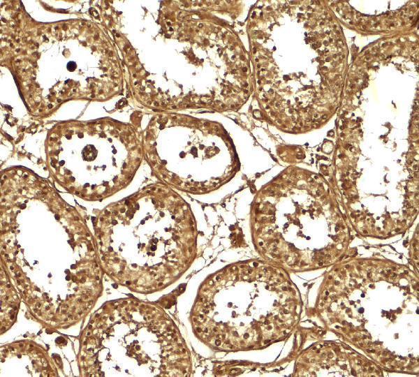
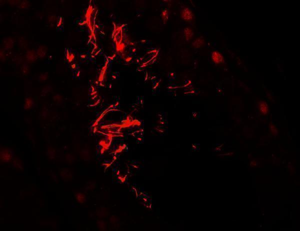
The IHC-validated antibody has real IHC and IF images from human testis tissue (catalog image captions); its listed reactivity is Human (catalog: A07362).

Real IHC data Immunohistochemistry of AKAP4 in human testis tissue with AKAP4 antibody at 5 μg/ml.
Anti-A-kinase anchor protein 4 AKAP4 Antibody
Cat # A07362

A07362 is listed for IHC-P and IF in Human samples (catalog: A07362 applications and reactivity). Its images show IHC and IF staining in human testis tissue (catalog: A07362 image captions).

Which to pick: Choose A07362 for paraffin-section IHC because IHC-P is listed and its own IHC image shows human testis tissue at 5 μg/mL (catalog: A07362 applications and IHC image caption); the fixative is unreported (catalog: A07362 IHC image caption). For IF, A07362 has a human testis tissue image at 20 μg/mL, but ICC validation is not listed (catalog: A07362 IF image caption and applications). Cross-species reactivity is unreported; the listed reactivity is Human (catalog: A07362 reactivity).

Each figure is that product's own IHC / IF validation image from its datasheet.

References

  1. UniProt Consortium. UniProt entry Q5JQC9 (AKAP4_HUMAN, A-kinase anchor protein 4).
  2. Human Protein Atlas. AKAP4 tissue IHC expression (reliability: Enhanced).
  3. Human Protein Atlas. AKAP4 subcellular location (ICC-IF): Localized to the principal piece..
  4. Human Protein Atlas. AKAP4 antibody validation summary (2 antibodies).
  5. The novel cancer-testis antigen A-kinase anchor protein 4 (AKAP4) is a potential target for immunotherapy of ovarian serous carcinoma. Oncoimmunology 2013 — PMC3667910.
  6. Evaluation of Culture Time and Media in an In Vitro Testis Organ Culture System. Birth defects research 2017 — PMC5462453.
  7. A novel cancer testis antigen, A-kinase anchor protein 4 (AKAP4) is a potential biomarker for breast cancer. PloS one 2013 — PMC3579772.
  8. Silencing of A-kinase anchor protein 4 inhibits the metastasis and growth of non-small cell lung cancer. Bioengineered 2022 — PMC8974088.
  9. PubMed PMID:9514854 — UniProt-cited evidence.
  10. PubMed PMID:9822690 — UniProt-cited evidence.
  11. PubMed PMID:15772651 — UniProt-cited evidence.