This website uses cookies to ensure you get the best experience on our website.


- Table of Contents
Plan AKR1A1 chromogenic IHC in paraffin sections using renal tubules as a high-staining reference (HPA tissue IHC). This guide covers fixation consistency, antibody titration within 1:50–1:200 (datasheet), and interpretation of cytoplasmic and nuclear staining (HPA tissue IHC).

Expected localisation, validated protocols, controls and antibodies — the at-a-glance facts below, then the full design guide.
| Expected localisation | Cytoplasmic and nuclear staining in renal tubules (HPA tissue IHC) | |
| Staining pattern | Cytoplasmic and nuclear signal across renal tubule cells (HPA tissue IHC) | |
| Antigen retrieval | Citrate pH 6.0 HIER, 95–98 °C, 20 min (rule: cytoplasmic / membrane antigen) | |
| Positive control | Kidney+4 more · see all | |
| Negative control | Caudate+4 more · see all |
| Fixation | Keep fixation consistent across paraffin sections. (standard IHC practice; not target-specific) | |
| Caveat | Staining and RNA show only medium consistency (HPA tissue IHC) | |
| Regulation | Regulation unspecified (UniProt) | |
| Isoform / epitope | No isoforms; chain 2–325; epitope location unreported (UniProt) |
Compare the catalog antibody’s IHC-P protocol (datasheet) with four published AKR1A1 paraffin-section protocols (PMC10837880; PMC5352327; PMC4834580; PMC6318002).
| Sample | Paraffin-embedded human tonsil carcinoma tissue; fixative not specified (datasheet A04855) |
| Fixation | Image fixative and duration unreported (datasheet A04855); verify before use. |
| Sectioning | 4–5 µm sections on charged slides (standard) |
| Deparaffinisation | Xylene, graded ethanol series to water (standard) |
| Antigen retrieval | Heat-induced epitope retrieval in citrate buffer, pH 6.0, 20 min at 95–98 °C (standard rule: cytoplasmic / membrane antigen) |
| Peroxidase block | 3% H2O2, 10 min, room temperature (standard) |
| Blocking | 10% normal serum of the secondary host, 30 min, room temperature (standard) |
| Primary antibody | Rabbit anti-AKR1A1, 1:50-1:200 (datasheet A04855) |
| Primary incubation | Overnight at 4 °C (standard) |
| Detection | HRP-polymer secondary, DAB chromogen 5–10 min (standard) |
| Counterstain | Hematoxylin, blue, dehydrate and mount (standard) |
| Expected result | AKR1A1-positive staining in cells in tubules of kidney (HPA tissue IHC: High). HPA tissue profile: Ubiquitous cytoplasmic and nuclear expression in renal tubules. No signal in the no-primary control. |
AKR1A1 is primarily cytosolic, with an apical cell membrane annotation and no transmembrane segment (UniProt P14550). In paraffin-section IHC, expect the clearest staining in kidney tubular cells, including cytoplasmic and nuclear staining; hepatocytes and several glandular or respiratory epithelial populations show weaker staining (HPA tissue IHC). HPA rates the tissue pattern Approved, with medium consistency between antibody staining and RNA expression (HPA tissue IHC).
| Kidney tubules show strong cellular staining, with visible cytoplasm and nuclei. | This matches the High tubular-cell signal and cytoplasmic and nuclear profile reported for kidney (HPA tissue IHC). Compare neighboring cell populations separately: the HPA observation identifies tubular cells and does not establish that every cell in the section should stain equally (HPA tissue IHC). |
| Staining is exclusively extracellular or forms a sharp membrane-only outline, without convincing intracellular signal. | That distribution needs investigation: cytosol is the primary location (UniProt P14550; HPA subcellular ICC-IF), and kidney IHC includes cytoplasm and nuclei (HPA tissue IHC). An apical membrane annotation exists, so focal apical staining alone should not be dismissed; assess the full cellular pattern and controls before calling artefact (UniProt P14550). |
| The strongest signal appears in a cell population listed as undetected, while expected positive cells are weak. | For example, HPA reports no detectable staining in heart cardiomyocytes or cervical squamous epithelial cells (HPA tissue IHC). Unexpected signal raises cross-reactivity or detection-background concerns; it does not prove either cause. Recheck cell identity, morphology and controls alongside a kidney tubular-cell section (HPA tissue IHC; general IHC practice). |
| Brown color spreads across stroma, empty spaces or most cells without readable cellular boundaries. | Diffuse signal cannot establish AKR1A1 localization. Review a no-primary control for detection background and assess blocking, washes and chromogen development (general IHC practice). The intended comparison is cellular staining in kidney tubules, rather than uniform color across the section (HPA tissue IHC). |
| Kidney tubular cells show no convincing signal. | This conflicts with the High tubular-cell staining reported by HPA, but one negative section cannot identify the failed step (HPA tissue IHC). Check tissue preservation, antibody preparation, retrieval settings and detection controls using the IHC-validated antibody's instructions (general IHC practice). Target-specific fixation sensitivity is unreported in the supplied sources. |
| Tissue and cell selection | Kidney tubular cells provide the strongest listed IHC reference (High); hepatocytes, bronchial respiratory epithelium and lung macrophages are Medium (HPA tissue IHC). HPA lists several undetected cell populations, so score the specified cells rather than declaring an entire organ positive or negative (HPA tissue IHC). |
| Compartment assignment | UniProt places AKR1A1 in the cytosol and at the apical cell membrane, with no transmembrane segment (UniProt P14550). HPA kidney IHC describes cytoplasmic and nuclear expression (HPA tissue IHC). These observations support checking intracellular signal while allowing nuclear and focal apical signal in the appropriate context. |
| Strength of tissue evidence | The tissue profile is Approved but has medium agreement between staining and RNA expression (HPA tissue IHC). Four listed antibodies have Approved IHC status (HPA antibodies). Treat this as support for the reported pattern, while retaining morphology and controls when interpreting an unexpected distribution (general IHC practice). |
| IF/ICC Q&A: Where should cellular fluorescence appear? | Mainly in cytosol, with additional nucleoplasmic signal (HPA subcellular ICC-IF). HPA lists A-431, U-251MG and U2OS ICC-IF images (HPA subcellular ICC-IF). This is localization context for the separate IF/ICC guide; the paraffin-section tissue pattern here comes from HPA tissue IHC. |
| Retrieval and detection checks | Use the IHC-validated antibody's documented paraffin-section workflow as the starting point (general IHC practice). Retrieval, blocking and detection settings can be checked against controls when a run fails (general IHC practice). The supplied UniProt and HPA records do not identify an AKR1A1-specific retrieval requirement or fixation effect. |
| Situation | Likely cause | Next action |
|---|---|---|
| Kidney tubular cells are negative across the section. | A failed staining run is possible; HPA reports High tubular-cell staining (HPA tissue IHC). | Check the IHC-validated antibody's instructions, reagent preparation, retrieval and detection controls (general IHC practice). Do not infer AKR1A1-specific fixation sensitivity from this result. |
| Nuclei stain along with tubular cytoplasm. | This can fit the reported kidney pattern, which includes nuclear expression (HPA tissue IHC). | Assess whether staining is confined to plausible cells and whether background controls are clean before treating nuclear signal as nonspecific (HPA tissue IHC; general IHC practice). |
| Color appears in no-primary controls or across acellular areas. | Detection background can produce color without target recognition (general IHC practice). | Review endogenous enzyme blocking where relevant to the chromogen, secondary detection, washes and development time (general IHC practice). Reassess AKR1A1 only after cellular boundaries become interpretable. |
| Only a continuous cell-surface rim is visible. | A membrane-only pattern does not reflect the mainly cytosolic localization, although UniProt also notes an apical membrane location (UniProt P14550; HPA subcellular ICC-IF). | Inspect the apical orientation and intracellular staining, then compare with kidney tubules and controls before assigning a compartment (HPA tissue IHC; general IHC practice). |
| A listed undetected cell type stains strongly. | The result conflicts with that specific HPA observation and could reflect cross-reactivity or detection background; its cause remains unproven (HPA tissue IHC; general IHC practice). | Confirm the cell type, examine a no-primary control and compare an expected positive population from the same run (HPA tissue IHC; general IHC practice). |
| A medium-level tissue looks faint beside kidney. | That difference may match HPA levels: kidney tubular cells are High, while hepatocytes and bronchial respiratory epithelial cells are Medium (HPA tissue IHC). | Score the named cell populations and compare sections processed together before changing staining conditions (HPA tissue IHC; general IHC practice). |
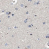
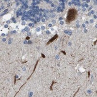
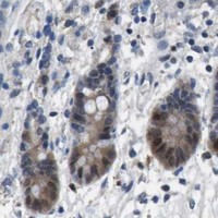


Comprehensive Human Protein Atlas IHC scoring per tissue (reliability: Approved — Medium consistency between antibody staining and RNA expression data.). Rows are taken directly from the HPA tissue chart — click any row's HPA link to view the source.
| Tissue | Cell type | Level | Evidence | Source |
|---|---|---|---|---|
| Kidney | Cells in tubules | High | Protein (IHC) | HPA → |
| Adrenal gland | Glandular cells | Medium | Protein (IHC) | HPA → |
| Bronchus | Respiratory epithelial cells | Medium | Protein (IHC) | HPA → |
| Duodenum | Glandular cells | Medium | Protein (IHC) | HPA → |
| Epididymis | Glandular cells | Medium | Protein (IHC) | HPA → |
| Tissue | Cell type | Level | Evidence | Source |
|---|---|---|---|---|
| Caudate | Glial cells | Not detected | Protein (IHC) | HPA → |
| Cerebellum | Cells in granular layer | Not detected | Protein (IHC) | HPA → |
| Cerebral cortex | Endothelial cells | Not detected | Protein (IHC) | HPA → |
| Cervix | Squamous epithelial cells | Not detected | Protein (IHC) | HPA → |
| Esophagus | Squamous epithelial cells | Not detected | Protein (IHC) | HPA → |
Troubleshoot AKR1A1 staining in paraffin sections by checking retrieval, controls, cellular localisation and scoring before drawing biological conclusions.
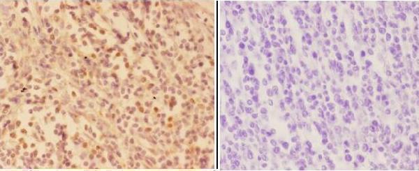
One AKR1A1 antibody has real IHC data from a human paraffin section (A04855 image caption); its listed reactivity also includes mouse and rat (catalog: A04855).
A04855 will render with an IHC image of a human paraffin section at 1:50, showing cytoplasmic and nuclear staining (A04855 image caption). Its listed applications are IHC and WB, and its listed reactivity is human, mouse and rat (catalog: A04855).
Which to pick: Choose A04855 for tissue IHC: it is a rabbit antibody with a listed IHC dilution of 1:50–1:200 and a paraffin-section image (catalog: A04855; A04855 image caption). For mouse or rat IHC, A04855 has listed species reactivity, but the supplied image shows only human tissue (catalog: A04855; A04855 image caption). No IF/ICC application or image is supplied for A04855, and the paraffin-section caption does not report the fixative (catalog: A04855; A04855 image caption).