This website uses cookies to ensure you get the best experience on our website.


- Table of Contents
Plan chromogenic ALDH7A1 IHC on paraffin sections with the catalog antibody at 0.5–1 μg/mL (datasheet PB10038). Assess cytoplasmic, mitochondrial-pattern staining using kidney proximal tubules or liver cholangiocytes as positive controls (HPA tissue IHC).

Expected localisation, validated protocols, controls and antibodies — the at-a-glance facts below, then the full design guide.
| Expected localisation | Cytoplasmic with a mitochondrial pattern (HPA tissue IHC) | |
| Staining pattern | Cytoplasmic, mitochondrial-pattern staining in most tissues (HPA tissue IHC) | |
| Antigen retrieval | Citrate pH 6 HIER, heat-mediated (datasheet PB10038) | |
| Positive control | Cerebellum+4 more · see all | |
| Negative control | Adipose tissue+4 more · see all |
| Fixation | Keep fixation conditions consistent across sections. (standard IHC practice; not target-specific) | |
| Caveat | Antibody staining and RNA have medium consistency (HPA tissue IHC) | |
| Regulation | Tissue-enhanced RNA in liver (HPA tissue RNA) | |
| Isoform / epitope | 3 isoforms; chain begins at residue 27; check epitope coverage (UniProt) |
The catalog antibody’s IHC-P protocol (datasheet PB10038) is paired with four published ALDH7A1 tissue protocols (PMC3458927; PMC6264057; PMC7847681; PMC5341386).
| Sample | Paraffin-embedded human lung cancer tissues; fixative not specified (datasheet PB10038) |
| Fixation | Image fixative and duration unreported (datasheet PB10038); verify before use. |
| Sectioning | 4–5 µm sections on charged slides (standard) |
| Deparaffinisation | Xylene, graded ethanol series to water (standard) |
| Antigen retrieval | Heat retrieval: Citrate pH 6, 20 min (datasheet PB10038) |
| Peroxidase block | 3% H2O2, 10 min, room temperature (standard) |
| Blocking | 10% goat serum (datasheet PB10038) |
| Primary antibody | Rabbit anti-ALDH7A1, 0.5-1μg/ml (datasheet PB10038) |
| Primary incubation | Overnight at 4 °C (datasheet PB10038) |
| Detection | Streptavidin-biotin complex (SABC), DAB chromogen (datasheet PB10038) |
| Counterstain | Hematoxylin, blue, dehydrate and mount (standard) |
| Expected result | ALDH7A1-positive staining in bergmann glia - cytoplasm/membrane of cerebellum (HPA tissue IHC: High). HPA tissue profile: Cytoplasmic expression with a mitochondrial pattern in most tissues. No signal in the no-primary control. |
ALDH7A1 is mainly mitochondrial, with additional cytosolic localization (HPA: supported ICC-IF locations); UniProt also lists the nucleus and cell and Golgi membranes, with no transmembrane segment (UniProt P49419). In tissue IHC, expect cytoplasmic staining with a mitochondrial pattern, especially in kidney proximal tubules, liver cholangiocytes, placenta cytotrophoblasts, and cerebellar Bergmann glia (HPA: High). HPA rates tissue staining Enhanced, with medium consistency against RNA expression (HPA: tissue IHC).
| Granular cytoplasmic staining in kidney proximal tubules or liver cholangiocytes, with little signal in adjacent cells. | This fits the reported mitochondrial-pattern cytoplasm and High staining in those cell types (HPA: tissue IHC). Compare cell types within the section rather than treating all tissue as uniformly positive. |
| Predominantly nuclear staining, with little cytoplasmic signal. | This does not match the principal tissue IHC pattern (HPA: tissue IHC). Nuclear localization is listed by UniProt, so compartment alone cannot prove artefact; check whether known-positive cells also show the expected cytoplasmic pattern (UniProt P49419; HPA: tissue IHC). |
| Strong staining in adipocytes, lung alveolar cells, or ovarian follicle cells. | These cell types are Not detected in HPA tissue IHC (HPA: adipose tissue, lung, ovary). Consider nonspecific antibody binding or endogenous detection activity, especially if the expected positive cell types are absent (general IHC practice). |
| Diffuse color across tissue and blank areas, obscuring cell boundaries. | This is difficult to assign to the reported cell-specific cytoplasmic pattern (HPA: tissue IHC). Background can arise from nonspecific binding or detection chemistry; a negative reagent control helps separate it from cellular staining (general IHC practice). |
| No signal in kidney proximal tubules or liver cholangiocytes. | Both are reported High by HPA (HPA: kidney, liver tissue IHC). First assess tissue preservation and the run’s positive control, then review antibody and detection steps; a blank slide alone does not establish absent ALDH7A1 (general IHC practice). |
| Compartment and topology (UniProt P49419; HPA: subcellular ICC-IF) | Mitochondria are the supported main location, with cytosol also supported (HPA: subcellular ICC-IF). No transmembrane segment is annotated, so membrane-like staining alone should not replace the expected cytoplasmic assessment (UniProt P49419; HPA: tissue IHC). |
| Cell-type variation (HPA: tissue IHC) | HPA reports High staining in selected cells, Medium in others, and undetected staining in several cell types (HPA: tissue IHC). Score the named cells and compartment; whole-section intensity can hide this variation (general IHC practice). |
| Stress-linked localization (UniProt P49419) | UniProt reports translocation to membranes with cellular stress and AMPK phosphorylation (UniProt P49419). This may inform an unusual localization, but the supplied tissue IHC record does not show how often that pattern occurs in sections (HPA: tissue IHC). |
| Antibody evidence (HPA: antibody validation; tissue IHC) | Two listed rabbit polyclonal antibodies, HPA023296 and HPA053675, have Enhanced IHC validation (HPA: antibodies). HPA describes medium staining-to-RNA consistency, so validation supports the pattern without making every observed stain specific (HPA: tissue IHC). |
| IF/ICC Q&A: What localization should an IF image show? (HPA: subcellular ICC-IF) | Mainly mitochondria, with additional cytosol (HPA: supported ICC-IF locations). This is an IF/ICC interpretation cue; judge paraffin chromogenic IHC against the tissue cell-type pattern instead (HPA: tissue IHC). |
| Situation | Likely cause | Next action |
|---|---|---|
| Known-positive kidney or liver cells are blank (HPA: kidney, liver tissue IHC). | The run may have failed, or target detection may be inadequate; the slide does not distinguish these causes (general IHC practice). | Check the run’s positive control and section quality, then review primary antibody, retrieval, and detection steps against the established IHC procedure (general IHC practice). ALDH7A1-specific retrieval sensitivity is unreported here. |
| Color covers negative reagent controls as well as tissue. | Detection-system background or nonspecific deposition is possible (general IHC practice). | Review blocking, washes, and chromogen development with an appropriate negative control (general IHC practice); do not score diffuse color as the HPA cytoplasmic pattern (HPA: tissue IHC). |
| Strong nuclear signal dominates the section. | The result differs from HPA’s principal cytoplasmic mitochondrial-pattern IHC, although UniProt lists a nuclear location (HPA: tissue IHC; UniProt P49419). | Inspect whether HPA-positive cell types also retain cytoplasmic staining, and compare a negative reagent control before assigning the nuclear signal to ALDH7A1 (HPA: tissue IHC; general IHC practice). |
| Undetected reference cells stain as strongly as expected positive cells. | Nonspecific binding or endogenous detection activity may contribute (general IHC practice); HPA lists adipocytes and lung alveolar cells as Not detected (HPA: tissue IHC). | Check cell identity, compare controls, and inspect staining with the primary antibody omitted (general IHC practice). Reassess whether the expected positive cells retain the reported compartment pattern (HPA: tissue IHC). |
| Only faint, uniform cytoplasmic color is visible. | The expected cell-to-cell contrast may be obscured by background or weak detection (HPA: tissue IHC; general IHC practice). | Compare proximal tubules or cholangiocytes with nearby cells and the run controls (HPA: kidney, liver tissue IHC; general IHC practice). Adjust staining conditions only within a validated IHC workflow (general IHC practice). |
| A distinct membrane-like pattern appears without the usual cytoplasmic pattern. | UniProt reports stress-associated membrane translocation, but the tissue IHC profile emphasizes cytoplasmic staining (UniProt P49419; HPA: tissue IHC). | Record the compartment and cell type, review controls, and interpret cautiously; the supplied evidence does not establish that membrane-only chromogenic staining is a routine positive result (HPA: tissue IHC; UniProt P49419). |


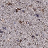

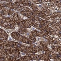
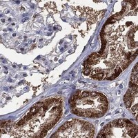


Comprehensive Human Protein Atlas IHC scoring per tissue (reliability: Enhanced — Medium consistency between antibody staining and RNA expression data.). Rows are taken directly from the HPA tissue chart — click any row's HPA link to view the source.
| Tissue | Cell type | Level | Evidence | Source |
|---|---|---|---|---|
| Cerebellum | Bergmann glia - cytoplasm/membrane | High | Protein (IHC) | HPA → |
| Kidney | Proximal tubules (cell body) | High | Protein (IHC) | HPA → |
| Liver | Cholangiocytes | High | Protein (IHC) | HPA → |
| Placenta | Cytotrophoblasts | High | Protein (IHC) | HPA → |
| Adrenal gland | Glandular cells | Medium | Protein (IHC) | HPA → |
| Tissue | Cell type | Level | Evidence | Source |
|---|---|---|---|---|
| Adipose tissue | Adipocytes | Not detected | Protein (IHC) | HPA → |
| Bone marrow | Hematopoietic cells | Not detected | Protein (IHC) | HPA → |
| Cervix | Squamous epithelial cells | Not detected | Protein (IHC) | HPA → |
| Duodenum | Endocrine cells | Not detected | Protein (IHC) | HPA → |
| Lung | Alveolar cells | Not detected | Protein (IHC) | HPA → |
Troubleshoot ALDH7A1 staining in paraffin sections by checking retrieval, cell identity, subcellular pattern and detection controls together.
PB10038 has IHC images from human lung cancer and rat brain paraffin sections, plus an IF/ICC image from U20S cells (PB10038 image captions).
The rendered SKU, PB10038, has IHC images from human lung cancer and rat brain paraffin sections (PB10038 IHC captions). Its IF/ICC image shows U20S cells, and its stated reactivity covers human, mouse and rat (PB10038 IF caption; PB10038 catalog data).
Which to pick: Choose PB10038 for paraffin-section tissue IHC: its captions document citrate retrieval at pH 6 for 20 minutes and 1 μg/ml primary antibody in human lung cancer and rat brain sections (PB10038 IHC captions). For IF/ICC, PB10038 has an image from U20S cells using 2 μg/ml primary antibody (PB10038 IF caption). For cross-species work, PB10038 lists human, mouse and rat reactivity, while its IHC-P details cover human and rat; the IHC captions do not report a fixative (PB10038 catalog data; PB10038 IHC captions).