ALDOA / Fructose-bisphosphate aldolase A · IHC design guide

Design Immunohistochemistry for ALDOA

This guide covers ALDOA staining in paraffin sections, where variable cytoplasmic staining is expected (HPA tissue IHC). Skeletal myocytes provide a high staining reference, but intensity varies across cell types (HPA tissue IHC).

Evidence assembled Oct 2026 · For research use; verify linked source records and product datasheet before use
Immunohistochemistry protocol sheet for ALDOA (IHC for ALDOA): expected localisation Cytoplasm in most cell types (HPA tissue IHC), antibody A05022-3, validated IHC image, and IHC protocol steps
Printable ALDOA IHC protocol sheet — expected localisation Cytoplasm in most cell types (HPA tissue IHC), antibody A05022-3, controls and protocol steps. Open the full ALDOA IHC guide →

ALDOA Immunohistochemistry Experimental Design Guide

Expected localisation, validated protocols, controls and antibodies — the at-a-glance facts below, then the full design guide.

Must know before staining
Expected localisation Cytoplasm in most cell types (HPA tissue IHC)
Staining pattern Variable cytoplasmic staining, including skeletal myocytes (HPA tissue IHC)
Antigen retrieval Citrate pH 6 HIER, heat-mediated (datasheet A05022-3)
Positive control ⓘ Cerebellum+4 more · see all
Negative control ⓘ Caudate+1 more · see all
Important caveats
Reasons your staining may differ from the expected pattern.
Fixation Keep fixation consistent across paraffin sections (standard IHC practice; not target-specific)
Caveat Antibody staining and RNA show medium consistency (HPA tissue IHC)
Regulation No expression regulator annotated (UniProt)
Isoform / epitope 2 isoforms; epitope differences unreported (UniProt)
Section 1

Recommended ALDOA IHC & IF Protocols

The catalog antibody protocol is paired with three published ALDOA IHC protocols for cervical, lung and renal tumors (PMC7419050; PMC12619220; PMC5338975).

Recommended immunohistochemistry (IHC-P) protocol parameters
SampleParaffin-embedded human Lung cancer tissues; fixative not specified (datasheet A05022-3)
FixationImage fixative and duration unreported (datasheet A05022-3); verify before use.
Sectioning4–5 µm sections on charged slides (standard)
DeparaffinisationXylene, graded ethanol series to water (standard)
Antigen retrievalHeat retrieval: Citrate pH 6, 20 min (datasheet A05022-3)
Peroxidase block3% H2O2, 10 min, room temperature (standard)
Blocking10% goat serum (datasheet A05022-3)
Primary antibodyRabbit anti-ALDOA, 0.5-1μg/ml (datasheet A05022-3)
Primary incubationOvernight at 4 °C (datasheet A05022-3)
DetectionStreptavidin-biotin complex (SABC), DAB chromogen (datasheet A05022-3)
CounterstainHematoxylin, blue, dehydrate and mount (standard)
Expected resultALDOA-positive staining in purkinje cells - cytoplasm/membrane of cerebellum (HPA tissue IHC: High). HPA tissue profile: Cytoplasmic expression at variable levels in most cell types, including skeletal myocytes. No signal in the no-primary control.
💡Decision noteStart with the catalog antibody’s heat-mediated citrate pH 6 retrieval (datasheet A05022-3); consider EDTA pH 9 for the published lung protocol (PMC12619220).
Section 2

What Is the Expected ALDOA Staining Pattern?

ALDOA is primarily cytosolic, with an additional sarcomeric location in skeletal muscle; it has no transmembrane segment (UniProt P04075). In paraffin-section IHC, expect variable cytoplasmic staining across many cell types, including skeletal myocytes (HPA: tissue IHC profile). HPA rates the tissue pattern Supported, with medium consistency between antibody staining and RNA expression; interpret individual cells and compartments in that context (HPA: tissue IHC reliability).

What am I looking at on my slide?
Skeletal myocytes show strong cytoplasmic staining; cerebellar Purkinje cells show cytoplasmic or membrane-associated staining.These are reported High signals (HPA: skeletal muscle myocytes; HPA: cerebellar Purkinje cells). Assess staining within the identified cells, because ALDOA expression varies across cell types (HPA: tissue IHC profile).
The dominant signal is nuclear or forms a sharp cell-surface rim without convincing cytoplasmic staining.A dominant nuclear or surface-only pattern conflicts with the expected cytosolic location and lack of a transmembrane segment (HPA: cytosol approved; UniProt P04075 topology). Review morphology and controls before calling it ALDOA; compartmental mislocalisation may reflect an artefact (general IHC practice).
Strong staining appears in a cell population reported as undetected, such as caudate glial cells or cardiomyocytes.HPA reports ALDOA as Not detected in those populations (HPA: caudate glial cells; HPA: heart muscle cardiomyocytes). Confirm cell identity and investigate cross-reactivity or endogenous chromogen activity; one discordant section does not establish a new expression pattern (general IHC practice).
Chromogen is spread across stroma, lumens, or the entire section, obscuring cell boundaries.This is difficult to score as the cellular cytoplasmic pattern reported for ALDOA (HPA: tissue IHC profile). Uneven background can arise from detection or blocking conditions; compare a no-primary control and inspect the counterstain (general IHC practice).
No convincing staining appears in a skeletal muscle positive-control section.Skeletal myocytes are reported as High (HPA: skeletal muscle myocytes). First verify tissue preservation, morphology, and the detection run; a blank known-positive control cannot support a negative call in the test section (general IHC practice).
💡Expected ALDOA appearanceCall a positive result when identifiable skeletal myocytes show strong cytoplasmic staining (HPA: skeletal muscle myocytes, High); isolated nuclear staining or diffuse acellular chromogen is suspect (HPA: cytosol approved; general IHC practice).
How each factor affects the staining
Tissue and cell selectionUse skeletal myocytes as a strong reference; Purkinje cells are also reported High, while several glandular and other cell populations are Medium or Low (HPA: tissue IHC). A weak cell is therefore not automatically a failed stain.
Compartment and topologyALDOA is cytosolic and has no transmembrane segment (HPA: cytosol approved; UniProt P04075 topology). UniProt also places it at the I band and M line in skeletal muscle; routine chromogenic IHC may be judged at the cellular level.
Antibody evidenceHPA lists IHC as Supported for HPA004177 and CAB006252; its overall tissue-pattern reliability is Supported with medium staining–RNA consistency (HPA: antibody validation; HPA: tissue IHC reliability). Treat unexpected cells as findings to verify.
IF/ICC expected pattern?For the separate IF/ICC application, HPA's approved main location is cytosol, with images from A-431, U-251MG, U2OS, RPTEC/TERT1, and serum-starved hTERT-RPE1 (HPA: subcellular ICC-IF). This does not define an IHC protocol.
Why is my staining missing, weak or wrong?
SituationLikely causeNext action
The skeletal muscle control is blank or much weaker than expected.The reported reference is High in myocytes (HPA: skeletal muscle myocytes); a blank control may reflect a failed staining or detection run (general IHC practice).Check section integrity, reagent sequence, antibody dilution, retrieval conditions, and chromogen development against the validated IHC procedure; repeat alongside a known-positive section (general IHC practice).
A test section is negative while its positive control stains correctly.ALDOA levels vary by cell type, and HPA reports Low or Not detected signals in some populations (HPA: tissue IHC profile).Confirm the intended cell population and compare its reported HPA level before scoring. Record the result as below detection in that specimen if morphology and controls remain satisfactory (general IHC practice).
Most nuclei stain more strongly than the surrounding cytoplasm.Dominant nuclear signal disagrees with the approved cytosolic localisation (HPA: subcellular ICC-IF) and reported cytoplasmic tissue pattern (HPA: tissue IHC profile).Compare a no-primary control, inspect counterstain and precipitate, and reassess the staining run before treating the nuclear signal as specific (general IHC practice).
Strong signal appears in cardiomyocytes or caudate glial cells.Those cell populations are reported Not detected (HPA: heart muscle cardiomyocytes; HPA: caudate glial cells); cross-reactivity or endogenous detection activity is possible (general IHC practice).Verify cell identity, run a no-primary control, and check endogenous enzyme blocking where relevant to the chromogen system (general IHC practice). Confirm discordant staining before reporting expression.
Diffuse brown background prevents cytoplasmic scoring.Section-wide deposit is inconsistent with a cell-localised cytoplasmic readout (HPA: tissue IHC profile); nonspecific detection or excessive development may obscure cells (general IHC practice).Inspect the no-primary control, blocking and washes, and chromogen development. Adjust the general IHC workflow only after confirming that the known-positive tissue remains readable (general IHC practice).
A low-staining tissue looks different from a strong reference section.HPA reports variable cytoplasmic levels; salivary glandular cells and cholangiocytes are Low, while skeletal myocytes are High (HPA: tissue IHC).Score the named cell population and compartment separately from overall tissue darkness. Compare sections stained in the same run, and avoid using the strong control's intensity as a universal cutoff (general IHC practice).

Sample controls for ALDOA IHC & IF

🧪Run skeletal muscle first: myocytes should stain (HPA: High in skeletal-muscle myocytes); use heart muscle cardiomyocytes as the tissue negative (HPA: Not detected in cardiomyocytes). Within the skeletal-muscle section, assess nonmyocyte cells that lack staining as internal negatives: they should show counterstain without specific chromogen, but their ALDOA-negative status needs independent validation because the supplied HPA row characterizes myocytes only (HPA: skeletal-muscle myocytes, High).
Positive control tissue: Cerebellum (Purkinje cells - cytoplasm/membrane, HPA High)
Negative control tissue: Caudate (HPA Not detected)
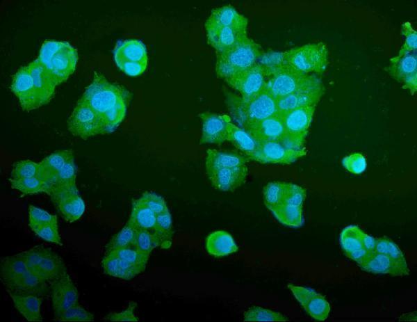
ICC-IF cell lines (HPA subcellular resource): HPA ICC-IF images show ALDOA in A-431, U-251MG, U2OS, RPTEC/TERT1, hTERT-RPE1 (serum starved), with annotated localisation: Cytosol (approved) (HPA subcellular).
Technical controls: Include a no-primary, secondary-only control and a concentration-matched rabbit IgG isotype control for the rabbit primary (selected-SKU caption: rabbit anti-ALDOA); use ALDOA-knockout material as a biological negative (standard IHC practice). For the skeletal-muscle positive, block endogenous peroxidase and check endogenous biotin background when using the caption’s biotin-based DAB detection (selected-SKU caption: biotinylated secondary, SABC and DAB).
⚠️Feasibility: A target-specific fixation window or fixation effect is unreported, and the selected-SKU paraffin-section caption does not state the fixative (selected-SKU caption: fixative unreported). The reported IHC conditions use citrate retrieval at pH 6 for 20 minutes, but the evidence does not establish whether ALDOA staining depends on retrieval (selected-SKU caption: heat-mediated citrate retrieval). Frozen-section performance and whether IF is easier are unreported; evaluate nonspecific chromogen in skeletal-muscle tissue with the negative controls (HPA: High in skeletal-muscle myocytes; standard IHC practice).

HPA tissue IHC evidence for ALDOA

Comprehensive Human Protein Atlas IHC scoring per tissue (reliability: Supported — Medium consistency between antibody staining and RNA expression data.). Rows are taken directly from the HPA tissue chart — click any row's HPA link to view the source.

Positive expression · recommended positive controls

TissueCell typeLevelEvidenceSource
Cerebellum Purkinje cells - cytoplasm/membrane High Protein (IHC) HPA →
Skeletal muscle Myocytes High Protein (IHC) HPA →
Adipose tissue Adipocytes Medium Protein (IHC) HPA →
Adrenal gland Glandular cells Medium Protein (IHC) HPA →
Appendix Glandular cells Medium Protein (IHC) HPA →

Undetected expression · recommended negative controls

TissueCell typeLevelEvidenceSource
Caudate Glial cells Not detected Protein (IHC) HPA →
Heart muscle Cardiomyocytes Not detected Protein (IHC) HPA →
Section 3

Advanced ALDOA IHC Tips

Troubleshoot ALDOA staining in paraffin section IHC using the catalog antibody’s reported conditions, expected cytoplasmic localisation, and cell type specific controls.

How should I adjust antigen retrieval when ALDOA staining is weak?
Start with heat mediated antigen retrieval in citrate buffer at pH 6 for 20 minutes (datasheet A05022-3). Keep section thickness, heating, cooling, and detection conditions consistent while comparing retrieval runs (standard IHC practice). The reported paraffin section workflow then used 1 µg/mL primary antibody overnight at 4°C, so confirm that baseline before changing retrieval (datasheet A05022-3). If staining remains weak, compare a longer citrate retrieval on adjacent sections while watching for tissue damage (standard IHC practice). Include a skeletal muscle section as a positive reference, because myocytes show high ALDOA staining (HPA: high in skeletal myocytes).
Could fixation explain weak or patchy ALDOA staining in paraffin sections?
The selected paraffin section caption does not state a fixative, and target specific fixation sensitivity is unknown (datasheet A05022-3). Record the actual fixative, fixation duration, processing history, and section age for each sample before comparing staining intensity (standard IHC practice). Test matched sections with the same citrate pH 6 retrieval for 20 minutes and the same detection conditions to isolate a fixation related difference (datasheet A05022-3; standard IHC practice). Check morphology and internal staining patterns before increasing primary concentration, since damaged or unevenly processed areas can give misleading chromogenic signal (standard IHC practice). Do not infer ALDOA fixation sensitivity from its cytoplasmic location or phosphorylation sites (UniProt P04075: localisation and modified residues).
Where should convincing ALDOA staining appear within cells and muscle fibres?
Expect predominantly cytoplasmic staining in paraffin sections, consistent with the tissue IHC profile and the approved cytosolic location (HPA: tissue profile; HPA: subcellular localisation). In skeletal muscle, ALDOA is reported at the sarcomeric M line and I band, so a structured fibre pattern can be biologically plausible (UniProt P04075: subcellular localisation). High staining in skeletal myocytes offers a useful reference, while glial cells in caudate and cardiomyocytes were reported as not detected in the cited tissue survey (HPA: tissue IHC). Review any apparent membrane accentuation against cell boundaries and morphology: ALDOA has no annotated transmembrane segment (UniProt P04075: topology). Score the compartment actually stained rather than treating every dark region as equivalent (standard IHC practice).
Could ALDOA isoforms or modified residues alter antibody staining?
ALDOA has 2 annotated isoforms and multiple modified residues, including phosphorylation and lysine acetylation sites (UniProt P04075: isoforms and modified residues). The supplied caption does not identify the antibody epitope, so its isoform coverage and sensitivity to those modifications cannot be established from this evidence (datasheet A05022-3). Check the immunogen or mapped epitope against the isoform sequences before interpreting a missing compartment or cell population as isoform specific (standard antibody validation practice). Compare adjacent sections processed together and, where available, use an independent antibody with a documented distinct epitope (standard IHC practice). Keep retrieval at citrate pH 6 as the initial condition during that comparison (datasheet A05022-3).
How can I assess ALDOA by IF alongside a cell type marker?
For a separate IF/ICC experiment, pair ALDOA with a marker identifying the expected cell type, such as a skeletal myocyte marker when examining muscle (HPA: high in skeletal myocytes; standard IF practice). Choose spectrally separated fluorophores and place the weaker signal in a channel with low tissue autofluorescence, checking single stain and unstained controls (standard IF practice). Because ALDOA is cytosolic and has no transmembrane segment, use a compatible permeabilisation step when the antibody must reach an intracellular epitope (HPA: approved cytosolic location; UniProt P04075: topology; standard IF practice). Optimise fixation and permeabilisation directly for IF/ICC; the paraffin section IHC caption does not report fixation or establish an IF/ICC protocol (datasheet A05022-3).
What should I check when ALDOA DAB staining is diffuse or excessive?
First compare primary omitted and secondary only controls with the stained section to locate nonspecific detection signal (standard IHC practice). The reported paraffin section method used 10% goat serum blocking, biotinylated secondary antibody, a streptavidin biotin complex, and DAB (datasheet A05022-3). Check endogenous biotin and include a peroxidase blocking step as appropriate for that detection system; these are general chromogenic IHC controls, not ALDOA specific findings (standard IHC practice). Then titrate the primary below the reported 1 µg/mL condition and shorten chromogen development if specific staining is obscured (datasheet A05022-3; standard IHC practice). Widespread cytoplasmic signal alone is insufficient evidence of background because expression varies across many cell types (HPA: tissue profile).
How should I quantify ALDOA staining across paraffin sections? ⚠ ANSWER MARKED FOR VERIFICATION
Define the cell population and cytoplasmic compartment before scoring, because ALDOA staining varies among cell types (HPA: tissue profile; HPA: approved cytosolic location). For chromogenic IHC, report an H score from 0–300, combining the percentage of viable target cells at each intensity, or report percentage positive cells with a prespecified threshold (standard IHC practice). If spatial abundance matters, count positive cells per mm² of viable tissue and normalise to the area or number of eligible target cells (standard IHC practice). Apply identical retrieval, imaging, colour separation, and scoring rules across sections; the catalog condition uses citrate pH 6 for 20 minutes (datasheet A05022-3; standard IHC practice). Record compartment and cell type alongside every numerical score (standard IHC practice).
How can I distinguish true ALDOA staining from section artefacts?
Prioritise staining in intact cytoplasm and plausible cell populations: ALDOA is cytosolic, and skeletal myocytes show high tissue staining (HPA: subcellular localisation; HPA: tissue IHC). Treat isolated nuclear signal or apparent surface only staining cautiously, since no transmembrane segment is annotated for ALDOA (UniProt P04075: topology). Exclude section edges, folds, necrotic areas, and precipitate from scoring, and compare those regions with primary omitted controls (standard IHC practice). Check residual endogenous peroxidase or biotin signal when using the reported biotin based DAB workflow (datasheet A05022-3; standard IHC practice). Finally, interpret a negative field against cell type matched references rather than assuming uniform ALDOA expression across tissue (HPA: variable tissue profile).
Boster reagents

Best ALDOA / Fructose-bisphosphate aldolase A IHC Antibodies

Two catalog antibodies have IHC images from human paraffin sections and IF images from human cells; A05022-3 also lists mouse and rat reactivity (catalog applications/reactivity; catalog image captions).

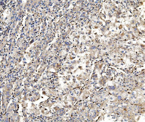
Real IHC data IHC analysis of ALDOA using anti-ALDOA antibody (A05022-3). ALDOA was detected in paraffin-embedded section of human Lung cancer tissues. Heat mediated antigen retrieval was performed in citrate buffer (pH6, epitope retrieval solution) for 20 mins. The tissue section was blocked with 10% goat serum. The tissue section was then incubated with 1μg/ml rabbit anti-ALDOA Antibody (A05022-3) overnight at 4°C. Biotinylated goat anti-rabbit IgG was used as secondary antibody and incubated for 30 minutes at 37°C. The tissue section was developed using Strepavidin-Biotin-Complex (SABC)(Catalog # SA1022) with DAB as the chromogen.
Anti-Aldolase/ALDOA Antibody ®
Cat # A05022-3
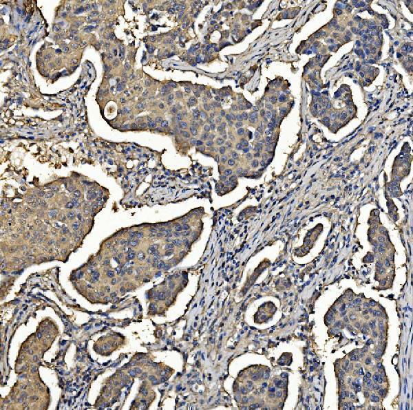
Real IHC data IHC analysis of Aldolase/ALDOA using anti-Aldolase/ALDOA antibody (M05022-2). Aldolase/ALDOA was detected in paraffin-embedded section of human liver cancer tissue. Heat mediated antigen retrieval was performed in EDTA buffer (pH8.0, epitope retrieval solution). The tissue section was blocked with 10% goat serum. The tissue section was then incubated with 2μg/ml mouse anti-Aldolase/ALDOA Antibody (M05022-2) overnight at 4°C. Biotinylated goat anti-mouse IgG was used as secondary antibody and incubated for 30 minutes at 37°C. The tissue section was developed using Strepavidin-Biotin-Complex (SABC) (Catalog # SA1021) with DAB as the chromogen.
Anti-Aldolase/ALDOA Antibody ® (monoclonal, 6H8)
Cat # M05022-2

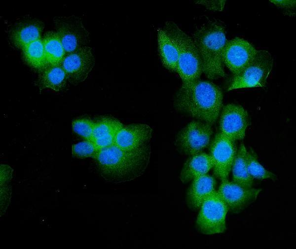
A05022-3 will render with IHC data from human lung cancer paraffin sections and has IF data from A431 cells (A05022-3 image captions). M05022-2 will render with IHC data from human liver cancer paraffin sections and has IF data from HEPG2 cells (M05022-2 image captions).

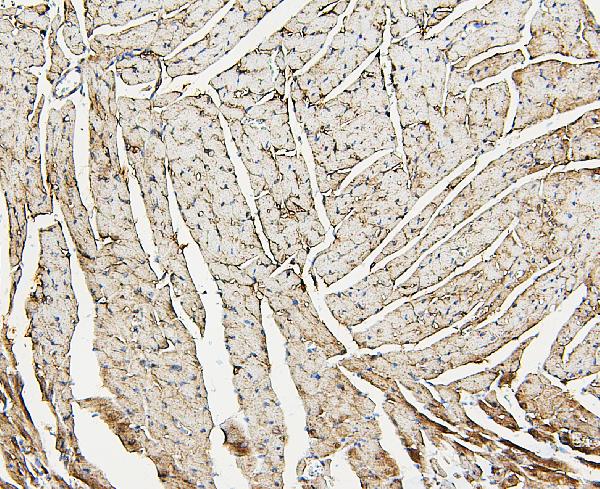
Which to pick: For human tissue IHC, choose A05022-3 for the illustrated citrate pH 6 retrieval or M05022-2 for the illustrated EDTA pH 8 retrieval; both captions describe paraffin sections, and neither reports the fixative (A05022-3 and M05022-2 IHC image captions). For IF/ICC, both have cell images: A05022-3 is rabbit derived and illustrated in A431 cells, while M05022-2 is mouse monoclonal clone 6H8 and illustrated in HEPG2 cells (catalog host/clone; IF image captions). For mouse or rat samples, choose A05022-3 based on its listed reactivity and mouse heart IHC image; M05022-2 lists human reactivity only (catalog reactivity; A05022-3 IHC image caption).

Each figure is that product's own IHC / IF validation image from its datasheet.

References

  1. UniProt Consortium. UniProt entry P04075 (ALDOA_HUMAN, Fructose-bisphosphate aldolase A).
  2. Human Protein Atlas. ALDOA tissue IHC expression (reliability: Supported).
  3. Human Protein Atlas. ALDOA subcellular location (ICC-IF): Localized to the cytosol..
  4. Human Protein Atlas. ALDOA antibody validation summary (2 antibodies).
  5. Knockout of the sulfide: quinone oxidoreductase SQR reduces growth of HCT116 tumor xenograft. Redox biology 2025 — PMC12433914.
  6. Aldolase A promotes epithelial-mesenchymal transition to increase malignant potentials of cervical adenocarcinoma. Cancer science 2020 — PMC7419050.
  7. Aldolase a in pan-cancer and lung squamous cell carcinoma: prognostic value and macrophage-driven immune suppression unveiled by multi-omics and cohort validation. Cancer cell international 2025 — PMC12619220.
  8. High expression of Aldolase A predicts poor survival in patients with clear-cell renal cell carcinoma. Therapeutics and clinical risk management 2017 — PMC5338975.
  9. PubMed PMID:3840020 — UniProt-cited evidence.
  10. PubMed PMID:3030757 — UniProt-cited evidence.
  11. PubMed PMID:3391172 — UniProt-cited evidence.