This website uses cookies to ensure you get the best experience on our website.


- Table of Contents
Plan chromogenic ALDOC IHC in paraffin sections around selective cytoplasmic staining in subsets of neurons and glia (HPA tissue IHC). Start with 2–5 μg/ml catalog antibody (datasheet A05296-1), and compare positive cerebellar white matter processes with negative adipocytes (HPA tissue IHC).

Expected localisation, validated protocols, controls and antibodies — the at-a-glance facts below, then the full design guide.
| Expected localisation | Cytoplasmic in subsets of neurons and glial cells (HPA tissue IHC) | |
| Staining pattern | Selective cytoplasmic staining in neurons and glia (HPA tissue IHC) | |
| Antigen retrieval | EDTA pH 8.0 HIER, heat-mediated (datasheet A05296-1) | |
| Positive control | Caudate+4 more · see all | |
| Negative control | Adipose tissue+4 more · see all |
| Fixation | Keep fixation consistent across sections (standard IHC practice; not target-specific); Matched tissue-IHC evidence does not establish the fixation claim. Validate the specimen-specific method before use. (selected-SKU IHC image A05296-1) | |
| Caveat | Adjacent cells may be negative because staining is subset restricted (HPA tissue IHC) | |
| Regulation | Brain and retina group enriched RNA (HPA tissue RNA) | |
| Isoform / epitope | No isoforms annotated; one 1–364 chain (UniProt) |
The catalog antibody protocol uses EDTA pH 8.0 retrieval (datasheet: A05296-1). The published IHC protocols below cover glioblastoma, colorectal cancer and gastric cancer tissue (PMC6770576; PMC12168302; PMC11565221).
| Sample | Paraffin-embedded rat brain tissue; fixative not specified (datasheet A05296-1) |
| Fixation | Image fixative and duration unreported (datasheet A05296-1); verify before use. |
| Sectioning | 4–5 µm sections on charged slides (standard) |
| Deparaffinisation | Xylene, graded ethanol series to water (standard) |
| Antigen retrieval | Heat retrieval: EDTA pH 8.0 (datasheet A05296-1); 20 min, 95–100 °C (standard) |
| Peroxidase block | 3% H2O2, 10 min, room temperature (standard) |
| Blocking | 10% goat serum (datasheet A05296-1) |
| Primary antibody | Rabbit anti-ALDOC, 2-5 μg/ml (datasheet A05296-1) |
| Primary incubation | Overnight at 4 °C (datasheet A05296-1) |
| Detection | HRP-conjugated secondary, DAB chromogen (datasheet A05296-1) |
| Counterstain | Hematoxylin, blue, dehydrate and mount (standard) |
| Expected result | ALDOC-positive staining in glial cells of caudate (HPA tissue IHC: Medium). HPA tissue profile: Selective cytoplasmic expression in subsets of neurons and glial cells. No signal in the no-primary control. |
In paraffin section IHC, expect selective cytoplasmic ALDOC staining in subsets of neurons and glial cells, with medium staining reported in specific brain cell populations and late spermatids (HPA tissue IHC: Enhanced reliability). ALDOC has no annotated transmembrane segment, consistent with a nonmembrane pattern (UniProt P09972 topology). Nuclear staining reported by ICC-IF needs separate interpretation (HPA subcellular ICC-IF).
| Selected neurons or glial cells show cytoplasmic staining; nearby cells remain weaker or unstained. | This fits the selective pattern reported in tissue sections (HPA tissue IHC: Enhanced reliability). Medium glial staining is documented in caudate, cerebral cortex and hippocampus; cerebellar white matter processes are also medium (HPA tissue IHC). Score the stained cell population and compartment, not just whether the whole section looks positive. |
| A paraffin section shows strong nuclear or membrane rim staining with little cytoplasmic signal. | This differs from the reported tissue IHC pattern and calls for a specificity check (HPA tissue IHC: selective cytoplasmic expression; UniProt P09972 topology: no transmembrane segment). Nuclear localisation is reported separately in ICC-IF, so nuclear signal alone is not proof of an ALDOC artefact across applications (HPA subcellular ICC-IF). |
| Cells outside the expected positive population stain as strongly as the selected brain cells. | Check for cross-reactivity or endogenous detection activity before assigning the signal to ALDOC (HPA tissue IHC: selective neuronal and glial expression; general chromogenic IHC practice). A useful contrast is adipocytes in adipose tissue or adrenal glandular cells, both reported as not detected; these are cell-specific observations, not claims that every cell in those tissues is negative (HPA tissue IHC). |
| Color spreads across most cells, extracellular spaces or the entire section without clear cell boundaries. | Treat this as background until a matched negative control and detection controls support specificity (general chromogenic IHC practice). Uniform haze does not reproduce the selective cytoplasmic pattern reported for tissue IHC (HPA tissue IHC). Inspect blocking, washes and detection chemistry before interpreting weak regional differences as ALDOC. |
| No staining appears in a section containing the reported positive cell population. | First confirm that the sampled area contains the relevant glial cells, white matter processes or late spermatids (HPA tissue IHC). A negative slide cannot establish absence of ALDOC if tissue sampling, antibody performance or the chromogenic detection run has failed (general IHC practice). Compare with a known-positive section processed in the same run. |
| Cell population and sampling | ALDOC tissue staining is selective, and the listed medium signals occur in specified glial cells, cerebellar white matter processes and elongated or late spermatids (HPA tissue IHC). Sample the relevant structures before treating a negative field as a failed stain. Small intestine enterocytes are listed as low, so they offer a weaker comparison (HPA tissue IHC). |
| IHC versus ICC-IF compartment | IF/ICC Q: Does nuclear signal contradict the tissue IHC pattern? A: HPA reports approved nucleoplasm and nucleoli fibrillar center localisation, with additional vesicle and cytosol localisation, in ICC-IF (HPA subcellular ICC-IF). Interpret that evidence within its application; the reported paraffin tissue pattern remains selective and cytoplasmic (HPA tissue IHC). |
| Antibody validation | HPA lists Enhanced IHC validation for HPA003282 and CAB020828, and Supported IHC validation for HPA067442 (HPA antibodies). Enhanced reflects agreement between staining and orthogonal evidence or independent antibodies; it supports the reported pattern but does not validate every new antibody, tissue preparation or detection run (HPA tissue IHC; HPA antibodies). |
| Protein topology and processing | ALDOC has no annotated transmembrane segment or signal peptide, and its annotated chain spans residues 1–364 (UniProt P09972 topology and processing). A membrane rim or secreted extracellular pattern therefore needs independent support. These annotations do not establish which epitope a particular antibody recognizes or how retrieval affects it. |
| Situation | Likely cause | Next action |
|---|---|---|
| The known-positive brain section has no visible signal. | The field may miss the reported positive cells or processes; an unsuccessful staining run is another possibility (HPA tissue IHC; general IHC practice). | Confirm the sampled region and cell identity, then check the run control and antibody and detection steps. Review antigen retrieval as a general paraffin IHC variable; no ALDOC-specific retrieval or fixation sensitivity is established by the supplied sources. |
| The slide has broad brown haze. | Nonspecific binding, insufficient washing or chromogenic detection background can obscure a selective pattern (general IHC practice). | Compare a negative control, review blocking and washes, and shorten or adjust detection development as appropriate. Accept ALDOC staining only where cell boundaries and cytoplasmic localisation can be judged against the HPA tissue pattern (HPA tissue IHC). |
| Many unexpected cells are positive. | Cross-reactivity or endogenous enzyme activity may contribute, depending on the chromogenic detection system (general IHC practice). | Check a negative control and an appropriate endogenous activity block for the detection chemistry. Compare the same cell type with HPA observations; its not-detected entries apply to named cells, such as adipocytes, rather than entire tissues (HPA tissue IHC). |
| Only nuclei or cell borders are strongly stained in paraffin IHC. | The result diverges from the selective cytoplasmic tissue IHC profile (HPA tissue IHC). ICC-IF nuclear localisation does not by itself validate this paraffin section result (HPA subcellular ICC-IF). | Review controls, tissue morphology and detection background, then repeat with an independently validated IHC antibody if available (HPA antibodies: two listed as IHC Enhanced). Record the application when comparing localisation results. |
| An expected negative comparison shows faint staining. | Weak background may overlap with a low signal; HPA reports low staining in small intestine enterocytes but no detection in several specifically named cell populations (HPA tissue IHC). | Compare intensity and compartment with the run controls and a known-positive population. Score the specified cell type rather than declaring the whole comparison tissue positive or negative (HPA tissue IHC; general IHC practice). |
| An ICC-IF image appears nuclear while the IHC section appears cytoplasmic. | The supplied application records report different patterns: approved nuclear locations in ICC-IF and selective cytoplasmic staining in tissue IHC (HPA subcellular ICC-IF; HPA tissue IHC). | Report each result with its application and controls. For the paraffin IHC decision, use the tissue IHC pattern; assess the ICC-IF image against its own nuclear, vesicle and cytosol annotations (HPA tissue IHC; HPA subcellular ICC-IF). |

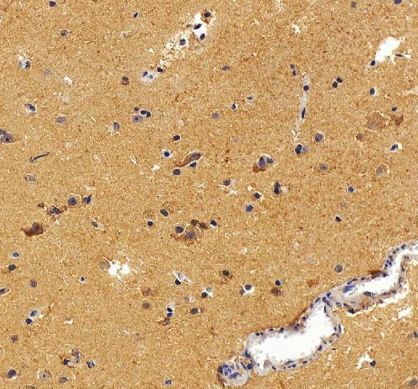
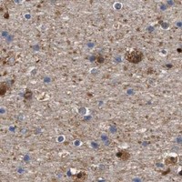


Comprehensive Human Protein Atlas IHC scoring per tissue (reliability: Enhanced — High consistency between antibody staining and RNA expression data.). Rows are taken directly from the HPA tissue chart — click any row's HPA link to view the source.
| Tissue | Cell type | Level | Evidence | Source |
|---|---|---|---|---|
| Caudate | Glial cells | Medium | Protein (IHC) | HPA → |
| Cerebellum | Processes in white matter | Medium | Protein (IHC) | HPA → |
| Cerebral cortex | Glial cells | Medium | Protein (IHC) | HPA → |
| Hippocampus | Glial cells | Medium | Protein (IHC) | HPA → |
| Testis | Elongated or late spermatids | Medium | Protein (IHC) | HPA → |
| Tissue | Cell type | Level | Evidence | Source |
|---|---|---|---|---|
| Adipose tissue | Adipocytes | Not detected | Protein (IHC) | HPA → |
| Adrenal gland | Glandular cells | Not detected | Protein (IHC) | HPA → |
| Appendix | Endocrine cells | Not detected | Protein (IHC) | HPA → |
| Bone marrow | Hematopoietic cells | Not detected | Protein (IHC) | HPA → |
| Breast | Adipocytes | Not detected | Protein (IHC) | HPA → |
Troubleshoot ALDOC staining in paraffin section IHC using the catalog antibody’s rat brain example and cell specific reference patterns (datasheet A05296-1; HPA tissue IHC).
A05296-1 has IHC images from paraffin-embedded rat and human brain sections (catalog IHC image captions). Its listed reactivity covers Human, Mouse and Rat (catalog reactivity).
A05296-1 will render with its own rat brain paraffin-section IHC figure (card image_alt). A separate caption documents human brain paraffin-section IHC; the application list includes IHC, and the reactivity list includes Human, Mouse and Rat (catalog IHC image_alts; catalog applications; catalog reactivity).
Which to pick: For tissue IHC, choose A05296-1: it is a rabbit polyclonal listed for IHC, with images from paraffin-embedded rat and human brain sections; the fixative is unreported (catalog host; catalog dilution_raw; catalog applications; catalog IHC image captions). There is no payload-supported IF/ICC pick because A05296-1 has no listed IF application or IF images (catalog applications; catalog if_image_alts). For cross-species planning, A05296-1 lists Human, Mouse and Rat reactivity, but its IHC images show rat and human brain only (catalog reactivity; catalog IHC image_alts).