ALK / ALK tyrosine kinase receptor · IHC design guide

Design Immunohistochemistry for ALK

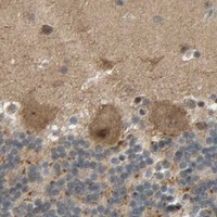
Plan ALK IHC in paraffin sections using cerebellar Purkinje cells as a positive reference (HPA tissue IHC). Compare staining with the expected neuronal cytoplasmic and nuclear pattern, while accounting for reported off-target staining (HPA tissue IHC).

Evidence assembled Oct 2026 · For research use; verify linked source records and product datasheet before use
Immunohistochemistry protocol sheet for ALK (IHC for ALK): expected localisation Cytoplasmic and nuclear staining in neurons (HPA tissue IHC), antibody M00301-5, validated IHC image, and IHC protocol steps
Printable ALK IHC protocol sheet — expected localisation Cytoplasmic and nuclear staining in neurons (HPA tissue IHC), antibody M00301-5, controls and protocol steps. Open the full ALK IHC guide →

ALK Immunohistochemistry Experimental Design Guide

Expected localisation, validated protocols, controls and antibodies — the at-a-glance facts below, then the full design guide.

Must know before staining
Expected localisation Cytoplasmic and nuclear staining in neurons (HPA tissue IHC)
Staining pattern Neuronal cytoplasmic and nuclear staining, most abundant in brain (HPA tissue IHC)
Antigen retrieval Citrate pH 6.0 HIER, 95–98 °C, 20 min (rule: cytoplasmic / membrane antigen)
Positive control ⓘ Cerebellum+4 more · see all
Negative control ⓘ Adipose tissue+4 more · see all
Important caveats
Reasons your staining may differ from the expected pattern.
Fixation Keep fixation consistent across sections. (standard IHC practice; not target-specific)
Caveat Presumed off-target staining was observed (HPA tissue IHC)
Regulation Transient in specific nervous-system regions (UniProt)
Isoform / epitope No isoforms listed; epitope side of membrane matters (UniProt)
Section 1

Recommended ALK IHC & IF Protocols

The catalog antibody protocol is followed by published ALK IHC methods from renal cell carcinoma, lung adenocarcinoma, and colorectal adenocarcinoma studies (PMC3830992; PMC5890087; PMC9440614).

Recommended immunohistochemistry (IHC-P) protocol parameters
SampleTissue sections; selected-image fixative not specified (standard IHC workflow)
FixationImage fixative and duration unreported (datasheet M00301-5); verify before use.
Sectioning4–5 µm sections on charged slides (standard)
DeparaffinisationXylene, graded ethanol series to water (standard)
Antigen retrievalHeat-induced epitope retrieval in citrate buffer, pH 6.0, 20 min at 95–98 °C (standard rule: cytoplasmic / membrane antigen)
Peroxidase block3% H2O2, 10 min, room temperature (standard)
Blocking10% normal serum of the secondary host, 30 min, room temperature (standard)
Primary antibodyRabbit anti-ALK, 1:200-1:1000 (datasheet M00301-5)
Primary incubationOvernight at 4 °C (standard)
DetectionHRP-polymer secondary, DAB chromogen 5–10 min (standard)
CounterstainHematoxylin, blue, dehydrate and mount (standard)
Expected resultALK-positive staining in purkinje cells of cerebellum (HPA tissue IHC: High). HPA tissue profile: Cytoplasmic and nuclear expression in several different tissue types, most abundant in neurons. No signal in the no-primary control.
💡Decision noteStart with citrate pH 6.0 HIER at 95–98 °C for 20 min (page retrieval rule); use T/E or ER2 retrieval when reproducing those studies (PMC5890087; PMC9440614).
Section 2

What Is the Expected ALK Staining Pattern?

ALK is a cell membrane receptor with an extracellular region, one transmembrane segment and a cytoplasmic kinase region (UniProt Q9UM73 topology). In tissue IHC, HPA reports cytoplasmic and nuclear staining most abundant in neurons; its IHC reliability is Approved, with low agreement between staining and RNA data and presumed off-target staining disregarded (HPA: tissue IHC). Interpret compartment and cell type together.

What am I looking at on my slide?
Strong staining in cerebellar Purkinje cells or neuronal cells of cerebral cortex and hippocampus (HPA: High in each).This fits the reported cell distribution (HPA: tissue IHC). Assess whether staining is cell-associated and reproducible; the tissue IHC profile includes cytoplasmic and nuclear staining, while receptor topology predicts a membrane-associated component (HPA: tissue IHC; UniProt Q9UM73 topology).
A nuclear-only pattern dominates without a convincing neuronal or membrane-associated pattern.Investigate possible artefact before calling it specific ALK: HPA reports some nuclear staining, so nuclear signal alone is not proof of error (HPA: tissue IHC). Compare morphology and controls, then reassess antibody and detection specificity (general IHC practice).
Prominent staining occurs in adipocytes or in liver cholangiocytes.These are discordant cell types: HPA reports ALK as not detected in adipose adipocytes and liver cholangiocytes (HPA: tissue IHC). Consider cross-reactivity or endogenous detection activity; inspect the negative reagent and detection controls (general IHC practice).
Diffuse chromogen covers tissue, including areas without defined cell borders.A widespread haze is difficult to score as cell-specific staining (general IHC practice). Check background in a negative reagent control and review blocking, washes and chromogen development before interpreting ALK distribution (general IHC practice).
No staining appears in Purkinje cells or cortical neurons on an otherwise evaluable section.These are HPA High cell populations (HPA: cerebellum and cerebral cortex). Check section integrity, retrieval, antibody incubation and detection controls (general IHC practice); absence alone cannot distinguish technical failure from specimen or antibody differences.
💡Expected ALK appearanceCall a positive result when identifiable Purkinje or other reported neuronal cells show strong, cell-associated staining (HPA: High in cerebellum, cortex and hippocampus); weigh membrane-associated signal against HPA's cytoplasmic and nuclear tissue profile (UniProt Q9UM73 topology; HPA: tissue IHC), and treat diffuse or unexpected-cell staining as suspect (general IHC practice).
How each factor affects the staining
Tissue and cell choiceUse cerebellar Purkinje cells or cortical or hippocampal neurons to assess an expected High pattern (HPA: tissue IHC). HPA reports adipose adipocytes and liver cholangiocytes as not detected, useful for checking unexpected staining (HPA: tissue IHC).
Epitope locationALK spans extracellular residues 19–1038, a transmembrane region at 1039–1059 and cytoplasmic residues 1060–1620 (UniProt Q9UM73 topology). Interpretation of a particular antibody's pattern depends on its mapped epitope; none is supplied here.
Evidence and antibody validationHPA010694 is IHC Approved and ICC Supported, but tissue staining has low agreement with RNA data and presumed off-target binding was disregarded (HPA: antibody validation; HPA: tissue IHC). Treat isolated unexpected staining cautiously.
IF/ICC Q: Where should ALK localise?A: HPA reports supported plasma membrane localisation in ICC-IF images from Rh30 and SH-SY5Y (HPA: subcellular). That observation helps assess localisation; it does not establish the appearance of chromogenic paraffin-section IHC.
Processing and modificationUniProt lists a signal peptide at residues 1–18 and 16 extracellular glycosylation sites (UniProt Q9UM73). Whether either affects this antibody's IHC signal is unknown without its epitope and assay evidence; do not infer a retrieval or fixation effect.
Why is my staining missing, weak or wrong?
SituationLikely causeNext action
High neuronal signal is absent in a positive-control section (HPA: cerebellar Purkinje cells, High).The assay may have failed at retrieval, primary incubation or detection; the absent stain alone does not identify which step (general IHC practice).Confirm tissue morphology and control staining, then review retrieval conditions, primary incubation and detection reagents in order (general IHC practice).
Adipocytes stain conspicuously (HPA: adipose adipocytes, Not detected).Cross-reactivity or background from the detection system is plausible; HPA also flags presumed off-target binding in its tissue assessment (HPA: tissue IHC).Compare an appropriate negative reagent control and a reported High neuronal control; evaluate cell borders and repeat with an independently validated reagent if needed (general IHC practice).
Brown deposit appears throughout the section, including tissue with no clear cell-specific pattern.Incomplete blocking, insufficient washing or excessive development can produce nonspecific chromogenic background (general IHC practice).Check the negative reagent control, review blocking and washes, and shorten development if controls support that adjustment (general IHC practice).
Staining is confined to nuclei.HPA observes nuclear as well as cytoplasmic tissue staining, so localisation alone cannot establish specificity (HPA: tissue IHC); a nuclear-only pattern still needs scrutiny.Check whether reported neuronal populations stain, compare controls and review the antibody's validation before assigning the nuclear signal to ALK (HPA: tissue IHC; general IHC practice).
A tissue section stains, but the expected neuronal cells do not.The signal may come from another cell population or background; HPA's tissue profile is most abundant in neurons and has low RNA agreement (HPA: tissue IHC).Score identified cell types separately, inspect morphology and negative controls, and avoid calling the section positive solely from diffuse or misplaced staining (general IHC practice).
An IF/ICC image shows mainly internal signal with little plasma membrane staining.HPA's supported ICC-IF localisation is plasma membrane (HPA: subcellular), although tissue IHC reports cytoplasmic and nuclear signal (HPA: tissue IHC).Review the IF/ICC guide's controls and image interpretation separately; do not transfer a paraffin IHC staining call directly to IF/ICC (general IHC practice).

Sample controls for ALK IHC & IF

🧪Run cerebellum first and look for ALK staining in Purkinje cells (HPA: High in Purkinje cells). Use adipose tissue as a negative comparator (HPA: Not detected in adipocytes); on the cerebellar slide, compare Purkinje cells with neighboring cells, but do not presume those cells are ALK-negative without validation (HPA: Purkinje-cell result only).
Positive control tissue: Cerebellum (Purkinje cells, HPA High)
Negative control tissue: Adipose tissue (HPA Not detected)
ICC-IF cell lines (HPA subcellular resource): HPA ICC-IF images show ALK in Rh30, SH-SY5Y, with annotated localisation: Plasma membrane (supported) (HPA subcellular).
Technical controls: Include a no-primary (secondary-only) control, an isotype control matched to the catalog antibody’s host species and class, and ALK-knockout tissue or cells as a biological negative (standard IHC practice). Block endogenous peroxidase for chromogenic detection; if examining cerebellum by IF, check unstained tissue for neuronal autofluorescence (standard IHC/IF practice).
⚠️Feasibility: No target-specific fixation window or fixation effect is reported, and the selected M00301-5 tissue-IHC caption does not state a fixative (selected-SKU caption). Optimize antigen retrieval empirically for paraffin sections; ALK-specific retrieval dependence and whether frozen sections or IF are easier are unreported (supplied evidence; standard IHC practice). Cerebellar neuronal pigment can complicate chromogenic interpretation, and lipofuscin can produce IF autofluorescence (standard IHC/IF practice).

HPA tissue IHC evidence for ALK

Comprehensive Human Protein Atlas IHC scoring per tissue (reliability: Approved — Low consistency between antibody staining and RNA expression data. Presumed off target binding observed and disregarded.). Rows are taken directly from the HPA tissue chart — click any row's HPA link to view the source.

Positive expression · recommended positive controls

TissueCell typeLevelEvidenceSource
Cerebellum Purkinje cells High Protein (IHC) HPA →
Cerebral cortex Neuronal cells High Protein (IHC) HPA →
Hippocampus Neuronal cells High Protein (IHC) HPA →
Caudate Neuronal cells Medium Protein (IHC) HPA →
Skin Keratinocytes Medium Protein (IHC) HPA →

Undetected expression · recommended negative controls

TissueCell typeLevelEvidenceSource
Adipose tissue Adipocytes Not detected Protein (IHC) HPA →
Appendix Glandular cells Not detected Protein (IHC) HPA →
Breast Adipocytes Not detected Protein (IHC) HPA →
Duodenum Glandular cells Not detected Protein (IHC) HPA →
Esophagus Squamous epithelial cells Not detected Protein (IHC) HPA →
Section 3

Advanced ALK IHC Tips

Troubleshoot ALK staining in paraffin sections by checking retrieval, controls, cellular location and scoring before interpreting chromogenic signal.

How should I retrieve ALK in paraffin sections when staining is weak?
Use citrate buffer at pH 6.0 for heat-induced retrieval at 95–98 °C for 20 min (page retrieval rule: cytoplasmic or membrane antigen). Let sections cool in the retrieval buffer, then compare a positive control and a negative control processed in the same run (standard IHC practice). If staining remains weak, test a longer retrieval interval on adjacent sections while watching for tissue damage and increased background (standard IHC practice). Record the buffer, temperature and duration with each result, since retrieval changes can alter both signal and morphology (standard IHC practice). Do not treat a retrieval-dependent nuclear signal alone as proof of ALK localisation (UniProt Q9UM73: cell membrane; HPA: low staining–RNA consistency).
Could fixation explain weak or uneven ALK staining?
Target-specific fixation sensitivity is unknown from the supplied evidence; the selected antibody’s tissue caption does not state a fixative (caption: M00301-5). For a paraffin-section investigation, document the fixative, fixation duration and tissue thickness for each specimen, then compare sections processed together (standard IHC practice). Uneven penetration or prolonged fixation can affect antigen accessibility in IHC generally, so assess morphology and retrieval performance before changing antibody conditions (standard IHC practice). Use a consistently processed positive control to distinguish a run problem from a specimen problem (standard IHC practice). Do not infer ALK-specific fixation behaviour from its membrane topology or the reported tissue staining pattern (UniProt Q9UM73 topology; HPA tissue IHC).
Where should ALK staining appear, and how should I assess nuclear signal?
Full-length ALK is a cell-membrane receptor with an extracellular region at residues 19–1038, a transmembrane segment at 1039–1059 and a cytoplasmic region at 1060–1620 (UniProt Q9UM73 topology). Supported IF localisation is at the plasma membrane, while HPA tissue IHC reports cytoplasmic and nuclear staining with low consistency against RNA data (HPA subcellular; HPA tissue IHC). Therefore, map chromogenic signal by cell type and compartment rather than calling any brown deposit ALK-positive (standard IHC practice). Check whether membrane or associated cytoplasmic staining repeats across sections and controls (UniProt Q9UM73; standard IHC practice). Treat isolated nuclear staining cautiously and inspect the negative control before interpreting it (HPA: presumed off-target binding; standard IHC practice).
How does epitope location affect ALK IHC interpretation?
Check the catalog antibody’s stated immunogen or mapped epitope before interpreting its stain; no epitope location is supplied here (catalog evidence supplied). ALK has an extracellular region at residues 19–1038 and a cytoplasmic region at 1060–1620, separated by a single transmembrane segment (UniProt Q9UM73 topology). Its extracellular region carries annotated glycosylation sites, while several annotated phosphotyrosines lie in the cytoplasmic region (UniProt Q9UM73 modifications). Those features make epitope identity relevant when comparing antibodies, but they do not establish this antibody’s binding or retrieval sensitivity (UniProt Q9UM73; catalog evidence supplied). The record lists 0 isoforms; do not assign staining differences to an isoform without separate evidence (UniProt Q9UM73 isoform record).
How should I adapt the ALK question for multiplex IF?
Treat IF/ICC as a separate validation task because this page supplies an IHC tissue caption, not an IF protocol for the catalog antibody (caption: M00301-5; application scope). Pair ALK with a neuronal lineage marker when examining the reported neuronal distribution, and verify that each channel retains its expected pattern in single-stain controls (HPA: strongest tissue staining in neurons; standard IF practice). Choose fluorophores after checking tissue autofluorescence and spectral overlap, using an unstained section and single-color controls (standard IF practice). If the antibody binds a cytoplasmic epitope, assess gentle permeabilisation; for an extracellular epitope, first assess staining without it (UniProt Q9UM73 topology; standard IF practice). The catalog epitope is unspecified, so confirm its location before choosing either condition (catalog evidence supplied).
What should I check when ALK chromogenic staining is diffuse?
Examine a no-primary control, reagent blanks and a known positive section in the same chromogenic run to locate background introduced by detection reagents (standard IHC practice). Adequate protein blocking, washing and an appropriate endogenous peroxidase block help limit nonspecific DAB deposition in paraffin sections (standard IHC practice). Compare the distribution with tissue morphology: staining concentrated at cut edges, folds or damaged areas warrants caution (standard IHC practice). HPA reports presumed off-target binding and low consistency between ALK staining and RNA expression, so diffuse cytoplasmic or nuclear color alone is weak evidence (HPA tissue IHC). Reassess antibody concentration against its documented instructions rather than inventing a dilution for this guide (standard IHC practice; catalog evidence supplied).
How should I score ALK staining across paraffin sections? ⚠ ANSWER MARKED FOR VERIFICATION
Define the eligible cell population and compartment before scoring, then keep those definitions fixed across specimens (standard IHC practice; UniProt Q9UM73: cell membrane). Report either the percentage of positive eligible cells or an H-score that combines percentage and intensity; record membrane and cytoplasmic signals separately if both are present (standard IHC practice; HPA tissue IHC: cytoplasmic staining reported). For spatial analyses, positive-cell density per mm² can be normalised to the area of viable tissue or the eligible compartment (standard IHC practice). Exclude folds, edges and necrotic regions by a prespecified rule, and apply the same threshold to matched controls (standard IHC practice). Flag nuclear-only scores separately because the tissue IHC profile has reported off-target concerns (HPA tissue IHC).
When does ALK staining support a true positive call?
A convincing call requires reproducible staining in intact, eligible cells that exceeds the negative control and fits the expected compartment (standard IHC practice; UniProt Q9UM73: cell membrane). ALK is reported in brain and CNS, while normal lymphoid cells lack reported expression; interpret cell identity and specimen context before assigning a signal to ALK (UniProt Q9UM73 tissue specificity). The selected antibody has a stained human non-hodgkin lymphoma image, but its caption does not report fixation or establish specificity for every pattern (caption: M00301-5). Reject color confined to edges, necrosis or endogenous enzyme activity when controls implicate artefact (standard IHC practice). Treat isolated nuclear staining cautiously given HPA’s presumed off-target binding and low staining–RNA consistency (HPA tissue IHC).
Boster reagents

Best ALK / ALK tyrosine kinase receptor IHC Antibodies

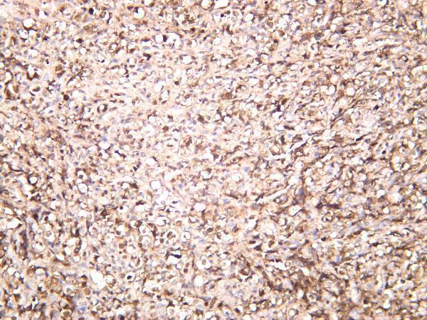
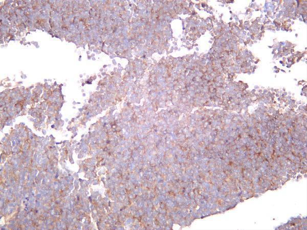
Anti-ALK antibodies have IHC images of human lymphoma and paraffin-embedded kidney; both list IF, and one lists mouse and rat reactivity (catalog images, applications and reactivity: M00301-5, M00301-3).

Real IHC data Human non-hodgkin lymphoma was stained with anti-ALK rabbit antibody
Anti-ALK Rabbit Monoclonal Antibody
Cat # M00301-5
Real IHC data Immunohistochemical analysis of paraffin-embedded human kidney, using ALK Antibody.
Anti-ALK Rabbit Monoclonal Antibody
Cat # M00301-3

M00301-5 lists human IHC and IF, with an IHC image caption describing human non-Hodgkin lymphoma (catalog applications, reactivity and IHC image: M00301-5). M00301-3 lists human, mouse and rat IHC, ICC and IF, with an IHC image caption describing paraffin-embedded human kidney (catalog applications, reactivity and IHC image: M00301-3).

Which to pick: For tissue IHC, choose M00301-5 when the human lymphoma image is most relevant (catalog IHC image: M00301-5) or M00301-3 for a paraffin-section example (catalog IHC image: M00301-3); the fixative is unreported in both captions (catalog IHC images: M00301-5, M00301-3). For IF/ICC, M00301-3 lists both applications and clone 20A85, while M00301-5 lists IF only (catalog applications and clone: M00301-3; catalog applications: M00301-5). For mouse or rat samples, select M00301-3 based on its listed reactivity; M00301-5 lists human reactivity only (catalog reactivity: M00301-3, M00301-5).

Each figure is that product's own IHC / IF validation image from its datasheet.

References

  1. UniProt Consortium. UniProt entry Q9UM73 (ALK_HUMAN, ALK tyrosine kinase receptor).
  2. Human Protein Atlas. ALK tissue IHC expression (reliability: Approved).
  3. Human Protein Atlas. ALK subcellular location (ICC-IF): Localized to the plasma membrane..
  4. Human Protein Atlas. ALK antibody validation summary (1 antibodies).
  5. ALK-Positive Renal Cell Carcinoma in a Large Series of Consecutively Resected Korean Renal Cell Carcinoma Patients. Korean journal of pathology 2013 — PMC3830992.
  6. Anaplastic lymphoma kinase (ALK)-expressing Lung Adenocarcinoma with Combined Neuroendocrine Component or Neuroendocrine Transformation: Implications for Neuroendocrine Transformation and Response to ALK-tyrosine Kinase Inhibitors. Journal of Korean medical science 2018 — PMC5890087.
  7. Inflammatory Myofibroblastic Tumor: An Updated Review. Cancers 2025 — PMC12026078.
  8. Colorectal Adenocarcinomas Harboring ALK Fusion Genes: A Clinicopathologic and Molecular Genetic Study of 12 Cases and Review of the Literature. The American journal of surgical pathology 2020 — PMC9440614.
  9. PubMed PMID:9174053 — UniProt-cited evidence.
  10. PubMed PMID:9053841 — UniProt-cited evidence.
  11. PubMed PMID:15815621 — UniProt-cited evidence.